Volume 1 6, Number 1 , June, 2013 doi:http://dx.doi.org/10.5397/CiSE.2013.16.1.40
Tumors of the scapula have low incidences.
Furthermore, primary malignant tumors are more common than benign tumors in the scapu- la.1,2)Benign tumors which are seen as osteolytic lesions in radiographs include solitary bone cysts, intraosseous ganglia, fibrous dysplasia and nonossifying fibromas. However, these oste-
olytic lesions are rare in the scapula, especially in the glenoid. We experienced three cases of symptomatic intraosseous osteolytic lesions of the glenoid and performed open curettage with or without bone grafting. Two cases were con- firmed as intraosseous ganglia and one case was fibrous dysplasia. We report these cases with a
※통신저자: 문 성 훈
강원도 춘천시 효자3동 17-1 강원대학교병원 정형외과학교실
Tel: 033) 258-9224, Fax: 033) 258-2149, E-mail: [email protected] 접수일: 2012년 11월 14일, 1차 심사완료일: 2013년 4월 17일, 게재 확정일: 2013일 6월 12일
증상이 있는 관절와의 양성 골내 골용해성 병변:
3예에 대한 증례보고
가천대학교 길병원 정형외과학교실, 강원대학교 정형외과학교실
김영규・조승현・문성훈*
Symptomatic Benign Intraosseous Osteolytic Lesions of the Glenoid:
Report of 3 cases
Young Kyu Kim, M.D., Seung Hyun Cho, M.D., Sung Hoon Moon, M.D.*
Department of Orthopaedic Surgery, Gacheon University, Gil Hospital, Incheon, South Korea Department of Orthopaedic Surgery, Kangwon National University Hospital, Chuncheon, South Korea*
Benign intraosseous osteolytic lesions of the glenoid are very rare. The present study reports on three cases of symptomatic intraosseous osteolytic lesions of the glenoid in which surgical interventions were made. Of the three, two cases presented with intraosseous ganglion and one case with fibrous dysplasia. In all the cases, the lesion was located at the posteroinferior portion of the glenoid, and it seems to be related to posterior shoulder pain. If intraosseous osteolytic lesions have symptoms or the risk for chondral defects or cortical breakage, surgi- cal intervention is needed and bone curettage with or without bone grafting will be a useful treatment option.
Key Words: Shoulder, Glenoid, Intraosseous ganglion, Fibrous dysplasia, Curettage, Bone graft
brief review of the literature.
Case Report
1. Case 1
A 51-year-old woman who had left shoulder pain for several years visited our clinic. She had no previous history of traumas, and her domi- nant side was right. The pain was dull and located at the posterior aspect of the shoulder.
The visual analog scale (VAS) score was 6 and the pain was aggravated during arm abduction with external rotation or extension.
On physical examination, she showed no atro- phy on the rotator cuff and periscapular mus- cles. There were no significant tender points.
The range of motion was 170°of active forward flexion, 70°of external rotation at the side, T8 level of internal rotation at the back, and 10 cm of cross-body adduction. This range of motion was not significantly different compared to the opposite side. She was negative for instability and rotator cuff-related tests except for pain on abduction with external rotation and extension.
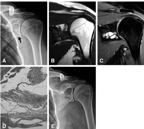
On simple radiographs, a radiolucent and well- defined multilobulated circular osteolytic lesion with mild sclerosis of the wall was found in the posteroinferior portion of the glenoid neck (Fig.
1A). It was seen as a multiseptated, lobulated lesion with high signal intensity on T2-weighted magnetic resonance (MR) images, and there was no cartilage or cortical breakage. No other lesions were found (Fig. 1B, C).
Under the diagnosis of an intraosseous gan- glion of the glenoid, open curettage with cyst removal was performed through the King’s sim- plified posterior approach. In the prone position, arm was abducted 90°and 10 cm vertical inci- sion was made from the posterior aspect of acromion. Retracting the posterior deltoid supe- riorly, the interval between the infraspinatus and teres minor was developed. The location of
lesion was based on preoperative MRI and radi- ographs, and targeted through fluoroscope.
After making a 0.5-cm square cortical window on the lesion, yellowish jelly-like material was evacuated from the lesion and curettage was performed. The biopsy result of the cystic mate- rial taken from the lesion was consistent with typical substance contained in ganglion cysts (Fig. 1D). After surgery, pain disappeared. A full range of motion was regained at 11 weeks after surgery. No findings suggestive of recurrence were seen in follow-up radiographs taken 1 year after surgery (Fig. 1E).
2. Case 2
A 51-year-old woman suffered from dull pain on the right posterior shoulder which started 1 year before this presentation. She had no previ- ous history of traumas but had a history of an operation for calcific tendinitis of the rotator cuff several years ago. Her right arm was domi- nant, and VAS score was 7. The range of motion was normal and tests for the rotator cuff and instability were negative. On simple radiographs, a radiolucent and multilobulated osteolytic lesion with a sclerotic rim was seen in the posteroinfe- rior portion of the glenoid (Fig. 2A). MR images confirmed that this lesion had no cartilage or cortical breakage. The lesion was multilobulated with high signal intensity on T2-weighted MR images and with low signal intensity on T1- weighted MR images (Fig. 2B, C).
Open curettage and allogenic bone grafting was performed for the treatment of the intraosseous lesion. During operation and biopsy, we con- firmed material that was typically seen in gan- glion cysts.
At the 1-year follow-up, she was satisfied and showed a full range of motion without pain. There was no recurrence or remaining intraosseous gan- glion on follow-up simple radiographs (Fig. 2D).
3. Case 3
A 40-year-old woman visited our clinic with a mass lesion in the glenoid of the left shoulder, which had been discovered at another clinic. She had a vague pain on the left posterior shoulder which was nondominant, and her VAS score was 4. There was no previous history of traumas or dislocation. She showed a full range of motion and negative for tests for the rotator cuff and instability.
On simple radiographs, a radiolucent unilobu- lated osteolytic lesion with a distinct sclerotic rim was seen in the inferior portion of the gle- noid neck and this lesion had heterogenous
intralesional radiopacity (Fig. 3A). A unilobulat- ed, well-circumscribed intraosseous lesion was seen in the inferior border of the glenoid neck with relatively high signal intensity on T2- weighted MR images, and, it showed dot-like dark signal intensity which was suggestive of calcification as seen in the simple radiographs (Fig. 3B, C).
Open curettage and allogenic bone grafting was performed, and as fibrous dysplasia was confirmed by pathological examination (Fig. 3D).
At the 11-week follow-up, she gained a full range of motion with pain free, and there was no evidence of recurrence in the simple radiographs taken at 1-year follow-up (Fig. 3E).
Fig. 1.(A) Radiograph of the left shoulder shows a well-defined and multilobular radiolucent osteolytic lesion at the inferior portion of the glenoid (arrow). (B, C) MR imaging shows a multisepted-lobulating lesion with low signal intensity on T1- weighted images and high signal intensity on T2-weighted images. Cortical expansion or breakage is not seen. (D) Photograph shows hypocellular fibrous tissue with myxoid changes in the wall of the cyst. There is no epithelial or synovial lining. (E) Fol- low-up radiograph taken 1 year after operation shows no findings of suggestive of recurrence of the osteolytic lesion.
A B C
D E
Discussion
Intraosseous ganglion is a benign and unilobu- lated or multilobulated lesion with extensive mucoid changes.3)It is a subarticular lytic lesion and most commonly affects bones around the knee.4) Intraosseous ganglion is not common compared to soft tissue ganglion. Furthermore, intraosseous ganglion in the glenoid is extremely rare and only a few cases have been reported.3,5) Clinical symptoms range from the absence of symptom to painful limitations of motion.
Radiographs of intraosseous ganglion show a unilobulated or multilobulated lesion located in subchondral bone adjacent to a joint and a well- defined osteolytic lesion with a surrounding area of sclerosis.3-5) This lesion can be seen as low
signal intensity equal to muscle on T1-weighted MR images and high signal intensity on T2- weighted MR images.5)
Many cases of intraosseous ganglion show no symptoms, progression or joint penetration.
However, when the mechanical stress of shoulder joint increase compression pressure, it can cause chondral defects or cortical breakage.6,7) In our cases, all of our patients with intraosseous gan- glion had pain in the affected shoulder.
Fibrous dysplasia is commonly located at the proximal femur and extremely rarely located at the glenoid. Cleeman et al.1) reported only 2 cases of fibrous dysplasia at the scapula among the 194 cases of tumors of the shoulder. One case was located at the inferior border of the scapula, and the other was located at the cora-
Fig. 2.(A) Radiograph shows a radiolucent multilobulated osteolytic lesion at the inferior portion of the glenoid (arrow). (B, C) In MR images, a multilobulated cystic lesion is seen with low signal intensity on T1-weighted images and high signal intensity on T2-weighted images. (D) In the follow-up radiograph taken 1year after operation, there are no significant recurrences or remaining osteolytic lesions.
A B
C D
coid process. These lesions show a characteristic ground-glass appearance and a lytic lesion in simple radiographs. The density of the lesion varies depending on the degree of mineralization of bony spicules in the lesion.8)In our third case, the lesion did not show a ground-glass appear- ance. As in intraosseous ganglion, symptoms of fibrous dysplasia are diverse. Fibrous dysplasia can weaken the structural integrity of the area, which can occasionally cause pain and even bony destruction.9,10)Our case of fibrous dysplasia had pain in the posterior shoulder.
All of our cases had intraosseous lesions in the posteroinferior portion of the glenoid. Therefore,
it is assumed that such a location can cause pain at the posterior aspect of the shoulder. This may explain the posterior shoulder pain during arm abduction or extension in the first case. All of our 3 cases were located at the posteroinferior portion of the glenoid. However, 2 cases of intraosseous ganglion were located adjacent to the articular cartilage involving subchondral bone, while 1 case of fibrous dysplasia did not involve subchondral bone. This difference in location may be useful for distinguish between intraosseous ganglion and fibrous dysplasia when the geometry of the lesions is difficult to distinguish. However, many cases will be needed to generalize this suggestion.
Fig. 3.(A) Radiograph shows a radiolucent unilobular osteolytic lesion with a distinct sclerotic rim in the inferior portion of the glenoid. This lesion shows dot-like heterogenous opacity suggestive of calcification (arrow). (B, C) MR imaging shows a unilobulated well-circumscribed cystic lesion at the inferior border of the glenoid neck with relatively low signal intensity on T1-weighted images and high signal intensity on T2-weighted images. The lesion shows dot-like low signal intensity sugges- tive of calcification. (D) Histological examination shows a large amount of marrow and curved or irregular-shaped trabeculae of woven bone which are embedded in a moderately cellular fibrous tissue. (E) Follow-up radiograph taken 1year after opera- tion shows complete regression of the osteolytic lesion.
A B C
D E
The symptoms of these 2 disease entities may dif- ferent, but if the symptoms or the risk for chon- dral defects or cortical breakage are present, sur- gical treatment can be considered. Several authors have suggested that bone curettage and bone grafting is necessary for the treatment of benign lytic lesions as in our cases.5,6,9-11) This treatment can alleviate motion-restricting pain and allow for a full athletic recovery.11) In our series, bone curettage and bone grafting was performed in 2 cases and bone curettage alone was performed in 1 case. All patients gained pain-free motion with no recurrence symptomatically and radiologically after surgery. This treatment can prevent pro- gression to lesions that cause chondral defects or cortical breakage that carry the risk of pathologic fractures.
Conclusion
Intraosseous ganglion and fibrous dysplasia of the glenoid are uncommon benign intraosseous osteolytic lesions. The symptoms of these 2 dis- ease entities may differ. When they are painful or located at the posteroinferior portion of the glenoid with size predisposing the lesions to pathologic fractures, appropriate surgical treat- ment should be performed.
REFERENCES
01) Cleeman E, Auerbach JD, Springfield DS. Tumors
of the shoulder girdle: a review of 194 cases. J Shouler Elbow Surg. 2005;14:460-5.
02) Samilson RL, Morris JM, Thompson RW. Tumors of the scapula. A review of the literature and an analysis of 31 cases. Clin Orthop Relat Res. 1968;
58:105-15.
03) Schajowicz F, Clavel Sainz M, Slullitel JA. Juxta- articular bone cysts (intra-osseous ganglia): a clini- copathological study of eighty-eight cases. J Bone Joint Surg Br. 1979;61:107-16.
04) Williams HJ, Davis AM, Allen G, Evans N, Mang- ham DC. Imaging features of intraosseous ganglia:
a report of 45 cases. Eur Radiol. 2004;14:1761-9.
05) Urayama M, Itoi E, Watanabe H, Sato K, Kamei J. Intraosseous ganglion of the glenoid. Orthopedics.
1999;22:705-6.
06) Murata K, Nakagawa Y, Suzuki T, Kobayashi M, Kotani S, Nakamura T. Intraosseous ganglion about to cause a fracture of the glenoid: a case report. Knee Surg Sports Traumatol Arthrosc. 2007;
15:1261-3.
07) Tudisco C, Bisicchia S. Intraosseous ganglion with impending fracture of the glenoid. Orthopedics.
2011;34:956-9.
08) Borys D, Canter R, James MA. Monostotic fibrous dysplasia of the distal phalanx: case report. J Hand Surg Am. 2010;35:1294-6.
09) Shih HN, Chen YJ, Huang TJ, Hsu KY, Hsu RW.
Treatment of fibrous dysplasia involving the proxi- mal femur. Orthopedics. 1998;21:1263-6.
10) Stephenson RB, London MD, Hankin FM, Kaufer H. Fibrous dysplasia. An analysis of options for treatment. J Bone Joint Surg Am. 1987;69:400-9.
11) Moretti VM, Slotcavage RL, Crawford EA, Lack- man RD, Ogilvie CM. Curettage and graft allevi- ates athletic-limiting pain in benign lytic bone lesions. Clin Orthop Relat Res. 2011;469:283-8.
초 록
관절와 내의 양성 골내 골용해성 병변은 매우 드물다. 저자들은 수술적 치료가 시행되었던 3예의 증상이 있는 양성 골내 골용해성 병변에 대하여 보고하고자 한다. 이 중 2예는 골내 결절종이었으며, 1예는 섬유이 형성증으로 진단되었다. 모든 예에서 병변의 위치는 관절와의 후하방이었으며, 이는 견관절의 후방 통증과 연관이 있는 것으로 생각된다. 골내 골용해성 병변이 증상을 보이거나, 연골 결손의 위험 또는 피질골의 파 괴의 위험이 있는 경우 치료가 필요하며 골 이식을 동반한 또는 동반하지 않은 단순 골 소파술이 유용한 치 료 방법으로 생각된다.
색인 단어: 견관절, 관절와, 골내 결절종, 섬유 이형성증, 소파술, 골 이식