INTRODUCTION
Malignant fibrous histiocytoma (MFH) is a rare primary neoplasm occurring in the bone; according to Mayo Clinic files, it accounts for less than 1% of primary bone tumors (1, 2).
The neoplasm is found most frequently in the long tubular bones of the lower extremities (1). Only rare cases arising in the skull have been published (3-6), and the majority of the patients had large lytic areas of bone destruction with exten- sions to the adjacent soft tissue, underlying dura, or brain pa- renchyma. We experienced a rare case of primary MFH involv- ing only a cranial vault with no extraosseous extensions in a 43-yr-old woman. This is an unusual presentation of MFH.
CASE REPORT
A 43-yr-old woman was admitted because of a palpable mass in the right parietal region, which had enlarged in size over the preceding 15 months. A physical examination revealed tender swelling in the right parietal region. She had a histo- ry of trauma in the same site 15 yr ago and no history of irradiation. There was no evidence of soft tissue mass or con- current illness. A plain skull radiograph showed a well-defined
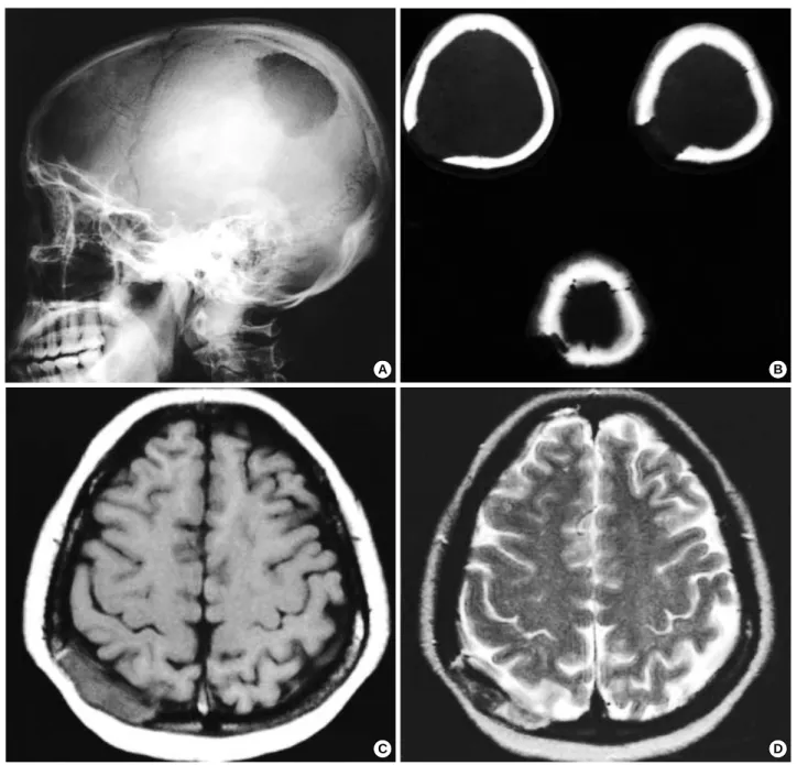
lytic defect, measuring 5 cm in diameter in the right parietal area (Fig. 1A). Computerized tomographic (CT) scanning de- monstrated an area of extensive lytic bony destruction with little discernible residual cortex and no significant periosteal reaction. The margins were well delineated, and there was no matrix formation within the lesion (Fig. 1B). T1-weighted magnetic resonance (MR) images showed a pancake-shaped, slightly low signal intensity mass with central nodularity, which exhibited heterogeneous and slightly high signal intensity on T2-weighted images and was well enhanced on contrast-en- hanced MR images. No evidence of extensions to the adjacent soft tissue and underlying dura mater was noted (Fig. 1C-F).
We suspected a benign osseous lesion, such as intraosseous meningioma or eosinophilic granuloma, because of the less aggressive radiological features, and a wide excision of the pari- etal bone was performed. On operative findings, the tumor was well distinguished from the nontumorous bone and was easily detached from the underlying dura or the adjacent muscle.
The resected specimen was a solid, disk-shaped lesion yel- lowish-brown in color and measuring 5×4×1 cm. It showed a more nodular round area, corresponding to the nodular signal pattern in the MR images. Histologically, the tumor consisted of atypical spindle cells arranged in a storiform or fascicular pat- tern (Fig. 2). The tumor cells showed high degree of pleomor-
Mee Joo, Ghi Jai Lee*, Young-Cho Koh�, O-Ki Kwon�, Yong-Koo Park�
Department of Pathology, Radiology*,
Neurosurgery�, Inje University, Seoul Paik Hospital, Seoul; Department of Pathology�, College of Medicine, Kyung Hee University, Seoul, Korea
Address for correspondence Mee Joo, M.D.
Department of Pathology, Seoul Paik Hospital, Inje University, 85, 2-ga, Jeo-dong, Jung-gu, Seoul 100-032, Korea
Tel : +82.2-2270-0155, Fax : +82.2-2270-0131 E-mail : [email protected]
609
Primary Intraosseous Malignant Fibrous Histiocytoma of the Skull : A Case Report
Malignant fibrous histiocytoma (MFH) is a rare primary neoplasm that constitutes less than 1% of the malignant tumors of bone, and involvement of the skull is very rare. We present a case of malignant fibrous histiocytoma of the skull, presenting an intraosseous lesion in a 43-yr-old woman. She had a rapidly growing, tender mass in the right parietal region. A plain radiograph showed an osteolytic lesion of the right parietal bone. Magnetic resonance imaging revealed that the lesion showed heterogeneous low signal intensity on T1-weighted images and slightly high signal intensity on T2-weighted images. No evidence of an extraosseous exten- sion to the adjacent dura and soft tissue was found, and a wide excision of the pari- etal bone was performed. Histologically, the tumor was a typical MFH displaying pleomorphic spindle cells in a storiform pattern. The results of immunohistochem- ical stainings revealed that the tumor cells were positive for vimentin, -1-antitryp- sin, and p53, and negative for smooth muscle actin, S100 protein, desmin, and MyoD1. Three months later, a mainly cystic, recurrent mass was developed at the previously operated site. Before the resection, we first performed the percutaneous aspiration cytology, revealing diagnostic multinucleated pleomorphic cells. There- after, she had to receive repetitive resections of recurrent or residual lesions, and she died of postoperative meningoencephalitis two years after the first operation.
Key Words : Histiocytic Disorders, Malignant; Skull Neoplasms; Parietal Bone
Received : 24 June 2002 Accepted : 22 August 2002
610 M. Joo, G.J. Lee, Y.-C. Koh, et al.
phism with abundant eosinophilic cytoplasm and occasional multinucleation, and abundant typical or atypical mitotic fig- ures (Fig. 3). To exclude the possibility of MFH-like osteosar- coma, we thoroughly searched for a tumor osteoid, but failed to find it. Immunohistochemical staining revealed positive reactivity for vimentin, -1-antitrypsin, and p53. There was no immunoreactivity for smooth muscle actin, S100 protein, desmin, MyoD1, and cytokeratin.
Postoperatively, the patient underwent radiotherapy of 6120
cGy. Three months later, a progressive swelling was palpable in the previously operated site. Follow-up MR images done at that time demonstrated a subcutaneous fluid collection with peripheral enhancement. A percutaneous needle aspiration was subsequently performed. The cytologic findings of the aspira- tion fluid obtained from the recurrent cystic lesion revealed some scattered pleomorphic large cells in the background of abundant inflammatory cells. These atypical cells frequently showed binucleated or multinucleated forms with abundant
Fig. 1.(A-D) Radiologic findings. (A) Plain skull lateral film shows a round lytic defect, measuring 5 cm in diameter in the right parietal bone.
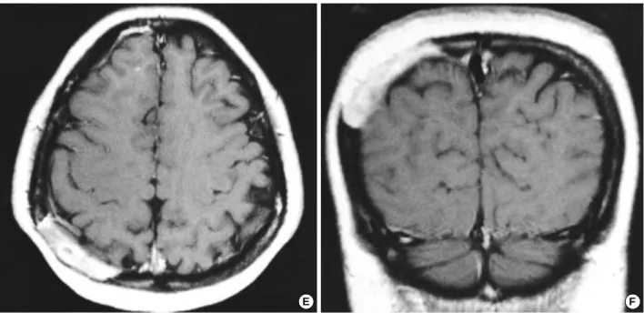
(B) Precontrast CT with bone setting shows a well-defined mass; the bony destruction is evident with little discernible residual inner and outer table and periosteal reaction is meager. (C-F) The tumor is confined to cranial vault showing slightly low signal intensity on T1-weight- ed axial MR image (C) and slightly heterogeneous signal intensity on T2-weighted axial image (D) comparing to the brain, and the tumor is enhanced well on contrast-enhanced T1-weighted axial (Fig. 1. continued next)
A
C
B
D
cytoplasm (Fig. 4). Thereafter, the patient received another operation, and the lesion was histologically confirmed to be the recurrent tumor.
DISCUSSION
Although many cases of MFH in soft tissues have been re- ported, the descriptions of their occurrences in bony areas are
rare (1, 2, 7-9), and extremely rare in the skull. The mandibu- lomaxillary area was the most common site for MFH in the skull (3-4), followed by the calvarium. We found eight cases of MFH in the calvarium (5-6): in the frontal bone and tempo- ral bone (two patients each); and in the frontotemporal bone, occipital bone, parietal bone, and clivus (one patient each). The patients’ ages ranged from 25 to 72 yrs old. There were five males and three females. Most of them had rapidly enlarging painless masses on the skull, as in the present case.
Fig. 2. Pleomorphic spindle cells are arranged in a storiform pat- tern (H&E, ×100).
Fig. 3.Tumor cells show high degree of pleomorphism with frequent multinucleation and mitotic figures (H&E, ×400).
Fig. 1.(Continued form the previous page) (E) and coronal (F) MR images. The involvement of adjacent scalp, dura, or brain parenchyme is not apparent.
E F
612 M. Joo, G.J. Lee, Y.-C. Koh, et al.
In addition to its unusual location, the radiologic findings of the present case were unique. At initial presentation, the lesion was confined to the cranial bone with no definite evi- dence of extraosseous (adjacent soft tissue, dura mater, or brain parenchyma) extensions. Typical radiologic findings of MFH include purely osteolytic, aggressive, destructive tumor growth with frequent soft tissue involvement, and periosteal reactions and expansile growth were rarely seen (10, 11). In all reported cases arising in the skull, the tumors involved all layers with inward and/or outward extensions and were sometimes pre- sented as an intracranial or a soft tissue mass (5, 6). In our case, some favorable findings to benign lesions were apparent; the tumor size was not small, but placed horizontally along the cranial vault, and it displayed good localization with no defi- nite extraosseous extensions. Considering the case retrospec- tively, extensive osteolysis with cortical destruction as well as an inhomogeneous nodular signal pattern on T2-weighted and contrast-enhanced T1-weighted MR images were suggestive of a malignant osseous lesion.
Before making a diagnosis of MFH, the exclusion of other types of pleomorphic sarcoma with dedifferentiated areas is mandatory because their histologic appearance is akin to MFH (1, 2, 12, 13). The marked pleomorphism with multinucleated giant cells and a storiform pattern observed in the present case were consistent with the characteristic histological findings of MFH. Moreover, differentiation to specific cells, such as smooth muscle cells, skeletal muscle cells, or fat cells, was not detected immunohistochemically, and there was no tumor osteoid suggestive of bone-forming tumor throughout the lesion. Naka et al. (13) reported that no overexpressions of the p53 protein were found in the MFH-like osteosarcoma, where-
as it tended to occur more frequently in MFH of the bone. In our case, a significant overexpression of the p53 protein was demonstrated.
At the first time of recurrence, the lesion was predominantly cystic with only peripheral enhancement. We thought that this lesion had to be discriminated from the retained hema- toma, and then we first performed the percutaneous aspiration cytology, which revealed diagnostic tumor cells. Multinucle- ated bizarre nuclei and abundant cytoplasm are thought to be characteristic cytologic findings for identifying the histiocytic features of this tumor. Although the final diagnosis of the re- current tumor relied on the histologic findings, the cytologic examination was helpful as an auxiliary study in our case.
All reported patients underwent gross total removal of the tumor; nevertheless, the prognosis is poor, similar to MFHs occurring in the other sites of the body (1, 2, 8, 12, 14). Our patient received wide resection of the affected skull bone with the adjacent dura and soft tissue followed by irradiation, and we expected a hopeful outcome considering the less aggressive presentation of the tumor. However, the patient had to receive repetitive resections of recurrent or residual lesions, and she died of postoperative meningoencephalitis two years after the first operation. We believe that adjuvant chemotherapy would have been necessary for this patient, as previous report stressed the usefulness of chemotherapy (14).
REFERENCES
1. Forest M, Tomeno B, Vanel D, Bullough PG. Orthopedic surgical pathology. In: Forest M, eds. Malignant Fibrous Histiocytoma. Chu- rchill Livingstone, Edinburgh 1997; 323-33.
2. Unni KK. Dahlin’s Bone Tumors: General Aspects and Data on 11,087 Cases, 5th eds. Lippincott-Raven, Philadelphia, 1996; 217-24.
3. Narvaez JA, Muntane A, Narvaez J, Martin F, Monfort JL, Pons LC. Malignant fibrous histiocytoma of the mandible. Skeletal Radiol 1996; 25: 96-9.
4. Besly W, Wiesenfeld D, Kleid S, Allan P, Poker I. Malignant fibrous histiocytoma of the maxilla: a report of two cases. Br J Oral Maxillofac Surg 1993; 31: 45-8.
5. Yoshida D, Harashima K, Node Y, Kojima T, Shimura T, Teramoto A. Malignant fibrous histiocytoma in the parietal bone. Neurol Med Chir (Tokyo) 1998; 38: 359-62.
6. Nakayama K, Nemoto Y, Inoue Y, Mochizuki T, Soares SB Jr, Ohata K, Katsuyama J, Onoyama Y, Wakasa K. Malignant fibrous histiocy- toma of the temporal bone with endocranial extension. Am J Neurora- diol 1997; 18: 331-4.
7. Dahlin DC, Unni KK, Matsuno T. Malignant (fibrous) histiocytoma of bone-fact or fancy? Cancer 1977; 39: 1508-16.
8. Capanna R, Bertoni F, Bacchini P, Bacci G, Guerra A, Campanacci M. Malignant fibrous histiocytoma of bone. The experience at the Riz- zoli Institute: report of 90 cases. Cancer 1984; 54: 177-87.
9. Huvos AG, Heilweil M, Bretsky SS. The pathology of malignant fibrous histiocytoma of bone: a study of 130 patients. Am J Surg Pathol Fig. 4.Bizarre multinucleated giant cells are scattered with many
inflammatory cells (Papanicolaou, ×400).
1985; 9: 853-71.
10. Feldman F, Lattes R. Primary malignant fibrous histiocytoma (fibrous xanthoma) of bone. Skeletal Radiol 1977; 1: 145-60.
11. Link TM, Haeussler MD, Poppek S, Woertler K, Blasius S, Lindner N, Rummeny EJ. Malignant fibrous histiocytoma of bone: conventional X-rayand MR imaging features. Skeletal Radiol 1998; 27: 552-8.
12. Nishida J, Sim FH, Wenger DE, Unni KK. Malignant fibrous histiocy- toma of bone: a clinicopathologic study of 81 patients. Cancer 1997;
79: 482-93.
13. Naka T, Fukuda T, Shinohara N, Iwamoto Y, Sugioka Y, Tsuneyoshi M. Osteosarcoma versus malignant fibrous histiocytoma of bone in patients older than 40 years. A clinicopathologic and immunohisto- chemical analysis with special reference to malignant fibrous histi- ocytoma-like osteosarcoma. Cancer 1995; 76: 972-84.
14. Bielack SS, Schroeders A, Fuchs N, Bacci G, Bauer HC, Mapeli S, Tomeno B, Winkler K. Malignant fibrous histiocytoma of bone: a retrospective EMSOS study of 125 cases. European Musculo-Skele- tal Oncology Society. Acta Orthop Scand 1999; 70: 353-60.