Copyrights © 2016 The Korean Society of Radiology
300
Case Report
pISSN 1738-2637 / eISSN 2288-2928 J Korean Soc Radiol 2016;75(4):300-303 http://dx.doi.org/10.3348/jksr.2016.75.4.300
Angiosarcomas are the most common sarcomas of the breasts but they are extremely rare, accounting for only 0.04% of pri- mary breast tumors (1). Because of its rarity, this malignant tu- mor can be easily misdiagnosed; moreover, this tumor has a high mortality rate. Additionally, radiologic findings are typically not specific; therefore, an accurate diagnosis by a radiologist is dif- ficult (2-4).
A few cases of angiosarcoma of the breast have been reported;
however, most reported cases are neither bilateral nor primary (5). We report herein a case of primary bilateral angiosarcoma of the breast in a young woman. She was treated with a right mastectomy and left wide local excision followed by six cycles of adjuvant chemotherapy. However, multiple metastatic lesions were found upon follow-up radiologic evaluation 3 years later.
CASE REPORT
A 34-year-old woman with a nonspecific medical history inci-
dentally identified a palpable mass in the upper portion of her right breast and visited a medical clinic. She did not complain of tenderness or pain. She was transported to our hospital for re- moval of angiosarcoma of the right breast, which was diagnosed using a core needle biopsy.
Upon physical examination, a large palpable mass was identi- fied in the upper inner portion of the right breast. The lesion did not show regional skin change. On mammography, both breasts were found to be extremely dense, which lowers the sensitivity of mammography. Real-time gray scale and color Doppler ul- trasonography showed a 3.9 × 1.5 × 2.6 cm irregular, circum- scribed, hypoechoic mass at the 12 o’clock position in the right breast. The mass was located in the glandular layer, and increased vascularity was not found around or within the mass (Fig. 1A, B). Significantly enlarged lymph nodes were not observed in axillary areas.
Contrast-enhanced breast magnetic resonance imaging (MRI) was performed to evaluate the extent of this mass prior to sur-
A Case of Primary Bilateral Angiosarcoma of the Breast
양측 유방에서 발생한 일차성 혈관 육종의 증례 보고Kiwook Lee, MD
1, Young Sook Kim, MD
1*, Hyungwoo Oh, MD
1, Eunju Yoon, MD
1, Mija Lee, MD
2Departments of 1Diagnostic Radiology, 2Pathology, College of Medicine, Chosun University, Gwangju, Korea
Angiosarcoma is a malignant tumor of endovascular origin that can occur in any part of the body including the breast; however, angiosarcoma of the breast is quite rare.
There are two sub-types of breast angiosarcoma. One of the subtypes, primary breast angiosarcoma, is very rare; only a few cases have been reported to date. Bilateral pri- mary breast angiosarcoma is even rarer, and several cases have been reported in post- menopausal women. We report a case of a 34-year-old woman with primary bilateral angiosarcoma. She had no other significant medical history, such as breast surgery or radiotherapy, which can lead to secondary angiosarcoma of the breast.
Index terms Breast
Breast Neoplasms Angiosarcoma
Magnetic Resonance Imaging Mammography
Received April 5, 2016 Revised May 13, 2016 Accepted July 3, 2016
*Corresponding author: Young Sook Kim, MD Department of Diagnostic Radiology, College of Medicine, Chosun University,
365 Pilmun-daero, Dong-gu, Gwangju 61453, Korea.
Tel. 82-62-220-3559 Fax. 82-62-228-9061 E-mail: [email protected]
This is an Open Access article distributed under the terms of the Creative Commons Attribution Non-Commercial License (http://creativecommons.org/licenses/by-nc/3.0) which permits unrestricted non-commercial use, distri- bution, and reproduction in any medium, provided the original work is properly cited.
301
Kiwook Lee, et al
jksronline.org J Korean Soc Radiol 2016;75(4):300-303 gery. A 4.3 × 3.6 × 4.6 cm sized heterogeneously enhancing, ir- regular mass was found in the right breast (Fig. 1C). The mass showed iso signal intensity compared to the breast parenchyma on T1-weighted images, with high signal intensity on T2-
weighted images. Some areas within the mass showed high sig- nal intensity on T1-weighted images and low signal intensity on T2-weighted images. Another 9 × 6 × 7 mm oval, well en- hancing mass was detected in the outer portion of the left breast.
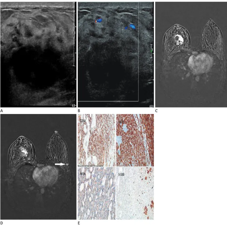
Fig. 1. A 34-year-old woman with primary bilateral angiosarcoma of the breast presenting with a palpable mass in the right breast.
A. Ultrasonography shows a circumscribed hypoechoic mass at the 12 o’clock position in the right breast.
B. On a color Doppler image, no increased vascularity is apparent around or within the mass.
C. Axial contrast-enhanced T1 fat suppression magnetic resonance imaging (MRI) shows an irregular mass in the upper central portion of the right breast (60s after gadolinium injection). Multiple unenhanced portions are apparent in the mass.
D. The lower level of the axial contrast-enhanced T1 fat suppression MRI scan shows a 9 mm enhancing oval shaped mass (arrow) in the lateral portion of the left breast.
E. Tumor cells show immunoreactivity for CD34, CD31, and actin, but not for S-100 (400 × magnification).
A
D
B C
E
302
A Case of Primary Bilateral Angiosarcoma of the Breast
jksronline.org
J Korean Soc Radiol 2016;75(4):300-303 This mass showed high signal intensity compared to the breast
parenchyma on T1- and T2-weighted images (Fig. 1D).
Because the lesion was pathologically diagnosed as an angio- sarcoma via a core needle biopsy and radiologic findings were strongly suggestive of malignancy, the attending surgeons per- formed a right mastectomy and local excision of the mass in the left breast.
Masses from both breasts were pathologically confirmed as angiosarcoma grade III after dissection. A vascular lesion was found in the central portion of the right breast mass. The resec- tion margin was free from carcinoma, and no lymph node me- tastasis was found. Immunohistochemical staining was positive for CD34, CD31, and actin, but it was negative for the S-100 protein (Fig. 1E). After surgery, a positron emission tomogra- phy (PET)-CT scan confirmed that there was no metastatic le- sion or residual malignancy.
Because of the high rate of recurrence, the patient was admin- istered six cycles of adjuvant chemotherapy, and radiologic eval- uations were performed annually. Three years following the first diagnosis, a metastatic lesion was found in the L1 vertebra via a bone scan and PET-CT. This lesion was treated with additional radiotherapy and chemotherapy; however, a follow-up exami- nation revealed an increase in the size and number of metastat- ic lesions (in both lungs, the lumbar spine, both clavicles, ribs, iliac bone, and the right pelvic peritoneum).
DISCUSSION
Angiosarcoma of the breast was first described by Schmidt in 1887. This malignant tumor has a vascular endothelial origin and it has a poor prognosis because of high rates of metastasis and local recurrence (6). The prognosis of angiosarcoma depends on its histological grade: low-grade angiosarcoma shows a better survival rate than high-grade angiosarcoma. The 5-year surviv- al rate is 76% for low-grade, 70% for intermediate, and 15% for high-grade angiosarcoma (7).
There are two types of angiosarcoma of the breast: primary and secondary. Secondary angiosarcoma is more common and it typically arises in an irradiated breast after breast conservation or lymphedema, mostly in postmenopausal patients. Primary angiosarcoma is rarer and it tends to sporadically arise in young- er women; the median age at diagnosis is the third and fourth
decades of life (1). Risk factors for primary angiosarcoma are not known.
Angiosarcomas arise at a young age and display a rapid growth pattern, and most patients identify an abnormality in their breast before a screening mammogram. Occasionally, discoloration of the skin may accompany the lesion because of vascularization (8). In the present case, the patient was a premenopausal 34-year- old woman, had no significant medical history, and presented with a palpable mass in a single breast without skin color change.
A radiologic diagnosis of primary angiosarcoma is difficult due to the young patient population and nonspecific findings.
Mammography for angiosarcoma shows typical circumscribed or obscured round, oval, lobulated margins (4, 9, 10). Addition- ally, because patients with angiosarcoma are young and typically have dense breasts, a mammogram is not appropriate for identi- fying this tumor. In our case, mammography also revealed dense breasts. Ultrasonography can provide a wide spectrum of find- ings: ultrasonography may show heterogeneous hypoechoic or hyperechoic masses with or without posterior acoustic shadow- ing. However, hypervascularity is usually present, which is in contrast to our case (4, 9, 10). Thus, owing to these challenges in radiologic diagnosis of angiosarcoma, MRI is thought to be the best modality for diagnosing angiosarcoma.
Many cases of angiosarcoma of the breast show low signal in- tensity on T1-weighted images and high signal intensity on T2- weighted images as well as early enhancement with prolongation on a dynamic study (3). In our case, the tumors were found to show iso- to high signal intensity on T1-weighted images, high signal intensity on T2-weighted images, and early enhancement with prolongation on a dynamic study. Only the enhancing pat- tern was similar to the previously reported findings.
The diagnosis of angiosarcoma of the breast is challenging for the radiologist. The young patient population, nonspecific radio- logical findings, and ordinary symptoms present challenges for the radiologist. Dynamic enhancing MRI and histologic exami- nation should be performed for a suspected angiosarcoma of the breast.
REfERENCES
1. Chen KT, Kirkegaard DD, Bocian JJ. Angiosarcoma of the breast. Cancer 1980;46:368-371
303
Kiwook Lee, et al
jksronline.org J Korean Soc Radiol 2016;75(4):300-303 2. Kaklamanos IG, Birbas K, Syrigos KN, Vlachodimitropoulos D,
Goutas N, Bonatsos G. Breast angiosarcoma that is not re- lated to radiation exposure: a comprehensive review of the literature. Surg Today 2011;41:163-168
3. Kikawa Y, Konishi Y, Nakamoto Y, Harada T, Takeo M, Ogata M, et al. Angiosarcoma of the breast - specific findings of MRI. Breast Cancer 2006;13:369-373
4. Liberman L, Dershaw DD, Kaufman RJ, Rosen PP. Angiosar- coma of the breast. Radiology 1992;183:649-654
5. Pai MR, Upadhyaya K, Naik R, Malhotra S. Bilateral angio- sarcoma breast diagnosed by fine needle aspiration cytol- ogy. Indian J Pathol Microbiol 2008;51:421-423
6. Sher T, Hennessy BT, Valero V, Broglio K, Woodward WA, Trent J, et al. Primary angiosarcomas of the breast. Cancer
2007;110:173-178
7. Rosen PP. Rosen’s breast pathology. Philadelphia: Lippin- cott Williams & Wilkins, 2001:839
8. Sener SF, Milos S, Feldman JL, Martz CH, Winchester DJ, Di- eterich M, et al. The spectrum of vascular lesions in the mammary skin, including angiosarcoma, after breast con- servation treatment for breast cancer. J Am Coll Surg 2001;
193:22-28
9. Schnarkowski P, Kessler M, Arnholdt H, Helmberger T. An- giosarcoma of the breast: mammographic, sonographic, and pathological findings. Eur J Radiol 1997;24:54-56 10. Yang WT, Hennessy BT, Dryden MJ, Valero V, Hunt KK,
Krishnamurthy S. Mammary angiosarcomas: imaging find- ings in 24 patients. Radiology 2007;242:725-734
양측 유방에서 발생한 일차성 혈관 육종의 증례 보고
이기욱
1· 김영숙
1* · 오형우
1· 윤은주
1· 이미자
2혈관 육종은 혈관내피 세포에서 기원하는 악성 종양이다. 이 종양은 유방을 포함한 우리 몸 어느 곳에서도 생길 수 있다. 유 방에서 발생한 혈관 육종은 일차성 혈관 육종과 이차성 혈관 육종으로 분류된다. 일차성 혈관 육종은 흔치 않으며 양측 유 방에서 기시하는 일차성 혈관 육종은 더 흔치 않아 폐경기 여성의 몇몇 증례만이 보고되었다. 저자들은 특이한 과거력을 가 지고 있지 않은 34세 여자 환자의 양측 유방에서 진단된 일차성 혈관 육종의 증례에 대해 보고하고자 한다.
조선대학교 의과대학 1영상의학과학교실, 2병리과학교실