Angioleiomyomas are a benign soft tissue tumor aris- ing from smooth muscle cells of blood vessels. It is also known as an angiomyoma, vascular leiomyoma or der- mal angioma. This tumor is relatively rare and typically presents as a small (maximum diameter < 2 cm), freely movable, painful mass (1, 2). Although angioleiomy- omas are most frequently reported in the lower extremi- ty and in middle-aged female patients, they can be
found throughout the body in male and female adults of all ages. We report a case of an angioleiomyoma of the left 3rd digit in a 31-year-old man.
Case Report
A 31-year-old man presented with a progressive en- larged palpable mass at the left 3rd finger. He reported that the mass was palpable for about 2 years, and pro- gressively increased in size. The man did not complain of any pain in the area. Upon physical examination, we discovered a soft, movable non-tender mass measuring approximately 3 × 2 cm and with a volar aspect of the proximal phalanx of left 3rd finger. Range of motion of the left 3rd finger was intact, though plain radiography demonstrated soft tissue swelling at the volar aspect of the left 3rd proximal phalanx without any bony abnor-
J Korean Soc Radiol 2011;64:599-602
─ 599 ─
Radiologic Findings of an Angioleiomyoma of the Finger: A Case Report1
Bo Seong Jeong, M.D., Jae Chan Shim, M.D., Ji Young Kim, M.D.
2, Yoon Kyeng Kang, M.D.
3, Jae Myeong Lee, M.D., Mi Young Nam, M.D., Ghi Jai Lee, M.D., Ho Kyun Kim, M.D.
1Department of Diagnostic Radiology, Seoul Paik Hospital, InJe University College of Medicine
2Department of Orthopedic Surgery, Seoul Paik Hospital, InJe University College of Medicine
3Department of Pathology, Seoul Paik Hospital, InJe University College of Medicine
Received December 7, 2010 ; Accepted April 29, 2011
Address reprint requests to : Jae Chan Shim, M.D., Department of Diagnostic Radiology, Inje University, Seoul Paik Hospital, 2-85 Jeo-dong, Jung-gu, Seoul 100-032, Korea.
Tel. 82-2-2270-0139 Fax. 82-2-2266-6799 E-mail: [email protected]
Angioleiomyomas are a rare benign smooth muscle tumor arising from vessel walls.
Although angioleiomyomas are most frequently reported in the lower extremities and in middle-aged female patients, they can be found throughout the body in male and fe- male adults of all ages. We report a rare case of an angioleiomyoma of the left 3rd digit in a 31-year-old man, which appeared as a small, well defined mass with multiple vas- cular structures on Doppler sonogram and MRI. The tumor was diagnosed by patholo- gy as an angioleiomyoma. Although angioleiomyomas are relatively infrequent, they should be considered in the differential diagnosis when multiple tortuous vascular structures are seen within a well demarcated mass in extremities on Doppler sono- gram and MRI.
Index words : Leiomyoma
Soft Tissue Neoplasms Extremities
Ultrasonography
Magnetic Resonance Imaging
malities. Sonography revealed a solid, oval and well de- fined mass about 2.5 cm in diameter. The mass was lo- cated just above the flexor tendon, but did not appear to be attached to the tendon. The mass showed a relatively heterogeneous, hypoechoic echo texture with posterior acoustic enhancement (Figs. 1A, B). Color Doppler eval-
uation revealed multiple vascular channels within the mass (Fig. 1C). On MRI, the mass showed homogeneous iso-signal intensity on T1 weighted image and heteroge- neous high signal intensity with a thin low signal cap- sule on a T2 weighted image, and multiple serpentine or linear low signal structures (Figs. 2A, B). The mass
Bo Seong Jeong,
et al : Radiologic Findings of an Angioleiomyoma of the Finger
─ 600 ─
A B
C
Fig. 2. The axial T1 weighted (A) and T2 weighted (B) images demonstrate well defined a mass with a thin capsule; curvilin- ear low signal structures are also noted within the mass (arrows in A and B). Gadolinium-enhanced T1 weighted image (C) demonstrate strong enhancement, especially peripherally.
A B
C
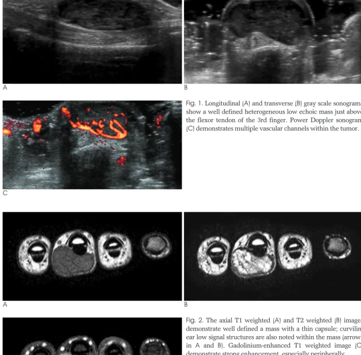
Fig. 1. Longitudinal (A) and transverse (B) gray scale sonograms
show a well defined heterogeneous low echoic mass just above
the flexor tendon of the 3rd finger. Power Doppler sonogram
(C) demonstrates multiple vascular channels within the tumor.
showed strong enhancement, especially peripherally on the gadolinium enhanced T1 weighted image (Fig. 2C).
A tumor resection was performed, and a well circum- scribed mass was identified in the subcutaneous tissue and over the flexor tendon of the 3rd digit. No invasion of the adjacent tendon was present and upon microscop- ic examination, the mass was demarcated by a fibrous thin capsule, and showed multiple tortuous small sized vessels surrounded by compact smooth muscle cells with dense fibrosclerotic tissue (Fig. 3). The pathologic diagnosis was a solid type angioleiomyoma.
Discussion
Angioleiomyomas are a rare form of leiomyoma.
Leiomyomas are a benign smooth muscle neoplasm that frequently occurs in extraskeletal sites such as the ovaries, uterus, bladder, lung, and gastrointestinal tract.
Occasionally, the skin and subcutaneous soft tissues are involved, as in cases of angioleiomyoma arising from the smooth muscle of small blood vessels.
Angioleiomyomas are sharply demarcated spherical masses, mostly measuring less than 2 cm in diameter (1, 2). The peak incidence is in the fourth to sixth decades of life (1). The lower extremity tumors occur in women twice as frequently as in men, and the upper extremity tumors occur more frequently in men than in women (1). Pain is experienced more often in tumors located in the lower extremity than in an upper extremity, the head or neck (1, 2).
Angioleiomyomas are classified into three histological
types: solid, venous and cavernous (3). Solid angi- oleiomyomas, described as closely compact smooth muscle and many small split-like vascular channels, oc- cur three times more in females than males and are gen- erally the most common type. Venous angioleiomy- omas, described as having thick and easily identifiable muscular walls, occur more commonly in males.
Cavernous angioleiomyomas, described as having dilat- ed vascular channels with less smooth muscle cells, are the least common type. Although angioleiomyomas are classified into three main types on pathologic examina- tion, the MRI features are similar to differentiate among the three types of angioleiomyomas (4).
Several studies have reported about the correlation be- tween pathology and MR findings. Hwang et al. (4) sug- gested that the smooth muscle and numerous vessels corresponded to the hyperintense areas, and the fibrous tissue appeared isointense on T2 weighted MR images.
In addition, a well defined peripheral, hypointense rim on T2 weighted images showed fibrous capsule and the interlacing isointense areas within the tumor, which were correlated with the various amounts of connective tissue and intravascular thrombus. Yoo et al. (5) suggest- ed that the presence of tortuous vascular channels sur- rounded by smooth muscle bundles and areas of myx- oid change explains the heterogenicity of signal intensity in the tumor on T2 weighted images. Some tumors showed predominant myxoid change and hyalization corresponding more so to the higher signal intensity on T2 weighted image than the remaining part of mass. On gadolinium-enhanced T1 weighted images, most of the tumors showed homogeneous high enhancement; how- ever, some only showed peripheral enhancement, which reflected the fewer vessels in the tumor tissue (5).
One case report of MRI findings described tortuous vas- cular channels surrounded by smooth muscle bundles that corresponded to tortuous low signal intensity on T1 and T2 weighted images, which was consistent with the diagnosis of angioleiomyoma (6).
In our case, the mass showed multiple vascular struc- tures on Doppler sonography, and serpentine or curvi- linear low signal intensity structures that were thought to be tortuous vascular channels seen on T1 and T2 weighted images. Upon microscopic examination, the low signal intensity rim on T2 weighted image was found to be well correlated with fibrous capsule and the heterogenicity on a T2 weighted image corresponded to multiple vascular channels and dense fibrosclerotic tis- sues.
J Korean Soc Radiol 2011;64:599-602
─ 601 ─ Fig. 3. Photomicroscopic image (H & E ×10) demonstrates compact smooth muscle cells surrounding the vessel wall.
Multiple small sized split-like vascular channels are also noted.
The differential diagnosis may include giant cell tu- mors of the tendon sheath, neurogenic tumor, and he- mangioma. Compared to an angioleiomyoma, a giant cell tumor of the tendon sheath frequently shows low signal intensity on T2 weighted images and an intimate relationship with the tendon sheath. Neurogenic tumors usually present as well demarcated masses of high sig- nal intensity on T2 weighted images, but do not show tortuous vascular structure. Hemangiomas can also at times show tortuous vascular channels and well defined nodular appearance, but usually show a lobulated con- tour, infiltrative margin, and relatively high signal inten- sity on T1 weighted images due to the fatty component within the mass (7). Internal calcification and saccular vascular structure with or without thrombosis are also helpful for differential diagnosis (7).
In conclusion, we suggest that the angioleiomyoma be considered when multiple vascular structures are noted in a well defined soft tissue mass arising from the subcu- taneous tissue of the extremities on MRI and Doppler
sonography.
References
1. Hachisuga T, Hashimoto H, Enjoji M. Angioleiomyoma: a clinico- pathologic reappraisal of 562 cases. Cancer 1984;54:126-130 2. Freedman AM, Meland NB. Angioleiomyomas of the extremities:
report of a case and review of the Mayo Clinic experience. Plast Reconstr Surg 1989;83:328-331
3. Morimoto N. Angioleiomyoma: a clinicopathologic study. Med J Kagoshima Univ 1973;24:663-683
4. Hwang JW, Ahn JM, Kang HS, Suh JS, Kim SM, Seo JW. Vascular leiomyoma of an extremity: MR imaging-pathology correlation.
AJR Am J Roentgenol 1998;171:981-985
5. Yoo HJ, Choi JA, Chung JH, Oh JH, Lee GK, Choi JY, et al.
Angioleiomyoma in soft tissue of extremities: MRI findings. AJR Am J Roentgenol 2009;192:291-294
6. Kinoshita T, Ishii K, Abe Y, NAganuma H. Angiomyoma of the lower extremity: MR findings. Skeletal Radiol 1997;26:443-445 7. Theumann NH, Bittoun J, Goettmann S, Le Viet D, Chevrot A,
Drape ′JL. Hemangiomas of the fingers: MR imaging evaluation.
Radiology 2001;218:841-847
Bo Seong Jeong, et al : Radiologic Findings of an Angioleiomyoma of the Finger
─ 602 ─
대한영상의학회지 2011;64:599-602
손가락에서 발생한 맥관평활근종의 영상의학적 소견:
증례 보고11