J Korean Soc Radiol 2016;75(5):404-409 http://dx.doi.org/10.3348/jksr.2016.75.5.404
INTRODUCTION
Infantile myofibromatosis (IM) is a rare mesenchymal tumor- ous disorder characterized by benign myofibroblastic prolifera- tion in the skin, subcutaneous tissue, muscle, bone and visceral organs (1-3). Most of the patients (88%) present within the first 2 years (1-3). IM may occur in a solitary form (myofibroma) or mul- ticentric form (myofibromatosis) (2). In the multicentric form of IM, the bone is often involved (57%), but only 9% of cases may occur in the bone as the solitary form of IM. The most common location of the solitary bone lesion is craniofacial bones (1-6).
There are only a few case reports describing the magnetic reso- nance (MR) imaging features of solitary IM of the skull (4-6). We report the MR imaging findings of solitary IM arising in the tem- poral bone with literature review.
CASE REPORT
A 10-month-old boy had a painless palpable lump in the right temporal region. The lump persisted unchanged for 4 months since the time when his mother had incidentally found it. He had no significant medical or family history. On physical examina- tion, the lesion was firm and non-tender. No other mass or lymph- adenopathy was present. The laboratory findings were unre- markable.
Radiographs of the skull demonstrated a 2.5 cm sized well-de- fined expansile osteolytic lesion with a thin sclerotic rim in the right temporal bone (Fig. 1). MR images showed a well-margin- ated, biconvex, oval-shaped mass in the diploic space of the tem- poral bone which showed intermediate signal intensity on both T1-weighted images [repetition time (TR)/echo time (TE), 450/12 ms] and T2-weighted images (TR/TE, 5740/122). The inner and outer cortical tables of the skull were relatively intact and intra- cranial abnormality or soft tissue invasion was not present (Fig.
Magnetic Resonance Imaging Findings of Solitary Infantile Myofibromatosis of the Skull: A Case Report
두개골에 발생한 영아 근섬유종증의 자기공명영상 소견: 증례 보고
Seung Eun Lee, MD
1, Kil Ho Cho, MD
1*, Jang Ho Suh, MD
1, Joon Hyuk Choi, MD
2Departments of 1Radiology, 2Pathology, College of Medicine, Yeungnam University, Daegu, Korea
Infantile myofibromatosis is a rare, benign mesenchymal disorder of early childhood characterized by solitary or multiple benign myofibroblastic tumors. The tumors may involve the skin, subcutaneous tissue, muscle, bone and visceral organs. We re- port magnetic resonance imaging findings of solitary infantile myofibromatosis arising in the temporal bone of a ten-month-old boy, and the diagnosis was con- firmed by surgical excision and histopathological examination.
Index terms Myofibromatosis Skull
Neoplasms
Magnetic Resonance Imaging
Received October 12, 2015 Revised March 28, 2016 Accepted May 26, 2016
*Corresponding author: Kil Ho Cho, MD
Department of Radiology, College of Medicine, Yeungnam University, 170 Hyeonchung-ro, Nam-gu, Daegu 42415, Korea.
Tel. 82-53-620-3045 Fax. 82-53-653-5484 E-mail: [email protected]
This is an Open Access article distributed under the terms of the Creative Commons Attribution Non-Commercial License (http://creativecommons.org/licenses/by-nc/3.0) which permits unrestricted non-commercial use, distri- bution, and reproduction in any medium, provided the original work is properly cited.
2A, B). On gadolinium enhanced T1-weighted images, the lesion showed intense heterogeneous enhancement (Fig. 2C, D).
Initially, Langerhans cell histiocytosis arising in the skull was considered radiologically before surgery.
Surgical excision of the tumor was performed. The tumor was attached to the dura mater with a granulation tissue-like mem- brane and tumor invasion was not seen. On gross examination, a well-demarcated, grayish-white solid tumor was present, mea- suring 2.5 × 2.4 × 1.5 cm (Fig. 3). Microscopically, low-power magnification showed a nodular appearance. The tumor showed spindle cell proliferation with a fascicular pattern (Fig. 4A). The tumor cells had elongated nuclei with eosinophilic cytoplasm (Fig. 4B). A thin-walled, branching, hemangiopericytoma-like vascular pattern was also present (Fig. 4C). On immunohisto- chemical staining, the tumor cells were positive for smooth mus- cle actin (Fig. 4D) and negative for desmin, S100 protein and CD34. Necrosis was absent and there was no evidence of malig- nancy. The final histopathological diagnosis was solitary IM of the skull.
DISCUSSION
Although relatively rare overall, IM is the most common fi- brous neoplasm of infancy and childhood (1). Stout first de- scribed the disorder in 1954 using the term ‘congenital general- ized fibromatosis’ (1-3) In 1981, Chung and Enzinger (2) introduced the term ‘IM’, and they classified the disorder into two groups; solitary form and multicentric form. In the solitary form, the most commonly affected site is the head and neck re- gion, followed by the trunk and extremities (2). These tumors are usually found in the soft tissues (skin, muscle, and subcutaneous tissue), but they are also found in the bones (9%), as seen in our case. The multicentric form is found in the soft tissues and bones, but it also involves the visceral organs (2, 3).
Most of the cases (88%) are diagnosed in the first 2 years (2).
Clinically, the patients usually present with a palpable, painless mass in the soft tissues. Visceral lesions cause different symptoms according to the organs involved (1-3).
The etiology is unknown. Congenital cases have been reported with an autosomal dominant and an autosomal recessive inheri- tance pattern. Another hypothesis is the intrauterine estrogen
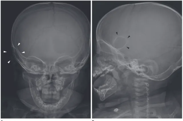
Fig. 1. The AP (A) and lateral (B) radiographs of the skull demonstrate a well-defined expansile osteolytic lesion with a thin sclerotic rim in the right temporal bone (arrowheads).
A B
stimulation (1, 3, 6).
The radiographic finding of the bone lesion of IM is a well- marginated osteolytic lesion with or without sclerotic rims (3-7).
Skull lesions tend to be round in shape and 1–4 cm in size, and they usually involve the temporal and parietal bones. They can expand the inner and outer cortical tables, but periosteal reaction
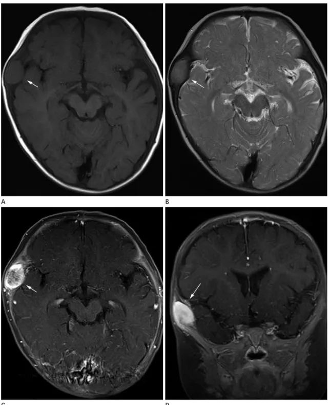
Fig. 2. MR images of solitary infantile myofibromatosis of the skull (arrows).
A well-marginated oval mass in the right temporal bone shows intermediate signal intensities on both T1-weighted image [repetition time (TR)/
echo time (TE), 450/12 ms] (A), and T2-weighted images (TR/TE, 5740/122) (B) (arrows). Contrast enhanced T1-weighted (TR/TE, 440/12) axial (C) and coronal (D) images demonstrate a mass with heterogeneous enhancement (arrows).
MR = magnetic resonance A
C
B
D
is rare (4-6). On computed tomography (CT) scans, the lesions appear hypodense or isodense to the brain parenchyma with marked and homogeneous or heterogeneous enhancement (4-6).
A few case reports have described the MR imaging features of IM arising in the skull (4-6). In the previously reported MR images, IM arising in the skull usually appeared as a soft tissue mass in
the skull with low signal intensity on T1-weighted images, high signal intensity on T2-weighted images and intense homoge- neous or heterogeneous enhancement (5, 6). Okamato et al. (4) reported IM in the skull which showed intermediate signal inten- sity on both T1- and T2-weighted images and it enhanced heter- ogeneously. The MR appearance was similar to that in the author’s
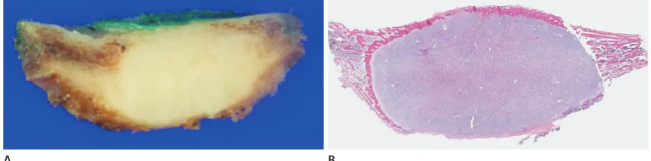
Fig. 3. Gross findings of the skull mass and whole-mount section.
A. The excised specimen shows a well-demarcated, grayish-white, solid tumor.
B. Whole-mount section shows cortical destruction caused by the tumor.
A B
Fig. 4. Microscopic findings.
A. Low power magnification shows a nodular appearance (hematoxylin-eosin stain, × 40).
B. Spindle-shaped tumor cells are arranged in a fascicular pattern (hematoxylin-eosin stain, × 100).
C. Thin-walled, branching blood vessels are surrounded by the tumor cells (hematoxylin-eosin stain, × 200).
D. The tumor cells are positive for smooth muscle actin (immunohistochemical stain, × 200).
A
C
B
D
case. They assumed that the considerable amount of collagen and absence of necrosis in the tumor resulted in decreased signal in- tensity on T2-weighted images. In our case, the skull lesion showed intermediate signal intensity on both T1- and T2- weighted images and heterogeneous enhancement, more intense in the peripheral area, on gadolinium-enhanced T1-weighted images. Microscopically, there was fibrosis with abundant colla- gen component in the tumor and absence of necrosis. These pathologic findings supported the assumption made by Okamato et al. (4). IM has two distinctive microscopic components, spin- dle cell proliferation with a fascicular pattern and a hemangio- pericytoma-like vascular pattern (1, 3). Heterogeneous arrange- ment of the two components might have resulted in heterogeneous enhancement. In our case, there were slightly more vascular structures in peripheral distribution, and it might have had an ef- fect on the peripheral intense enhancement. The histologic fea- tures of the lesions arising in both soft tissue and bone are said to be similar (1). Murphey et al. (3) and Koujok et al. (7) reported the imaging findings of IM arising in the soft tissue. The MR ap- pearance of the soft tissue lesion often showed low to intermedi- ate signal intensity on T1-weighted image, central high signal in- tensity on T2-weighted images and peripheral enhancement. The central non-enhancing focus with high signal intensity on T2- weighted image was associated with central necrosis, pathologi- cally (3, 7). Regressing IM with central necrosis is characterized by a central focus of T2 prolongation in both soft tissue and bone lesion.
The radiologic differential diagnosis includes Langerhans cell histiocytosis (eosinophilic granuloma), intraosseous meningio- ma and metastatic neuroblastoma. On MR images, most of the lesions usually appear as an osteolytic soft tissue calvarial mass with low signal intensity on T1-weighted images, high signal in- tensity on T2-weighted images, and enhancement on contrast enhanced T1-weighted images (8, 9). It might be difficult to dif- ferentiate IM from other diseases because the MR imaging find- ings are nonspecific. However, unlike the high signal intensity on T2-weighted images observed in other diseases, IM usually shows intermediate to low signal intensity on T2-weighted images due to its collagen component. Langerhans cell histiocytosis usually occurs in child and young adults (4) and the peak prevalence is between 1 and 4 years of age (8). Extensive osseous destruction is often seen in MR images (8). These lesions can show beveled
edge appearance on a radiograph and CT due to asymmetric de- struction of the inner and outer tables (8, 9). Intraosseous menin- gioma is rare in children. In MR images, these lesions often show the dural tail sign and homogeneous enhancement (9). However, Okamoto et al. (4) reported a case of IM in the skull with the du- ral tail sign. Therefore, the dural tail sign is not a pathognomonic finding to distinguish between intraosseous meningioma and IM. Metastatic neuroblastoma is the most common malignant metastatic skull lesion in children (10). These lesions can appear as single or multiple osteolytic lesions and show more aggressive imaging features with extension to produce epidural deposits and scalp invasion which is better demonstrated with MR images (8, 10). Thickened bone, hair-on-end periosteal reaction, and sepa- ration of the sutures are also the possible radiologic findings (10).
The MR imaging findings of IM in the skull are not pathogno- monic and overlap with those of other soft tissue calvarial mass- es. Histopathologic examination should be performed for a de- finitive diagnosis (4, 6).
The prognosis and treatment of IM depend on the type and the location of the disease (1-3). Surgical resection is usually rec- ommended for a solitary bone lesion and the recurrence rate is low (10%). The solitary form without visceral involvement usual- ly has a favorable prognosis and spontaneous regression is often observed (3-6).
In conclusion, solitary IM of the skull is a relatively rare entity, and MR imaging findings seem to be nonspecific and variable.
However, IM should be considered in the differential diagnosis of a case of a well-defined soft tissue mass of infancy arising in the skull.
REFERENCES
1. Inwards CY, Unni KK, Beabout JW, Shives TC. Solitary con- genital fibromatosis (infantile myofibromatosis) of bone.
Am J Surg Pathol 1991;15:935-941
2. Chung EB, Enzinger FM. Infantile myofibromatosis. Cancer 1981;48:1807-1818
3. Murphey MD, Ruble CM, Tyszko SM, Zbojniewicz AM, Pot- ter BK, Miettinen M. From the archives of the AFIP: mus- culoskeletal fibromatoses: radiologic-pathologic correla- tion. Radiographics 2009;29:2143-2173
4. Okamoto K, Ito J, Takahashi H, Emura I, Mori H, Furusawa
두개골에 발생한 영아 근섬유종증의 자기공명영상 소견: 증례 보고
이승은
1· 조길호
1* · 서장호
1· 최준혁
2유아의 근섬유종증은 소아에서 발생하는 드문 중간엽 세포 기원의 양성 질환으로 근섬유아세포가 증식하여 단발성 혹은 다발성의 종괴를 형성하며 피부, 피하조직, 근육, 뼈 혹은 내장 기관을 침범할 수 있다. 10개월 남아에서 수술로 확진된 두 개골의 유아 근섬유종증 1예의 자기공명영상 소견을 보고한다.
영남대학교 의과대학 1영상의학과, 2병리과
T, et al. Solitary myofibromatosis of the skull. Eur Radiol 2000;10:170-174
5. Tsuji M, Inagaki T, Kasai H, Yamanouchi Y, Kawamoto K, Uemura Y. Solitary myofibromatosis of the skull: a case report and review of literature. Childs Nerv Syst 2004;20:
366-369
6. Merciadri P, Pavanello M, Nozza P, Consales A, Ravegnani GM, Piatelli G, et al. Solitary infantile myofibromatosis of the cranial vault: case report. Childs Nerv Syst 2011;27:
643-647
7. Koujok K, Ruiz RE, Hernandez RJ. Myofibromatosis: imag-
ing characteristics. Pediatr Radiol 2005;35:374-380 8. Morón FE, Morriss MC, Jones JJ, Hunter JV. Lumps and
bumps on the head in children: use of CT and MR imaging in solving the clinical diagnostic dilemma. Radiographics 2004;24:1655-1674
9. Yim Y, Moon WJ, An HS, Cho J, Rho MH. Imaging findings of various calvarial bone lesions with a focus on osteolytic lesions. J Korean Soc Radiol 2016;74:43-54
10. D’Ambrosio N, Lyo JK, Young RJ, Haque SS, Karimi S. Im- aging of metastatic CNS neuroblastoma. AJR Am J Roent- genol 2010;194:1223-1229