Hypertrophic osteoarthropathy represents a clinical syndrome consisting of clubbing of the digits of the hands and feet, enlargement of the extremities sec- ondary to periarticular and osseous proliferation, and painful and swollen joints. The condition has been di- vided into two categories, primary hypertrophic os- teoarthropathy, also known as “pachydermoperiosto- sis”, and secondary hypertrophic osteoarthropathy, fre- quently referred to as “hypertrophic pulmonary os- teoarthropathy” due to its association with a variety of pulmonary causes. Recent reports have described the findings of magnetic resonance imaging and bone scintigraphy(1-4). We report a case of pachydermope- riostosis, describing the imaging features apparent at plain radiography, bone scintigraphy, and magnetic res-
onance imaging.
Pachydermoperiostosis (Primary Hypertrophic Osteoarthropathy): Case Report1
Moon Hee Paik, M.D., Bae Young Lee, M.D., Kang Hoon Lee, M.D., Kyung Sup Song, M.D., Jin Wou Kim, M.D.2, Ki Ouk Min, M.D.3
We report a case involving a young male with the complete form of primary hyper- trophic osteoarthropathy. He presented with the typical features of the condition:
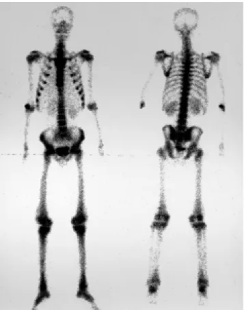
thickening and furrowing of the skin of the forehead and scalp, and digital clubbing of the hands and feet. Plain radiographs of the long bones of the extremities depicted bi- lateral irregular, shaggy, cortical diaphyseal thickening. T1- and T2-weighted magnetic resonance imaging(MRI) of the femur demonstrated low-signal-intensity cortical thickening. Bone scintigraphy revealed no photon uptake in the long bones.
Index words : Primary hypertrophic osteoarthropathy, MR Primary hypertrophic osteoarthropathy, bone scan
1Department of Diagnostic Radiology, St. Paul’s Hospital, The Catholic University of Korea
2Department of Dermatology, St. Paul’s Hospital, The Catholic University of Korea
3Department of Clinical Pathology, St. Paul’s Hospital, The Catholic University of Korea
Received July 23, 2002 ; Accepted September 18, 2002
Address reprint requests to : Bae Young Lee, M.D., Department of Diagnostic Radiology, The Catholic University of Korea, St. Paul’s Hospital, 620-56 Jeonnong-dong, Dongdaemun-gu, Seoul 130-709, Korea.
Tel. 82-2-958-2084 Fax. 82-2-957-2810 E-mail: [email protected]
Fig. 1. Photograph of the patient’s face shows furrowing and thickening of the skin of the forehead.
Case Report
A 28-year-old man presented with a ten year history of pain in both knees, not known to have occurred in other family members. During previous admission for the treatment of a duodenal ulcer, his morphologic features of thickening and furrowing of the skin of the forehead and scalp, and digital clubbing of hands and feet, which were specific features of primary hypertrophic os- teoarthropathy, were detected by a dermatologist.
At physical examination, the furrowing and thicken- ing of the skin of the forehead and scalp, and digital clubbing of the fingers, were confirmed(Fig. 1). The pa- tient’s height and weight were within normal limits, and endocrinological evaluation failed to establish any ab- normality, including thyroid acropathy. Laboratory tests, however, revealed that he was anemic(hemoglo- bin, 7.2 mg/dL).
Radiographs of the patient’s skull depicted a normal sella and mandibular angle, and the findings of plain chest radiography were also normal. Plain radiographs
A
Fig. 2. A. Plain radiograph of hands shows soft tissue swelling of the fingers (digital clubbing).
B. Plain radiograph of upper extremity shows irregular shaggy diaphyseal cor- tical thickening.
C. Plain radiograph of femurs shows bilateral irregular, shaggy diaphyseal cortical thickening. Bilateral symmetri- cal, well defined, scalloped intrame- dullary radiolucency is also seen in the diaphysis.
of the hands showed soft tissue thickening of the fingers without bony abnormality(Fig. 2A), but those of the ex- tremities revealed bilateral irregular shaggy diaphyseal cortical thickening of the tibias, fibulas, femurs, radii, ulnas, and humeri. In addition, the diaphysis of both fe- murs showed symmetrically well-defined, scalloped in- tramedullary radiolucency(Figs. 2B, C). At T1- and T2- weighted MRI, low-signal-intensity periosteal thicken- ing was seen in the diaphysis of the femur, and at T1- weighted and T2 spectral pre-saturation inversion recov- ery(SPIR) imaging, the intramedullary cavity of the dia- physis of the femur demonstrated higher signal intensity than the femoral head and neck. After the administra- tion of gadolinium, both femurs showed homogeneous diffuse enhancement, with the intramedullary cavity showing greater enhancement than the femoral head and neck(Figs. 3A, B, C). Bone scanning demonstrated showed no photon uptake in long bones(Fig. 4).
Biopsy of the skin of the forehead revealed the pres- ence of hyperkeratosis, acanthosis of the epidermis and hyperplastic sebaceous glands, and perivascular chronic
inflammatory cell infiltration and edematous change in the dermis(Fig. 5).
The patient was treated with nonsteroidal anti-inflam- matory drugs, resulting in symptomatic relief of arthral- gia.
Discussion
Early descriptions of the clinical entity known as hy- pertrophic osteoarthropathy are credited to Friedreich (1868) and Marie(1890). On the basis of the underlying etiology, the condition has been divided into two cate- gories, primary(hereditary or idiopathic) and secondary hypertrophic osteoarthropathy. The former, also known as “pachydermoperiostosis”, is inherited in an autoso- mal dominant pattern, with variable expressivity.
Secondary hypertrophic osteoarthropathy is frequently referred to as “hypertrophic pulmonary osteoarthropa- thy” due to its association with a variety of pulmonary causes such as bronchogenic carcinoma, mesothelioma, bronchiectasis, and pulmonary abscess(5).
A
C
B
Fig. 3. A-C. Coronal T1 weighted (A), T2 SPIR weighted (B), and contrast enhanced T1 weighted images (C).
MRI of femur shows low signal intensity, periosteal thickening in the diaphysis of the femur on T1 and T2 weighted images.
The intramedullary cavity of the diaphysis of the femur demon- strated higher signal intensity than head and neck of femur on T1 and T2 weighted images. After administration of gadolini- um, both femurs showed homogenous diffuse strong enhance- ment. The intramedullary cavity showed more enhancement than the head and neck of femur.
Primary hypertrophic osteoarthropathy is far less common than the secondary form. In this disorder, vague bone and joint pains are reported in conjunction with progressive enlargement of the extremities, and
digital clubbing, coarsening of the skin(pachydermia) and excessive sweating(hyperhidrosis) may occur. In the extreme form of pachydermia, cutis verticis gyrata develops, characterized by massive thickening and fur- rowing of the skin of the forehead and scalp, which re- sembles the gyral pattern of the brain. The clinical mani- festations are somewhat variable, with affected patients demonstrating either the complete syndrome(pachyder- mia, periostosis, cutis verticis gyrata), the incomplete form(sparing the scalp), or the forme fruste(pachyder- mia with minimal or absent periostitis)(5).
The major pathologic finding in this disorder is perios- titis involving the long tubular bones of the extremities.
The initial phase of inflammatory periostitis is charac- terized by subperiosteal lymphocyte and plasma cell in- filtration followed by subperiosteal new bone forma- tion. With time, there is evolution of the periosteal con- figuration from an initial single layer to several layers and finally to an irregular wavy appearance (6).
Eventually, a pseudocortex forms and ultimately fuses with the original cortex, resulting in increased shaft di- ameter(7). There is intermittent activity of the periosteal process, and correlation of radiologic patterns with du- ration of disease suggests that thicker, more extensive periosteal reaction is indicative of long-standing disease (6).
The radiologic findings typify the skeletal changes and type of periosteal reaction that develop in patients with long-standing disease, in which there is diaphyseal, metaphyseal, epiphyseal, and interosseous membrane involvement. The role of magnetic resonance imaging in the detection and characterization of periosteal new bone formation has recently been emphasized, and MR imaging can identify subradiographic cellular periosteal proliferation and allow assessment of the morphology and chronicity of periostitis(8). According to Moore(8), the MR appearance of periosteal reaction can be charac- terized according to the underlying histology. The initial phase of periostitis is termed “cellular” periosteal reac- tion and is characterized by thickening and differentia- tion of the periosteum into two histological layers. At T1-weighted MR imaging, a curvilinear layer of inter- mediate signal intensity superficial to cortical bone is ob- served, and at T2-weighted imaging, signal intensity in- creases. With maturation, single- or multiple-layer pe- riosteal reaction is seen: one or more sheets of new wo- Fig. 4. Bone scintigraphy shows no photon uptake in bones.
Fig. 5. Photomicrograph of forehead skin shows hyperplastic
not contiguous with the underlying cortical bone is seen (2). In our case, T1- and T2- weighted images were of low signal intensity, and periosteal reaction was ob- served. According to the explanation contained in the lit- erature mentioned above, this appears to be the matura- tion, rather than the active phase.
In our case, an additional finding at plain radiography of femurs was symmetrical scalloped intramedullary ra- diolucency in the diaphysis. At T1- weighted and T2- SPIR MR imaging of the femur, the intramedullary cavi- ty of its diaphysis demonstrated higher signal intensity than its head and neck of femur, but after the adminis- tration of gadolinium, both femurs showed homogenous diffuse enhancement and the intramedullary cavity showed more enhancement than the femoral head and neck. Unfortunately biopsy of the intramedullary cavity was not performed, so this bone lesion could not be proven histopathologically.
The reported bone scintigraphy findings of primary hypertrophic osteoarthropathy are of two types.
Characterically, one is normal despite the presence of periosteal reaction at radiographic examination(9), and the other shows increased pericortical uptake. The fea- tures of pachydermoperiostosis include consistently symmetrical increased pericortical linear deposition of the tracer, especially along the distal ends of the tibia, fibula, ulna, and radius. In addition, increased trans- verse diameter of the long bones, especially at their dis- tal ends is noted(4). One of the explanations given for this pericortical uptake is increased blood flow during the active stage of the disease and decreased flow during the quiescent stage(4). Jajic et al. stated that bone scintigraphy revealed increased pericortical uptake when the disease was active. During remission, the find- ings of scintigraphy were negative and not useful in the diagnosis of periostosis(3). In our case, bone scanning demonstrated no photon uptake in the long bones, de- spite the periosteal reaction observed at plain radiogra- phy. This suggests that bone scans were obtained during the remission stage of the disease.
The differential diagnosis includes osteomyelitis, thy- roid acropathy, hypervitaminosis A, infantile cortical hyperostosis, fluorosis, and venous stasis. In differential diagnosis, the superficial nature, symmetry, and distal diaphyseal location of the periosteal reaction of hyper- trophic osteoarthropathy are clearly distinguishable
from actual infection of the bone at those locations. The distribution of hypertrophic osteoarthropathy is quite different from that of thyroid acropathy, which predom- inantly affects metacarpals, metatarsals, and phalanges.
The accentuated enthesial reaction of occurring in hy- pervitaminosis A, the predominantly mandibular, clav- icular, scapular and costal involvement found in infan- tile cortical hyperostosis (Caffey’s disease), and the en- thesial calcification, trabecular distortion, and osteoscle- rosis occurring in fluorosis are also easily distinguished from the osseous lesions of hypertrophic osteoarthropa- thy. Periosteal reaction secondary to venous stasis oc- curs only in the lower extremity and is limited to the tib- ia and fibula (10).
In conclusion, when periosteal reaction in long bones, with specific clinical features of digital clubbing and pachydermia, is apparent, the possibility of pachyder- moperiostosis should be considered. It is likely that both bone scintigraphy and MR reflect the stage of the dis- ease and the chronicity of periostitis.
References
1. Dermirpolat G, Sener RN, Stun EE. MR imaging of pachyder- moperiostosis. J Neuroradiol 1999;26:61-63
2. Lorendo R, Pathria MN, Salonen D, Resnick D. Magnetic reso- nance imaging in pachydermoperiostosis. Clinical Imaging 1996;
20:212-218
3. Jajic Z, Jajic I, Nemcic T. Primary hypertrophic osteoarthropathy:
clinical, radiologic, and scintigraphic characteristics. Arch Med Res 2001;32:136-142
4. Bomannji J, Nagaraj N, Jewkes R, Fields M, Maini RN.
Pachydermoperiostosis: Technetium-99m-methylene diphospho- nate scintigraphic pattern. J Nucl Med 1991;32:1907-1909
5. Resnick D. Diagnosis of bone and joint disorders, 3rd ed. Philadel- phia: WB Sauders,1995:4421-4434
6. Pineda C, Martinez-Lavin M, Goobar J, Sartoris D, Clopton P, Resnick D. Periostitis in hypertrophic osteoarthropathy: relation- ship to disease duration. AJR Am J Roentgenol 1987;148:773-778 7. Gall E, Bennett GA, Bauer W. Generalized hypertrophic os-
teoarthropathy: a pathologic study of seven cases. Am J Pathol 1951;27:349-381
8. Moore SG. Magnetic resonance imaging and computed tomography of cortical bone. In Bloem JL, Sartoris DJ (eds): MRI and CT of the Musculoskeletal system, A Text-Altas. Baltimore: Williams &
Wilkins, 1992:139-152
9. Juhl JH, Crummy AB, Kuhlman JE. Paul and Juhl’s essentials of ra- diologic imaging, 7th ed. Lippincott-Raven, 1998:122
10. Rothschild BM, Rothschild C. Recognition of hypertrophic os- teoarthropathy in skeletal remains. J Rheumatology 1998;25:2221- 2227
대한방사선의학회지 2002;47:533-538
일차성 비후성 골관절증: 증례 보고1
1가톨릭대학교 의과대학 진단방사선과
2가톨릭대학교 의과대학 피부과
3가톨릭대학교 의과대학 임상병리과
백문희・이배영・이강훈・송경섭・김진우2・민기옥3
일차성 비후성 골관절증은 경피증과 골막반응을 특징으로 하는 드문 질환이다. 본 저자는 양측 슬관절통을 호소하는 28세 남자의 일차성 비후성 골관절증 증례를 보고하고자 한다. 임상적으로 경피증이 이마와 두피에서 보였고, 수지 곤 봉상이 관찰되었다. 단순촬영에서 대퇴골, 경골, 비골, 상완골, 척골과 요골에서 골막반응이 관찰되었다. 자기공명영상 에서 T1과 T2 강조영상에서 저신호강도의 골막반응이 보였다. 골 스캔에서는 이상 소견이 없었다.