Chondromas of the soft tissue are rare benign tumors that usually arise in the hands and feet. On rare occa- sions, this tumor arises from the capsule or paraarticular soft tissue of large joints, such as the knee (1). These le- sions should be differentiated from synovial chodro- matosis, localized pigmented villonodular synovitis and other calcified lesions occurring about the joints. We re- port here on a case of intracapsular and paraarticular chondroma of the knee joint in 38-year-old man, and we also include a review of the previous literature.
Case Report
A 36-year-old man visited our hospital and he had a 6- month history of pain and swelling of the left knee.
There was no history of trauma. Plain radiographs re- vealed ill defined soft tissue opacity with joint effusion (Fig. 1). MRI (Philips Gyroscan T5-NT) was then per-
formed, and it demonstrated a 3×2×1 cm sized well defined ovoid-shaped solid mass in the infrapatellar area. The T1 weighted sagittal image (TR/TE 625/20, spin echo) showed iso-signal intensity muscle with a subtle high signal intensity area in its central portion (Fig. 2A). The sagittal T2 weighted image (TR/TE 1800/
J Korean Radiol Soc 2004;51:449-452
─ 449 ─
Intracapsular and Paraarticular Chondroma of the Knee: Case Report1
Ji Chang Kim, M.D., Yeon Soo Lee, M.D., Jong Hun Ji, M.D.2, Eun Hee Lee, M.D.3, Si Won Kang, M.D.
We report here on a case of intracapsular and paraarticular chondroma of the left knee in a patient with a 6-month history of knee pain and swelling. Magnetic reso- nance image (MRI) revealed a well-defined solid mass with central hemorrhagic necro- sis in the infrapatellar area of the knee.
Index words :Chondroma Knee
Magnetic resonance (MR)
1Department of Diagnostic Radiology, Dae Jeon St Mary’s Hospital, Catholic University of Korea
2Department of Orthopedic Surgery, Dae Jeon St Mary’s Hospital, Catholic University of Korea
3Department of Clinical Pathology, Dae Jeon St Mary’s Hospital, Catholic University of Korea
Received October 27, 2003 ; Accepted August 17, 2004
Address reprint requests to : Ji Chang Kim, M.D., Department of Diagnostic Radiology, Dae Jeon St Mary’s Hospital, Catholic University of Korea, Dea Jeon 301-723, Korea.
Tel. 82-42-220-9645 Fax. 82-42-257-0511 E-mail: [email protected]
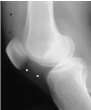
Fig. 1. Lateral radiograph of the knee shows ill-defined soft tis- sue opacity in the infrapatellar area (white arrows) and bulging of the suprapatellar bursa due to joint effusion (black arrows).
80, spin echo) demonstrated peripheral high signal in- tensity with a central low signal intensity area (Fig. 2B).
The area of low signal intensity on the long TR images corresponded to hemorrhagic necrosis. Arthroscopy re- veals that the mass was in an intracapsular location and partially attached to the synovial membrane adjacent to the anterior cruciate ligament. Arthroscopic removal was done. The removed mass measured 3×2×1 cm in size and the surface was white and glistening. Central necrosis was seen on the cut section. On histologic ex- amination, the tumor consisted of hyaline cartilage and central hemorrhagic necrosis, and this was surrounded by a thin synovial membrane (Fig. 3). The mass was di- agnosed as an intracapsular and paraarticular chondro- ma.
Discussion
There are three known variants of extraskeletal chon- dromas: synovial chondromatosis, para-articular chon- dromas, and soft tissue chondromas. These last two variants are very rare and may show atypical features (2). Intracapsular and paraarticular chondromas have been variously named capsular osteomas, osteochon- dromas or chondromas depending on the relative pro- portion of bone and cartilage. The terms “intracapsular and paraarticular chondroma” was firstly used by Jaffe in 1958. This rare, benign cartilaginous tumor arises from the capsule or the paraarticular connective tissue of the large joints, and the knee is the most frequently
Ji Chang Kim, et al: Intracapsular and Paraarticular Chondroma of the Knee
─ 450 ─
A B
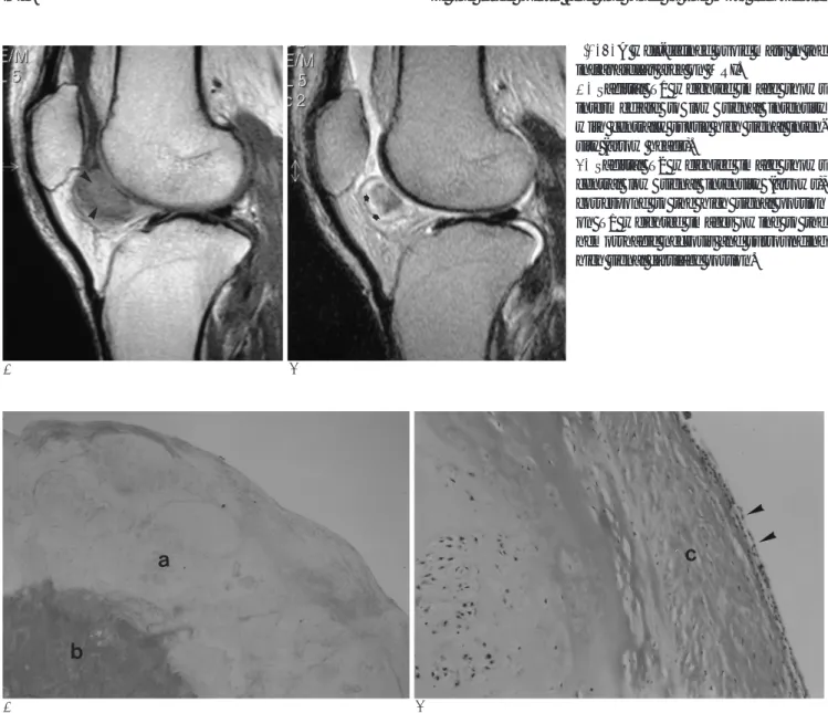
Fig. 2. A well-defined ovoid mass in the infrapatellar area on MRI.
A. Sagittal T1 weighted image shows intermediate to low signal intensity with centrally subtle high signal inten- sity (arrow heads).
B. Sagittal T2 weighted image shows central low signal intensity (arrows), correspond to the high signal portion on T1 weighted images owing to the hemorrhagic necrosis and surrounding high signal cartilage portion.
A B
Fig. 3. Photomicrograph of specimen.
A. Low power field (Hematoxylin-eosin stain, ×40) reveals hyaline cartilage (a) and hemorrhagic necrosis (b).
B. High power field (Hematoxylin-eosin stain, ×200) reveals peripheral thin synovial membrane (arrow heads) and fibrous tissue (c).
involved joint (3). The precise pathogenetic mechanism of these tumors is still controversial. It has been pro- posed that they originate due to cartilaginous metaplasia of the connective tissue following ossification (4). These tumors are found in wide age range of patients and there is no preponderance for either gender. Clinically, these tumors present as slowly growing masses that oc- casionally cause pain or tenderness of the involved joints. The characteristic plain radiographic appearance of this lesion is the presence of a well defined mass about the knee, usually at the infrapatellar area, which contains variable amounts of chondroid matrix calcifica- tion or ossifications. In some instances, the tumors are essentially radiolucent (1, 5, 6), and our case is included in this instance. In our case, there was no chondroid cal- cification or ossified area in the tumor; therefore, it was difficult to detect on plain radiograph (Fig. 1). The MR findings vary according to the degree of ossification or calcification. The cartilagenous portion of the tumor shows low signal intensity on T1 weighted images and bright signal intensity on T2 weighted images. The cen- tral ossified area shows the radiologic features of fatty tissue due to the high proportion of fatty bone marrow among the bone trabeculae (6). In our case, the central portion showed high signal intensity on T1 weighted im- ages and low signal intensity on T2 weighted images;
this suggested hemorrhagic necrosis that may have been due to repeated minor trauma (Fig. 2A, B).
Radiologically, intracapsular and paraarticular chon- droma should be differentiated from calcified soft tissue lesions about the joints such as calcifying bursitis, old hematoma, tumoral calcinosis, periosteal chondroma, calcified synovial sarcoma, primary synovial chondro- matosis and synovial chondrosarcoma. In our case, lo- calized pigmented villonodular synovitis (PVNS) and synovial chondromatosis were considered in the differ- ential diagnosis. PVNS shows the characteristic hemo- siderin deposition, and this shows as dark signal intensi- ty on the T1 and T2 weighted MR images. Hence PVNS can be differentiated by the signal intensity from intra- capsular and paraarticular chondroma. Synovial chon- dromatosis presents as intra- or extra-articular multiple osteocartilagenous nodules arising from the synovium.
Conversly, intracapsular and paraarticular chondromas are solitary masses that originate from the capsular and the paraarticular connective tissue. However, synovial
chodromatosis can present as a single giant intracapsu- lar nodule on rare occasion. Histologically, synovial chondromatosis differs from intracapsular and para-ar- ticular chondroma because synovial chondromatosis is composed of small hyaline nodules that are arranged in characteristic chondrocyte clusters with slight atypia and focal endochondral ossification. In contrast, intracp- sular chondroma shows large masses of cartilage with prominent endochondral ossification (5-7).
Soft tissue chondromas are subdivided into two broad categories. Two thirds of these lesions are composed predominantly of mature viable hyaline cartilage. They may contain focal area of fibrosis, hemorrhage, necrosis, calcifications, ossifications or granuloma formation. One third of soft tissue chondromas are characterized by the presence of immature chondroblasts (8). Malignant transformation has not been reported. The treatment of choice is excision or marginal resection. Recurrence is infrequent and this may result from the inadequacy of excision (1, 5, 6).
In conclusion intracapsular and paraarticular chondro- ma of the knee should be radiographically differentiated from other tumors and non-tumorous conditions such as those mentioned above. MRI is useful to detect this tu- mor and to radiologically diagnose it.
References
1. Steiner GC, Meushar N, Norman A, Present D. Intracapsular and paraarticular chondromas. Clin Orthop 1994;303:231-236
2. Marcial-Seone RA, Marcial-Seone MA, Ramos E, Marcial-Rojas RA. Extraskeletal chondromas. Bol Assoc Med PR 1990;82:394-402 3. Jaff HL. Tumors and tumorous conditions of the bones and joints.
Philadelphia: Lea & Febiger, 1958:567-569
4. Rodriguez-Peralto JL, Lopez-Barea F, Gonzalez-Lopez J.
Intracapsular chondroma of the knee: an unusual neoplasm. Int J Surg Pathol 1997;5:49-54
5. Nuovo MA, Desai P, Shankman S, Present D. Intracapsular and paraarticular chondroma of the knee. Bull Hosp Jt Dis Orthop Inst 1990;50:189-95
6. Gonzalez-Lois C, Garcia-de-la-Terre JP, SantosBriz-Terron A, Vila J, Manrique-Chico J, Martinez-Tello FJ. Intracapsular and para-ar- ticular chondroma adjacent to large joints: report of three cases and review of the literature. Skeletal Radiol 2001;30:672-676 7. Edeiken J, Edeiken BS, Ayala AG, Raymond AK, Murray JA, Guo
SQ. Giant solitary synovial chondromatosis. Skeletal Radiol 1994;
23:23-9
8. Bansal M, Goldman AB, DiCarlo EF, McCormack R. Soft tissue chondromas: diagnosis and differential diagnosis. Skeletal Radiol 1993;22:309-15
J Korean Radiol Soc 2004;51:449-452
─ 451 ─
Ji Chang Kim, et al: Intracapsular and Paraarticular Chondroma of the Knee
─ 452 ─
대한영상의학회지 2004;51:449-452
슬관절 관절강내 및 관절주위 연골종: 증례 보고1
1가톨릭의대 대전 성모병원 영상의학과
2가톨릭의대 대전 성모병원 정형외과
3가톨릭의대 대전 성모병원 임상병리과
김지창・이연수・지종훈2・이은희3・강시원
관절내 및 관절강내 연골종은 매우 드문 양성 종양으로 주로 슬관절에 발생한다. 6개월간의 좌측 슬관절 동통과 종창 을 주소로 내원한 38세 남자가 자기공명영상에서 슬관절 내부에 경계가 분명하며 출혈성 괴사를 동반한 고형 종괴가 관찰되었고 병리학적으로 관절강내 및 관절주위 연골종으로 진단 되었기에 문헌 고찰과 함께 보고하고자 한다.