J Korean Soc Radiol 2017;76(5):346-353 https://doi.org/10.3348/jksr.2017.76.5.346
INTRODUCTION
Bladder urothelial cell carcinoma is the most common malig- nancy among urothelial origin neoplasms. An accurate evalua- tion of the local extent of lesions (so called T-stage) is important to determine optimal therapeutic strategy and to predict treat- ment outcomes. Various image modalities can be used for the diagnosis and staging of urothelial cell carcinoma such as con- ventional cystoscopy, ultrasonography (US), computed tomog- raphy (CT) and magnetic resonance image (MRI). Routine two- dimensional (2D) gray scale US has been used as a screening modality of bladder disease due to its non-invasiveness, no radi- ation hazard, and easiness to apply. However, accuracies for T- staging of routine 2D US have been reported from 62% to 92%, showing lower accuracies to be associated with deeper tumors (1). Recent advances in image reconstruction and display tech- nology have made three-dimensional (3D) volumetric US pos-
sible. This has provided additional information including a 3D impression of the pathological structure, unlimited opportunity to view in multiple planes and increased certainty of diagnosis with decreased subjectivity. It has also offered a more delicate an- atomical delineation of small lesions which enables distinction of the superficial disease from the infiltrative disease (2).
3D volumetric US has become widely available in various ul- trasound machines. In prior studies, 2- to 5-MHz convex or cur- vilinear transducers were used (3-5). Patients were asked to drink about 500 mL to 1000 mL of water and not to void for an hour before the US examination in order to perform it with the blad- der fully distended (3, 4). 2D US examinations were performed prior to 3D US to optimize the images and to adjust the region of interest. There are four main types of 3D US data acquisition systems: 1) tracked freehand systems, 2) untracked freehand sys- tems, 3) mechanical assemblies, and 4) 2D arrays (6). The ac- quired 3D volume data sets display 3D images depending on
Application of Three-Dimensional Volumetric Ultrasonography in Patients with Bladder Cancer and Its Mimickers: A Pictorial Essay
방광암과 유사 질환의 감별에 있어 3차원 초음파의 적용: 임상 화보Sujin Ko, MD, Seong Sook Hong, MD*, Jiyoung Hwang, MD, Hyun-Joo Kim, MD
Department of Radiology, Soonchunhyang University Seoul Hospital, Seoul, Korea
Various diseases of the urinary bladder can be demonstrated as being polypoid, a nodu- lar bladder mass or as focal bladder wall thickening. This includes malignant or benign neoplasms, urinary stones, or other inflammatory bladder conditions. In daily practice many of these bladder diseases are easily confused with bladder cancer. On the other hand, ultrasonography (US) is safe and can be easily applied as a screening modality or an initial evaluating tool for urinary bladder disease. Furthermore, additional three-di- mensional (3D) volumetric techniques can support more delicate delineation of these lesions. This study presents a 3D volumetric US for bladder lesions, and demonstrates various pathological conditions of the urinary bladder ranging from bladder cancer to other benign lesions.
Index terms Bladder Cancer
Three-Dimensional Imaging Ultrasonography
Received August 9, 2016 Revised September 5, 2016 Accepted October 12, 2016
*Corresponding author: Seong Sook Hong, MD Department of Radiology, Soonchunhyang University Seoul Hospital, 59 Daesagwan-ro, Yongsan-gu, Seoul 04401, Korea.
Tel. 82-2-709-9396 Fax. 82-2-709-3928 E-mail: [email protected]
This is an Open Access article distributed under the terms of the Creative Commons Attribution Non-Commercial License (http://creativecommons.org/licenses/by-nc/4.0) which permits unrestricted non-commercial use, distri- bution, and reproduction in any medium, provided the original work is properly cited.
rendering techniques such as surface rendering, multiplanar re- formatting, and volume rendering techniques (6).
The purpose of this article is to present 3D volumetric US for bladder lesions and demonstrate various pathological conditions of the urinary bladder, ranging from bladder cancer to other le- sions.
Superficial Polypoid Bladder Tumor
Superficial polypoid bladder tumor is defined as a tumor con- fined to the mucosa and lamina propria with a polypoid appear- ance, which embraces benign bladder lesions such as benign papillomas and bladder malignancy including superficial urothe- lial cancer (7, 8).
On CT and MRI, superficial polypoid bladder tumor may ap- pear as focal bladder wall thickening or an enhancing mass pro-
jecting toward the lumen without evidence of muscular or peri- vesical invasion (1, 9). Current CT and MRI techniques cannot accurately resolve the various bladder wall layers, thereby accu- rate T staging of a superficial bladder tumor is limited (1, 9).
2D US can demonstrate intraluminal polypoid bladder lesion or focal bladder wall thickening. With the application of addi- tional 3D volumetric US techniques, a small superficial bladder mass can be shown more clearly (Fig. 1). A previous study dem- onstrated that 3D volumetric US is a valuable method in a dis- tinction between superficial bladder cancer (pTa) and muscle-in- vasive bladder cancer (pT1) (2). In the case of multiple bladder lesions, 3D volumetric US can distinguish each bladder nodule separately (Fig. 2).
There are limitations in detecting small and flat lesions or le- sions located within the dome of the urinary bladder due to gas shadowing from interposed bowel loops, therefore convention-
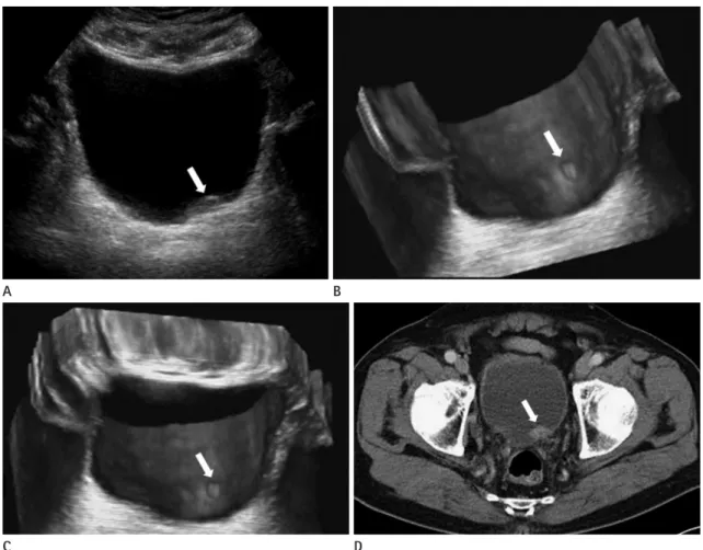
Fig. 1. Superficial polypoid bladder tumor.
A. 2D US reveals a tiny echogenic elevated bladder lesion (arrow) with indistinct margin.
B, C. 3D US demonstrates the bladder nodule (arrow) with a clearer demarcation.
D. Enhanced CT scan shows a well-enhanced polypoid bladder nodule (arrow) at the bladder base.
CT = computed tomography, US = ultrasonography, 2D = two-dimensional, 3D = three-dimensional
C D
A B
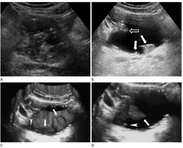
Fig. 2. Multiple superficial polypoid bladder tumors.
A. 2D US of a collapsed bladder showing unclear polypoid bladder nodules.
B. 2D US after bladder filling, three polypoid bladder nodules (arrows) are noted.
C, D. 3D US clarified three bladder masses (arrows) at a similar site. An additional bladder nodule (arrowheads) is newly identified, which was not detected on the 2D US. The new bladder nodule was confirmed by conventional cystoscopy.
US = ultrasonography, 2D = two-dimensional, 3D = three-dimensional
A B
C D
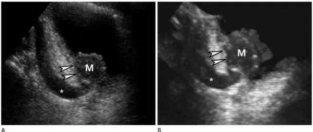
Fig. 3. Bladder mass with indwelling urinary catheter.
A. 2D US reveals an echogenic bladder mass (arrows) in a matching site, but contour of the bladder tumor is ambiguous, owing to the urinary catheter (C).
B. 3D US provides clearer information about the margin and size of the bladder tumor (arrows) and clarifies the relationship between the tumor and the catheter (C).
C. Enhanced CT scan shows a well enhancing bladder mass (arrows) along the left lateral wall of the urinary bladder, which is adjacent to the uri- nary catheter (C).
CT = computed tomography, US = ultrasonography, 2D = two-dimensional, 3D = three-dimensional
A B C
al cystoscopy remains necessary (4). However, detection accu- racies by US have known to range from 80% to 95% (10), and 3D volumetric US was more sensitive than 2D US in diagnosing bladder tumors in prior studies (3, 11). Thus it could be predicted that this technique would play an important role in the detec- tion of small lesions and could be performed when convention- al cystoscopy is not suitable, such as in patients with severe ure- thral strictures (4).
Bladder Mass with an Indwelling Urinary Catheter
Patients with urinary tract neoplasm should maintain a urinary catheter due to a variety of reasons. However, a urinary catheter adjacent to a bladder mass may cause blurring of its boundary or the reverberation artifact, which results in decreased diagnostic performance of imaging modalities. In this case, 3D volumetric US can provide additional information of spatial structures and play a role in distinguishing a bladder lesion from the adjacent urinary catheter (Fig. 3).
Invasive Bladder Cancer
Invasive bladder cancer is defined as a malignant bladder tu-
mor which invades muscularis propria and beyond (stage T2 or higher) (1, 9, 12). Accurate staging of invasive bladder cancer is important for proper tumor management. Due to the fact that invasive bladder cancer is usually solid and pathologically poorly differentiated (13), it is not a good candidate for bladder pre- serving therapy (2).
CT and MRI are helpful in the diagnosis of invasive bladder cancer when perivesical fat invasion (T3) and direct invasion to adjacent organs, abdominal wall and pelvic wall (T4) are identi- fied (9). However, in consideration of stage T2 tumors, current CT and MRIs have limitations in detecting and evaluating le- sions due to poor resolution of the bladder’s wall layers. On CT, retraction of the outer bladder wall at the site of a tumor may suggest deep muscle invasion (T2b); on MRI, T2a and T2b tu- mors can occasionally be differentiated using a combination of T2-weighted and contrast-enhanced T1-weighted sequences, be- cause T2-weighted hypointensity of the bladder muscle is pre- served in T2a tumors (1).
It is difficult to estimate the depth of invasion or to evaluate extravesical structures by US. US has also shown lower accura- cies in T-staging in association with the deeper tumors (1, 9), and current US still has a limitation in differentiating bladder wall layers (1). However, US is a real-time frontline examination with- out radiation hazards or invasiveness. Administration of 3D
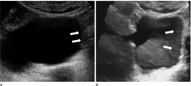
Fig. 4. Infiltrative bladder cancer in UVJ with hydroureter.
A. At the sagittal plane of a 2D US, a round hypoechoic bladder mass (M) is identified at the left ureterovesical junction with an obstructive left hydroureterer (*). The outer contour of the urinary bladder (arrowheads) looks smooth, which suggests it has not yet progressed to a stage T2 bladder tumor .
B. 3D US revealed that the outer wall (arrowheads) of the urinary bladder was irregular just beside the bladder mass (arrowheads) with an ob- structive left hydroureter (*). These findings are suggestive of perivesical invasion (T3). The lesion was confirmed by surgery and pathologically re- ported as a perivesical invasion.
US = ultrasonography, UVJ = ureterovesical junction, 2D = two-dimensional, 3D = three-dimensional
A B
volumetric techniques provides a higher sensitivity when screen- ing for bladder diseases and offers virtual sonographic cystos- copy with less invasiveness when compared with conventional cystoscopy (4). Sometimes, bladder cancer near the vesicoure- teric orifices may cause ureteric obstruction presenting hydro- ureter or hydroureteronephrosis (1), which can be identified during a US exam (Fig. 4).
Thus, US with a 3D volumetric technique may play a role in evaluating the local extent of bladder lesions when considering stage T2 or less invasive bladder cancer, even if current US tech- niques have limitations in differentiating bladder wall layers and evaluation of extravesical structures (1).
Direct Invasion of Bladder by Malignancy from Adjacent Pelvic Organs
Direct bladder invasion by advanced cervical cancer, which is classified as FIGO stage IV and is not suitable for local treatment, is reported in about 11.5% of patients (14). An MRI has high accuracy in detection and quantification of bladder invasions, but it is expensive and not available in all medical institutions.
A can US provide similar information about the evaluation of a bladder wall invasion when compared to that of an MRI (14).
US features in each sequential stages of a bladder wall invasion is presumed to be a disruption of the endopelvic fascia without in-
volvement of the inner bladder wall, thickened bladder wall and changes in bladder mucosa, or interruption of entire bladder wall (Fig. 5).
About 5% to 10% of colorectal cancers extend to the adjacent organ; urinary bladder is the most commonly involved (6). As described above, US features demonstrate sequential stages of in- vasion, ranging from disruption of mesorectal fascia to interrup- tion of the entire bladder wall with mass formation (15).
Metastatic Cancer
Metastatic tumors of the urinary bladder from distant primary malignant foci are rare, and account for only 1.5% of all bladder tumors. The most common primary sites are gastric carcinoma, malignant melanoma, breast carcinoma, and lung carcinoma (16). Metastatic bladder tumors show either polypoid lesions similar to that of typical urothelial cell carcinoma, or focal wall thickening of the urinary bladder (Fig. 6) (16). As bladder me- tastasis is a late manifestation of malignancy, evidence of inva- sive primary neoplasm or other signs of a distant primary neo- plasm is commonly identified on imaging studies (7).
Non-Tumorous Bladder Conditions
There are many non-tumorous bladder conditions mimick- ing bladder cancer due to the manifestation of focal bladder wall
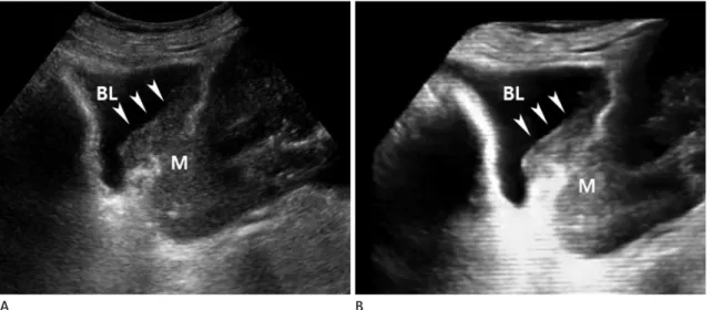
Fig. 5. Direct invasion of the urinary bladder by advanced cervical cancer.
A. 2D US shows a large ill-defined uterine cervical mass (M) extending to the bladder (BL) wall (arrowheads).
B. 3D US demonstrates the margin of bladder mass more clearly than a conventional 2D US technique (arrowheads).
US = ultrasonography, 2D = two-dimensional, 3D = three-dimensional
A B
thickening (17), these include infections (cystitis, tuberculosis, acute schistosomiasis, malacoplakia, cystica glandularis), amy- loidosis, endometriosis, and even benign bladder wall trabecu- lation. Flat bladder lesions can be easily missed on US but even when seen the appearances are often equivocal. In these cases, a sonographic differential diagnosis must be considered and fur- ther work up, including conventional cystoscopy, should be recommended.
Focal cystitis shows nonspecific bladder wall thickening and nodularity, therefore sometimes it is misdiagnosed as transitional
cell carcinoma (Fig. 7).
A urinary stone at the ureterovesical junction (UVJ) with sur- rounding focal inflammatory changes due to irritation by the stone is shown as focal UVJ thickening, mimicking bladder can- cer. In this case, a US examination will help to find any evidence of urinary stones, demonstrating echogenic structures with distal posterior acoustic shadowing (Fig. 8). Recent studies report that a transvaginal US in females is helpful in the detection of urinary stones (18).
Fig. 6. Metastatic bladder adenocarcinoma from advanced gastric cancer. 2D US and 3D US show diffuse infiltrative masses (arrowheads), con- firmed to be have metastasized from gastric cancer to the lateral wall of the urinary bladder.
US = ultrasonography, 2D = two-dimensional, 3D = three-dimensional
Fig. 7. Focal cystitis.
A. 2D US reveals multiple elevated nodular bladder lesions (arrows) with an indistinct margin.
B. 3D US more clearly demonstrates bladder mucosal nodularity (arrows) than 2D US.
US = ultrasonography, 2D = two-dimensional, 3D = three-dimensional
A B
CONCLUSION
Various diseases of urinary bladder (including bladder cancer, benign neoplasm, inflammatory condition, and even urinary tract stones) may be demonstrated as a polypoid, nodular blad- der mass or as focal wall thickening of the urinary bladder; they are often easily confused with each other. In consideration of bladder cancer, a conventional cystoscopy is mandatory when attempting to determine a diagnosis. In addition a CT or MRI should be performed during an evaluation of extravesical struc- tures because a US has limitations in the differentiation of blad- der wall layers and the evaluation of a perivesical invasion.
Nevertheless, application of 3D volumetric US in this article has shown good depiction of bladder lesions with better spatial resolution. Especially when comparing other image modalities such as 2D gray scale US, CT, or MRI in cases of superficial and small lesions of the bladder. US is also well known to be a safe, widely available and less time-consuming imaging modality.
Application of 3D volumetric US provides additional infor- mation in the distinction and differentiation of various bladder diseases, including bladder cancer, with no harm to the patients.
REFERENCES
1. Ng CS. Radiologic diagnosis and staging of renal and blad-
der cancer. Semin Roentgenol 2006;41:121-138
2. Wagner B, Nesslauer T, Bartsch G Jr, Hautmann RE, Gott- fried HW. Staging bladder carcinoma by three-dimensional ultrasound rendering. Ultrasound Med Biol 2005;31:301- 305
3. Silva-Ramos M, Louro N, Versos R, Cavadas V, Marcelo F.
Does 3D ultrasound enhance the diagnosis of bladder tu- mours in patients with haematuria? ISRN Urol 2012;2012:
158437
4. Kocakoc E, Kiris A, Orhan I, Poyraz AK, Artas H, Firdolas F.
Detection of bladder tumors with 3-dimensional sonogra- phy and virtual sonographic cystoscopy. J Ultrasound Med 2008;27:45-53
5. Moon MH, Kim SH, Lee YH, Cho JY, Jung SI, Park SH, et al.
Diagnostic potential of three-dimensional ultrasound-based virtual cystoscopy: an experimental study using pig blad- ders. Invest Radiol 2006;41:883-889
6. Downey DB, Fenster A, Williams JC. Clinical utility of three-dimensional US. Radiographics 2000;20:559-571 7. Wong-You-Cheong JJ, Woodward PJ, Manning MA, Sester-
henn IA. From the Archives of the AFIP: neoplasms of the urinary bladder: radiologic-pathologic correlation. Radio- graphics 2006;26:553-580
8. Pashos CL, Botteman MF, Laskin BL, Redaelli A. Bladder cancer: epidemiology, diagnosis, and management. Cancer Fig. 8. A left UVJ stone.
A. 2D US shows a small echogenic urinary stone (arrowhead) at the left ureterovesical junction with swelling of the surrounding soft tissue (ar- rows).
B. 3D US demarcates the urinary stone (arrowhead) and swelling of left UVJ (arrows) more clearly.
US = ultrasonography, UVJ = ureterovesical junction, 2D = two-dimensional, 3D = three-dimensional
A B
Pract 2002;10:311-322
9. Vikram R, Sandler CM, Ng CS. Imaging and staging of tran- sitional cell carcinoma: part 1, lower urinary tract. AJR Am J Roentgenol 2009;192:1481-1487
10. Cochlin DL, Dubbins PA, Goldberg BB, Halpern EJ. Urogenital Ultrasound: a text atlas. 2nd ed. London: Taylor and Francis, 2006:147-150
11. Park HJ, Hong SS, Kim JH, Kwon SB, Kwon KH, Choi DL, et al. Tumor detection and serosal invasion of bladder cancer:
role of three-dimensional volumetric reconstructed US. Ab- dom Imaging 2010;35:265-270
12. Lee R, Droller MJ. The natural history of bladder cancer. Im- plications for therapy. Urol Clin North Am 2000;27:1-13, vii.
13. O’Brien T, Cranston D, Fuggle S, Bicknell R, Harris AL. Dif- ferent angiogenic pathways characterize superficial and
invasive bladder cancer. Cancer Res 1995;55:510-513 14. Huang WC, Yang JM, Yang YC, Yang SH. Ultrasonographic
characteristics and cystoscopic correlates of bladder wall invasion by endophytic cervical cancer. Ultrasound Obstet Gynecol 2006;27:680-686
15. Nyam DC, Seow-Choen F, Ho MS, Goh HS. Bladder in- volvement in patients with colorectal carcinoma. Singa- pore Med J 1995;36:525-526
16. Kim HC, Kim SH, Hwang SI, Lee HJ, Han JK. Isolated blad- der metastases from stomach cancer: CT demonstration.
Abdom Imaging 2001;26:333-335
17. GM Baxter, PS Sidhu. Ultrasound of the urogenital system.
New York: Thieme, 2006:137-138
18. MI Resnick, RA Older. Diagnosis of genitourinary disease.
New York: George Thieme Verlag, 1997:588
방광암과 유사 질환의 감별에 있어 3차원 초음파의 적용: 임상 화보
고수진 · 홍성숙* · 황지영 · 김현주
방광의 다양한 질환은 결절 형태의 종괴나 국소적인 벽 두께 증가로 나타날 수 있는데, 양성 및 악성 종양, 요관 결석 그리 고 다양한 염증성 질환 등을 포함한다. 이러한 질환들은 감별이 중요한 방광암과 혼돈될 수 있다. 초음파검사는 방광 질 환의 선별 검사 또는 일차적 진단 방법으로서 안전하며 쉽게 적용될 수 있다. 게다가 추가적인 3차원 초음파 기법(three- dimensional volumetric technique)은 병변을 더 자세히 구분하고 분석하는 데 추가적인 정보를 줄 것이다. 저자들은 방광 암 및 방광의 다양한 양성 질환들을 구분하기 위해 3차원 초음파검사의 기법을 적용하고자 하였다.
순천향대학교 서울병원 영상의학과