신성선종(nephrogenic adenoma)은 이형성(metaplasia)의 특성을 가진 비뇨기계의 드문 종양으로 방광에 호발한다. 요 로에 대한 수술을 받은 과거력이 있거나 요로염증을 앓았던 환자에서 발생되나 이러한 요로 자극요소들과 신성선종 간의 명확한 관련여부는 아직까지 정확하게 밝혀진 바가 없다 (1).
조직학적 특성은 입방형 상피세포와 이형성 관 구조물들로 구 성되는 것이며 신성 이형성증(nephrogenic metaplasia) 또는 선종성 이형성증(adenomatous metaplasia) 이라고도 불리우 고 있다 (2). 현재까지 내시경소견 혹은 병리조직소견이 보고 된 바 있으나 방사선학적 소견에 대한 보고는 드물다 (3, 4).
저자들은 8세 여아의 방광에 발생된 신성선종 1예를 경험 하였기에 초음파소견, 전산화단층촬영 (이하 CT로 약함)과 MRI 소견을 문헌고찰과 함께 보고한다.
증례 보고
8세 여아가 배뇨장애를 주소로 내원했다. 환아는 3년전 교 통사고로 인한 치골 골절, 방광파열, 요도 및 질 손상을 입고 타병원에서 방광과 요도의 일차 봉합 등의 수술 치료를 받았 다. 입원 중 요도협착, 급성 신우신염, 폐쇄성 신병증, 방광요 관역류증 등으로 장기간동안 폴리카테터를 삽입하였으며 보존 적 치료를 받던 중 배뇨장애가 악화되어 본원으로 전원되었다.
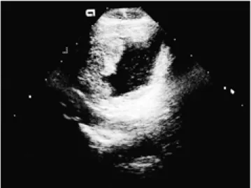
입원 후 시행한 초음파검사상 심한 수신증과 요관 확장이 있 었고 방광벽이 불균질한 저에코로 광범위하고 불규칙적으로 비후되었으며 특히 우측벽에는 유두상의 종괴가 관찰되었고 (Fig. 1) 도플러검사에서는 이 부위에 혈류신호가 증가되었다.
CT상 비후된 방광벽은 불균질하게 조영증강되고 우측의 유두 상의 비후부위에서는 보다 강하게 조영증강되었다 (Fig. 2). 방
광경에서 우측 방광벽의 다발성 유두상 종괴는 부드럽고 유동 성이 있으며 요도를 간헐적으로 막고 있었다. 조직검사상 신 성선종으로 판명되었고 환아는 치골상방광루조성술 (supra- pubic cystostomy)을 시행받았다. 1년 6개월후의 추적검사중 치골상방광루 부위의 복벽이 충혈되고 종창이 심해져 시행한 MRI에서는 T1강조영상에서 방광벽은 전반적으로 비후되고 불 균질한 저신호강도이며 (Fig. 3A) T2강조영상에서는 불균질 한 고신호강도이며 유두상 종괴가 방광의 우측 전상벽에서 관 찰되었다. 조영증강 T1강조영상에서 비후된 방광벽은 불균질 하고 강하게 조영증강되며 상부 방광벽에는 유두상의 종괴가 관찰되었고 관상스캔에서는 방광삼각부 침범 및 요관의 확장 도 관찰되었다 (Fig. 3B). 또한 치골상방광루 부위의 복벽을 따라 농양이 형성되었고 이는 비후된 방광벽과 연결되었다. 방 광 부분절제 및 회맹장도관(ileocecal conduit)을 시행하였고 조직병리학적 검사에서 유두상의 관조직(papillary tubules)이 편평 혹은 입방형 세포로 둘러싸여 있고 방광의 점막층과 일 부 점막하층을 침범한 신성선종으로 진단되었다 (Fig. 4).
고 찰
신성선종은 1950년 Friedman과 Kuhlenbeck (5)에 의해 처 음 보고되었으며 요로에 생기는 매우 드문 종양이다. 발생기 전은 명확히 규명된 바 없으나 만성 또는 반복적인 요로감염 등의 염증이나 요로에 대한 외과적 시술, 요로결석이나 이물 질 등에 의한 물리적 자극에 대한 이형성반응 때문으로 알려 져왔고 (1) 신성 이형성증 또는 선종성 이형성증(adenoma- tous metaplasia)으로 불리워지기도 한다. 조직학적 특성은 입 방형 세포와 이형성 관 구조물로 구성된 것이다 (2). 요로계 어느 부분에서나 발생할 수 있고 방광이 호발부위이나 이는 방광종양의 약 1% 미만을 차지하고 드물게는 요로게실이나 회장도관(ileal conduit)에서 발생된 증례도 보고된 바 있다 (6, 대한방사선의학회지 2001;44:377-379
─ 377 ─
방광에 발생한 신성선종: 1예 보고1
정 선 희・이 선 화・한 운 섭2
신성선종은 이형성 특성을 보이는 비뇨기계의 양성질환으로 방광에 호발하나 소아에서는 매 우 드문 방광종양이다. 저자들은 3년전 교통사고로 요도, 질 및 방광이 파열되고 요도협착 및 방광요관역류증 등의 후유증이 발병된 8세 여아에서 발생된 방광의 신성선종 1예를 경험하였 기에 이의 초음파소견, 전산화단층촬영 (CT) 및 자기공명영상 (MR) 소견을 보고한다.
1이화여자대학교 의과대학 진단방사선과학교실
2이화여자대학교 의과대학 해부병리학교실
이 논문은 2000년 11월 2일 접수하여 2001년 1월 16일에 채택되었음.
7). 신성선종은 성인에서 호발되며 평균연령이 40-50 세로 알려져있고 Oliva와 Young (7)은 여자에서 그 빈도가 2배 이 상 높은 것으로 보고한 반면, Vorreuther 등 (8)은 성인에서 는 남자, 소아의 경우는 여아에서 발생빈도가 더 높은 것으로 보고하였다. 주된 임상증상은 혈뇨나 배뇨통 등이다.
신성선종의 방광경소견이 비뇨기과영역의 논문에서 널리 보 고된 반면 이에 대한 방사선학적 영상진단소견에 관한 보고는 드물다 (3, 4). 일부연구에서 초음파소견은 저에코의 방광벽 비후 또는 방광 내부로 돌출되는 저에코 종괴로 보고되었으며 (3) 저자들의 경우에도 유사한 소견이 관찰되었다. CT소견은 방광 내부의 종괴와 방광벽의 비후로 악성종양과 유사한 것으 로 보고되었으며 (4) 본 증례의 경우도 전반적인 방광벽의 비 후와 국소적 종괴형성으로 관찰되었다. 또한 강한 조영증강 소
견을 보여 악성종양과 유사하였다. 그러나, 인접조직의 침윤 및 임파절 종대의 소견은 없었다. MR소견은 요도게실에 발생된 신성선종 1예에서 보고되었고 이는 T1 및 T2강조영상에서 액 체의 신호강도를 보이는 다방성 낭종으로 보고되었다 (6). 저 자들의 경우는 T1강조영상에서는 불균질한 저신호강도, T2강 조영상에서는 불균질한 고신호강도였고 불균질한 강한 조영증 강이 관찰되었다. 본 증례의 경우 CT 혹은 MR로서 신성선종 이 침범된 부위가 정상부위와 감별되며 방광벽의 전층이 침범 된 것으로 수술전 진단하였으나 병리적으로 점막 및 점막하층 의 침범이 확인되었다. 신성선종과 감별진단을 요하는 질환으 로는 국소성 방광염으로 인한 위종양이나 이행상피세포암 (transitional cell carcinoma), 선암 (adenocarcinoma)과 횡문 근육종 (rhabdomyosarcoma) 등이 있으며 악성 종양의 경우 정선희 외 : 방광에 발생한 신성선종
─ 378 ─ Fig. 1. Transverse sonographic scan of the urinary bladder shows irregular thickening and polypoid projection along the right lateral wall.
Fig. 2. CT image demonstrates heterogeneous and intense en- hancement at the right lateral wall of the urinary bladder (ar- rows).
A B
Fig. 3. MR performed 1 1/2 years after initial US and CT. T1-weighted axial scan (A) shows diffuse and irregular thickening of the urinary bladder wall with heterogeneous low signal intensity. Gd-enhanced T1-weighted coronal scan (B) reveals irregular thicken- ing with polypoid projections(white arrows) at the heterogenously enhanced urinary bladder wall . The bladder is distorted and the right distal ureter is dilated (black arrows).
는 불분명한 변연, 주위침습 혹은 림프절종대등의 소견을 흔 히 보인다 (3, 6).
방광의 신성선종에 대한 치료는 방광경을 통한 외과적 절제 술이 널리 이용되나 본 증례와 같이 광범위한 경우는 방광절 제술도 시행한다. 재발율은 소아의 경우 75%, 성인 38%로 보 고된 바 있고 재발을 진단하기 위해서 정규적인 방광경검사가 권장되고 있다 (2). 저자들의 증례경우 첫 방광경진단 1년6개 월후 추적검사에서 방광벽의 침범이 더 진행되었으며 또한 이 러한 평가에 초음파검사가 선별검사로서 간편하고 유용하였다.
결론적으로 소아의 방광벽이 불규칙적으로 비후되거나 유두
상의 종괴가 관찰될 경우 특히 요로계의 손상 혹은 물리적 자 극을 받았던 과거력이 있는 경우에는 신성선종의 가능성도 고 려해야 할 것으로 생각된다.
참 고 문 헌
1. Cremer H. Adolphs HD. The natural history of nephrogenic ade- noma of the urinary bladder. Z Krebsforsch Klin Onkol Cancer Res Clin Oncol 1978;91:49-53
2. Husan AN, Armin AR, Schuster GA. Nephrogenic metaplasia of urinary tract in children: report of three cases and review of the lit- erature. Pediatr Pathol 1988;8:293-300
3. Jequier S, Bugmann P, Brundler MA. Nephrogenic adenoma of the bladder: ultrasound demonstration. A case report. Pediatr Radiol 1999;29:185-187
4. Zingas AD, Kling GA, Crotte E, Shumaker E, Vazquez PM.
Computed tomography of nephrogenic adenoma of the urinary bladder. J Comput Assist Tomogr 1986;10(6):979-82
5. Friedman NB, Kuhlenbeck H. Adenomatoid tumor of the bladder reproducing renal structures (nephrogenic adenomas). J Urol 1950;
64:657-670
6. Carl GK, Ece IA, Jeffrey JB. Nephrogenic adenoma arising from a urethral diverticulum: magnetic resonance features. Urology 1995;
45:323-325
7. Olivia E, Young RH. Nephrogenic adenoma of the urinary tract: a review of the microscopic appearance of 80 cases with emphasis on unusual features. Mod Pathol 1995;8:722-730
8. Vorreuther R, Naval W, Hake R, Engelmann U. Nephrogenic ade- noma of the bladder. Urol Int 1994;53:227-275
대한방사선의학회지 2001;44:377-379
─ 379 ─ Fig. 4. Photomicrograph shows many tubules with lining of cuboidal to flattened epithelium in small aggragated pattern and dilated lumens (H&E stain, ×250).
J Korean Radiol Soc 2001;44:377-379
Address reprint requests to : Sun Wha Lee, M.D., Department of Diagnostic Radiology, Ewha Womans University, Mokdong Hospital 911-1, Mok-dong, Yangcheon-gu, Seoul 158-710, Korea.
Tel. 82-2-650-5174 Fax. 82-2-2644-3362 E-mail: [email protected]
Nephrogenic Adenoma Arising from the Urinary Bladder:
A Case Report
1Sun Hee Chung, M.D., Sun Wha Lee, M.D., Woon Seup Han, M.D.2
1Department of Diagnostic Radiology, College of Medicine, Ewha Womans University
2Department of Anatomical Pathology, College of Medicine, Ewha Womans University
Nephrogenic adenoma is a benign metaplastic lesion of the urinary tract occurring most frequently at the urinary bladder. It is very rare, especially in children. We describe the US, CT and MRI findings of nephro- genic adenoma arising from the urinary bladder in an 8-year-old girl who suffered rupture of the bladder, ure- thra and vagina after a traffic accident and whose condition was complicated by urethral stricture and vesi- coureteral reflux.
Index words :Bladder, US Bladder neoplasms Bladder, MR
─ 380 ─