: Vol. 35, No. 3, 2005
MMP-2, MMP-8 Expression in gingival tissue of Chronic Periodontitis associated to
Type 2 Diabetes Mellitus
Min-Gu Kang Hyun-Jeong ChaㆍSun-Hee Songㆍ Jin-woo ParkㆍJo-Young SuhㆍJae-Mok Lee
Department of Periodontology, College of Dentistry, Kyungpook National University
Ⅰ. Introduction
Diabetes mellitus(DM) in itself does not cause periodontitis. However, there are epi- demiologic evidences that diabetes predis- poses and accelerates the irreversible perio- dontal destruction. Severe periodontitis has been associated with an increased risk of poor glycemic control and, in turn, un- treated advanced periodontal disease can deteriorate the metabolic control of dia- betes1-4).
Accumulation of plaque is not related di- rectly to the early onset and rapid pro- gression of periodontal tissue destruction in diabetes. Various pathogenic factors have been suggested to explain the increased prevalence and severity of periodontitis in diabetes. An exaggerated inflammatory re- sponse to potential periodontopathogenic
bacterial virulence factors, such as lipopoly- saccharide(LPS), may influence the perio- dontitis in diabetes. Moreover, an impaired recruitment and cellular function of poly- morphonuclear leukocytes(PMNLs) has been linked to extensive periodontal tissue de- struction1-3).1)
Structual studies on diabetic patients have revealed vascular changes in gingiva and other tissues, a thickening of subendotherial basement membrane and, paradoxically, an increased permeability of basement mem- brane to virulent pathogenic microflora and to its potential vilurent factors5-7). Futher- more, a prolonged exposure to hyperglycemia can be primary factor responsible for the for- mation of non-enzymatic advanced glyco- sylation end-products(AGEs), demonstrated recently in periodontal tissues of diabetic patients. AGEs are further responsible for Correspondence author: Jae-Mok Lee, Department of Periodontology, College of Dentistry, Kyungpook National University, 50 Samduk-Dong, Jung-Gu, Daegu, 700-422, South Korea
diabetic collagen cross-links, which again lead to the above-mentioned microvascular complications such as vascular hardening and basement membrane disintegration2,8,9). AGEs also bind to macrophage receptors and induce a cycle of cytokine(Interleukin-1:IL -1, Tumor Necrosis Factor-α:TNF-α) upregu- lation. These can, in turn, induce an ele- vated expression of matrix metalloprotein- ases(MMPs) in diabetic periodontal tissue
10-12).
MMPs are a family of at least 16 secreted and membrane-bound zinc- and calcium- de- pendent protease which function in the deg- radation and remodeling of extracellular ma- trix proteins during different developmental processes such as organ morphogenesis, bone formation, angiogenesis, and remodel- ing during reproductive processes, as well as in pathological processes such as inflamma- tion, chronic degenerative disease and tumor invasion13).
In summary, they are responsible for deg- radation of extracellular matrix components such as collagen and proteoglycans. MMPs, produced by both infiltrating and resident cells of the periodontium, play a role in physiologic and pathologic events. It is rec- ognized that an imbalance between activated MMPs and their endogenous tissue inhibi- tors of metalloproteinases(TIMPs) leads to pathologic breakdown of the extracellular matrix during chronic periodontitis14-17).
Of all MMPs, matrix metalloproteinase-2 (MMP-2, 92 kDa gelatinase A) is the most widely distributed. MMP-2 has been identi- fied in fibroblast, keratinocytes, endothelial cells, monocytes/macrophages, osteoblast,
odontoblast and chondrocytes. And ex- pression of MMP-2 in periodontium is lo- calized mainly in fibroblast and granulation tissue of periodontal connective tissue(CT).
In human periodontium, MMP-2 affects in- tegrity of basement membrane and attach- ment of junctional epithelium and elevated MMP-2 expression level in chronic perio- dontitis is already reported in several inves- tigations15-20). These results suggest the in- volvement of MMP-2 in periodontal destruc- tion.
The major collagenase species detected in inflamed human periodontium is inductive collagenase-2 or MMP-8 rather than con- stitutively expressed fibroblast-type MMP-1, which is more associated with normal tissue remodeling.8,13) MMP-8 is mainly a polymor- phonuclear leukocyte-(PMN-) specific matrix metalloproteinase stored in specific granules of PMNs.13,17)When neutrophils are recruited to a site of inflammation, they release large quantities of MMP-8 stored in specific gran- ules, and the inflammatory response is sus- tained by recruitment of new cells rather than by the local synthesis of MMP-8. MMP -8 activity and release are regulated by fac- tors such as cytokines(TNF-α or IL-1β) and various bacterial virulence factors that affect MMP-8 release by PMN degraulation follow- ing PMN infiltration to the site of inflam- mation.2)During active phase of peridontitis, MMP-8 levels in GCF & inflammed gingival tissue are pathologically elevated, and may have a key role of periodontal destruc- tion.13,14)On the other hands, several studies show evidences that type 2 diabetes mellitus induces dysregulation of the MMP and
Tissue Inhibitors of Metalloproteinase (TIMP) system in human gingival tissue
3,8,21). The effect of type 2 diabetes on MMP and TIMP expression effectively amplifies the degradation arm of the MMP/TIMP sys- tem that could lead to net activation of ma- trix breakdown.
Because investigations to examine MMP- 2, MMP-8 expression in diabetic gingival tissue related to periodontitis are limited, we investigated the effect of type 2 diabetes mellitus on MMP-2, MMP-8 production in human gingival tissue with chronic perio- dontitis. The purpose of this study is to quantify and to compare relation of the ex- pression of MMP-2, MMP-8 in the gingival tissue of patients with type 2 DM and sys- temically healthy adults with chronic perio- dontitis.
Ⅱ. Materials and Methods
1. Study population and Tissue sampling
Study population comprised 8 patients with type 2 diabetes mellitus aged 36-68 years and 16 non-diabetic control subjects of the similar age group. Before surgery, pa- tient's systemic condition(age, sex, blood glucose level) and clinical criteria of gingi- va(Sulcus Bleeding Index according to Muhlemann and Son22):SBI, pocket depth) were recorded. Avoiding local anesthetic in- filtration into biopsy site, marginal gingival tissue samples were obtained during perio- dontal surgery or tooth extraction following informed consent.
Each gingival samples were divided into
three groups based on the patient's systemic condition & clinical status of gingiva (gin- gival color, gingival bleeding, probing dep- ths, and radiographic evidence of bone re- sorption).
Group 1(normal, n=8, mean age 41 [30 to 60], 5 males and 3 females) is clinically healthy gingiva obtained from 8 systemically healthy adults. Gingival samples were ob- tained during crown lengthening procedure from tooth with sulcus depth ≤3 mm, no loss of attachment and no inflammatory signs. Mean glucose level was 110.87 mg/dl.
Group 2(chronic periodontitis, n=8, mean age 40 [33 to 48], 5 males and 3 females) is inflammed gingiva from patients with chron- ic periodontitis. The diagnosis of chronic pe- riodontitis was established on the basis of clinical and radiographic criteria(bone re- sorption) according to the classification sys- tem for periodontal diseases and condi- tions23). All patients in group 2 were sys- temically healthy and had more than one periodontal pocket of ≥5 mm depth and at least one pocket with ≥4 mm loss of attachment. All gingival samples were ob- tained during periodontal flap surgery, gin- givectomy or extraction from teeth with probing depth ≥5 mm, swelling of the mar- ginal gingiva and bleeding corresponding to Sulcus Bleeding Index 3. Mean glucose level was 103.25 mg/dl and this value was slight- ly lower than that of group 1. Group 3 (chronic periodontitis associated to type 2 DM, n=8, mean age 55 [36 to 71], 6 males and 2 females) is inflammed gingiva from type 2 diabetic patients with chronic periodontitis. Patients in group 2 and 3 had
similar periodontal conditions, but systemi- cally, patients in group 2 were healthy and patients in group 3 were suffering from type 2 diabetes mellitus. Mean glucose level was 163.57 mg/ dl in FBS and 228.57 mg/dl in 2PP. Diabetic control was performed by in- sulin medication in all 8 patients and, addi- tionally, diet control was also performed in 3 of them. Gingival samples were obtained by similar way described above.
The sample cohort consisted of 8 clinically healthy, 8 inflammed and 8 diabetic pa- tients' inflammed samples from total of 24 subjects. Following surgery, excised tissue specimens were immediately placed on liquid nitrogen and subsequently frozen(-70℃).
2. Tissue preparation, Protein Isolation and Western blotting
For Western blotting, as previously de- scribed technique by Cho,24) frozen tissues were homogenized in RIPA lysis buffer(10 mM phosphate buffer, pH 7.4, 10% glycer- ol, 1% NP-40, 0.1% SDS, 4 mM EDTA, 0.15 M NaCl) with 1:30 diluted protease in- hibitor cocktail(Roche). The lysates were so- nicated 3 times for 10 seconds and centri- fuged at 12,000 g for 15 minutes. Protein concentration of supernatant were routinely determined by a Bradford protein assay us- ing Bio-Rad protein assay reagent with BSA as a standard.
Lysates were boiled in SDS sample buffer (1M Tris-Cl(pH 6.8), 40% glycerol, 8% SDS, 2%-mercaptoethanol, 0.002% Bromophenole blue). Prepared samples were separated by 10% sodium dodecyl sulfate(SDS)-polya-
crylamide gels and transferred to a poly- vinylidene difluride membrane. The mem- branes were subsequently blocked in Tris- buffered saline(TBS) containing 5% pow- dered milk and 1% BSA for 1 hours, and then incubated with polyclonal anti-MMP-2 antibody(Sigma-Aldrich Corporation, ST.
Luis, U.S.A, prepared in rabbits, diluted 1:
1000 in TBS/1% BSA) for 1.5 hours at room temperature. The membrane was washed (five times for 5 minutes with Tween 20) and incubated with a horseradish peroxidase (HRP)-conjugated goat anti-rabbit secondary antibody(diluted 1: 1000 in TBS) for 1 hours at room temperature. After additional wash- ing(five times for 5 minutes with Tween 20) the Western blot procedure was completed with an ECL Advance development kit(Amer- sham, Beckinghamshire, U.K.)
The quantitative analysis(ratio of MMP-2, MMP-8/β-actin) of MMP-2 MMP-8 expre- ssion was performed using a densitometer (Image Gauge V 3.46, Koshin Graphic Sys- tems, FUJI PHOTO FILM CO, Japan). After normalization to β-actin(Abcam Ltd., Cam- bridge Science Park, UK) in each sample, level of MMP-2 was expressed as a ratio(ra- tio of MMP-2, MMP-8/β-actin) and the dif- ferences of densities among 3 groups were determined.
3. Statistical Analysis of the Western blot results
All data were presented as means ± standard deviation and results were statisti- cally analyzed. The relative MMP-2, MMP-8 levels among each 3 groups were compared
Group 1 Group 2 Group 3
1 2 3 4 1 2 3 4 1 2 3 4
MMP-2 B-actin
Figure 1. Western analysis of samples for MMP-2. MMP-2 corresponding to molecular weight 72 kDa was expressed in all samples including healthy gingiva, and expression of MMP-2 was increased in group 3 than in group 1 subjects. In order to quantify detected MMP-2, normalization to β-actin was performed.
Group 1 : healthy gingiva from systemically healthy patient
Group 2 : inflammed gingiva from systemically healthy patient with chronic periodontitis Group 3 : inflammed gingiva from type 2 diabetic patient with chronic periodontitis using one way ANOVA followed by Scheffe
test. The criterion for statistical significance was defined as P<0.05.
Ⅲ. Results
Antibodies to MMP-2, MMP-8 cross-re- acted with 72, 60kDa molecular weight of MMP-2, MMP-8 in 3 groups. In order to quantify detected MMP-2, MMP-8 normal- ization to β-actin was performed. The quan- tification(ratio of MMP-2, MMP-8/β-actin) of MMP-2, MMP-8 in each sample was per- formed using a densitometer. The average amounts of MMP-2, MMP-8 among 3 groups were compared in Table 1, 2 and Figure 1, 2, 3. Although age, glucose level and SBI value were similar within the same group, various amount of MMP-2, MMP-8 was de- tected in each samples.
As to mean MMP-2 levels in each 3 groups, the amount of MMP-2(ratio of MMP -2/β-actin) was 2.108, 2.560 and 3.266 in group 1, 2 and 3. There were some tenden- cies that mean amount of MMP-2 was rather
increased in inflamed gingiva compared to healthy gingiva and was more increased in type 2 diabetic gingival tissue. That is, the amount of MMP-2 was increased in group 2 compared to group 1. Also, increased amount of MMP-2 was seen in group 3 compared to group 2. But the differences among 3 groups were not statistically significant (P>0.05).
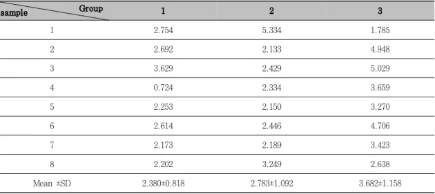
As to mean MMP-8 in each 3 group, the amount of MMP-8(ratio of MMP-2/β-actin) was 2.380, 2.783, 3.682 in group 1, 2, 3.
The amount of MMP-8 was increased in group 2 compared to group 1. Also, in- creased amount of MMP-8 was observed in group 3 compared to group 2. But the dif- ference among 3 groups was not statistically significant(P>0.05).
That is, the amount of MMP-2, MMP-8 was increased in group 2, 3 compared to group 1. Also, increased amount of MMP-2, MMP-8 was observed in group 3 compared to group 2. But there were no statistically significant difference in among 3 groups (P>0.05)
Figure 2. Mean amounts(ratio of MMP-2/β-actin) and standard deviation of MMP-2 in group 1, 2 and 3. In the inflamed gingival(with or without diabetes, group 2 and 3), MMP-2 was increased compared to healthy gingiva. But the differences were not statistically significant(P>0.05).
Table 1. The quantitative analysis(ratio of MMP-2/β-actin) of MMP-2 of each sample was per- formed using a densitometer and average amount of MMP-2 in 3 groups were identified. The differences between 3 groups were not statistically significant(P>0.05).
sample Group 1 2 3
1 2.210 1.422 3.619
2 2.676 0.690 2.019
3 2.413 3.821 2.958
4 1.829 4.867 6.071
5 2.116 2.290 2.713
6 1.332 2.730 3.047
7 1.984 2.368 3.046
8 2.304 2.296 2.655
Mean ±SD 2.108±0.407 2.560±1.304 3.266±1.219
Table 2. The quantification(ratio of MMP-2/β-actin) of MMP-8 of each sample was performed using a densitometer and average amount of MMP-8 in 3 groups were identified.
sample Group 1 2 3
1 2.754 5.334 1.785
2 2.692 2.133 4.948
3 3.629 2.429 5.029
4 0.724 2.334 3.659
5 2.253 2.150 3.270
6 2.614 2.446 4.706
7 2.173 2.189 3.423
8 2.202 3.249 2.638
Mean ±SD 2.380±0.818 2.783±1.092 3.682±1.158
Figure 3. Mean amounts(ratio of MMP-8/β-actin) and standard deviation of MMP-8 in group 1, 2 and 3. In the inflamed gingiva(with or without dia- betes, group 2 and 3), MMP-8 was increased compared to healthy gingiva.
In group 3, the increase of MMP-8 was statistically significant compared to group 1(P>0.05).
Ⅳ. Discussion
The purpose of this study was to inves- tigate the effect of type 2 diabetes mellitus on MMP-2, MMP-8 production in human gingival tissue with chronic periodontitis.
MMP-2, MMP-8 corresponding to molec- ular weight 72, 60kDa was expressed in most
samples. The quantitative analysis(ratio of MMP-2,-8/β-actin) of MMP-2, MMP-8 level in each sample was performed using a densi- tometer and we could find some tendencies that mean amount of MMP-2, MMP-8 was rather increased in inflamed gingiva com- pared to healthy gingiva and was more in- creased in type 2 diabetic gingival tissue.
Enhanced expression of MMP-2 in chronic periodontitis patient and its activation mechanisms were investigated in several studies15,17,20). For instance, Anne-Laure Ejeil15) et al studied the relation between degree of gingival inflammation and ex- pression of MMPs, and showed that active forms of MMP-2 are highly increased in in- flamed gingiva. About its overexpression mechanism, Chang et al25) studied the regu- lation of MMP-2 synthesis and secretion in human periodontal ligament fibroblast(PD- LF) and found that periopathogens, such as P. gingivalis and A actionomycetemcomitans, and IL-1α can elevate MMP-2 secretion in human PDLF. In periodontitis, PDL cells are a major group of cells highly affected by the P. gingivalis product. Pattamapon et al26) observed MMP-2 activation in human periodontal ligament fibroblast by adding P.
gingivalis supernants in vitro study and Grayson et al27) observed elevated MMP-2 levels in gingival crevicular fluid of chronic periodontitis patient and concluded that P.
gingivalis produces typsin-like proteases and these activated latent pro MMP-2 to active form in concentration- and time- dependent manner.
Therefore, it was suggested that enhanced expression of MMP-2 in periodontal disease is mainly related to increase of P. gingivalis, A. actinomycetemcomitans and IL-1 etc.
However investigations in chronic periodon- titis associated to type 2 DM are limited and, to our knowledge, no regulatory mecha- nisms of MMP-2 in type 2 diabetic gingival tissue were identified or suggested.
On the other hands, several studies dem-
onstrate that MMP-2 & MMP-9 are major MMPs highly affected by type 2 diabetes in other human tissue.5-7,28,29) Alison et al7) studied the regulation of MMP-2 synthesis and secretion in 2 key vascular cells, endo- therial cells and macrophages, and found that hyperglycemia can increase MMP-2 gene expression in these cell types thereby accelerating atherogenesis in diabetes. Wall et al28) incubated diabetic dermal fibroblast, assessed MMP-2 production by gelatin zy- mography and western blotting and con- cluded that human diabetic dermal fibro- blasts isolated from unwounded skin show significantly increased production of pro- MMP-2 when compared with their nondia- betic counterparts. Therefore type 2 DM-as- sociated overexpression of MMP-2 in perio- dontium can be also expected and this is supposed by the fact that type 2 DM can upregulate AGEs-induced IL-1 and can alter fibroblast in human periodontal connective tissue. In addition, increased susceptability to periopathogens, especially A. actionomy- cetemcomitans and P. gingivalis, in diabetes can be additional factor. In human perio- dontium, expression of MMP-2 is localized mainly in fibroblast of CT cells. Fibroblast in PDLs and CT are a major group of cells highly affected by P. gingivalis products and can elevate MMP-2 expression if affected.
MMP-8 is the major collagenase species detected in inflamed human periodontium, and MMP-8 present in gingival tissue in all subjects are predominantly derived from de- granulating triggered neutrophils.13,14,17,) To a minor extent they can be derived from the non-PMN lineage cellular sources such as
resident fibroblasts, endothelial and epi- thelial cells.30,31) During periodontitis, MMP -8 released from PMNs, in a latent, inactive proform becomes activated by the indepen- dent and/or combined actions of host- and microbial-derived proteases and reactive oxygen species(ROS) produced by triggered PMNs. MMP-8 synthesis by neutrophils is completed during granulocyte precursor cell differentiation in the bone marrow. When neutrophils are recruited to a site of in- flammation, they release large quantities of MMP-8 stored in specific granules, and the inflammatory response is sustained by re- cruitment of new cells rather than by the local synthesis of MMP-8.32)
In diabetes, neutrophils have been shown to exert various degrees of dysfunction re- garding chemotaxis, chemokinesis and deg- ranulation.33) This is suggested to explain the increased susceptibility of diabetic pa- tients to various infectious diseases includ- ing periodontitis. In our present study, al- though it is not statistically significant, the mean MMP-8 level was slightly higher in patients with type 2 diabetes mellitus than in control subjects. The results suggest that PMN dysfunction is not reflected in the gin- gival neutrophil degranulation product(i.e.
MMP-8), which released from neutrophils recruited to inflammatory gingiva.
Elevated degree of activation of MMP-8 has been observed in GCF, gingival tissue and mouthrinse samples during active phas- es of periodontitis,33) and suggested that may have a key role of periodontal destruc- tion.8,14) Also, previous studies have shown that diabetic patients with periodontitis
have elevated MMP-8 levels in GCF,14,30,31) and it seemed to be related to accelerated periodontal destruction in diabetes. The findings seen in human diabetes are strong- ly supported by observation in rat models of insulin-deficient diabetes.32,33) These animal model studies have shown reduced collagen soubility and turnover resulting evidently from the formation and action of elevated levels of advanced glycation end-products (AGEs). These AGEs act on the target cells through the cell surface receptors, that can induce the expression of MMPs, adhesion molecules, cytokines and prostaglandins.33) Elevated collagenase level in gingiva of rats with streptozocin induced diabetes strongly associate with non-enzymatic glycation of proteins, alveolar bone loss and other tissue destructive diabetic complications.
In these findings, elevated level of MMP-8 may related to MMP-2 expression each other and inflammation were in turn accelerated.
Although statistically not significant, the mean MMP-8 level was slightly higher in in- flammed gingiva than in healthy gingiva, and higher in patients with type 2 diabetes mellitus than in control subjects. Theses re- sults have shown type 2 diabetes may more induce MMP-2, MMP-8 expression.
Because MMP-2, MMP-8 affects integrity of basement membrane and attachment of junctional epithelium, increased MMP-2, MMP-8 production induced by type 2 dia- betes may have important consequences for periodontal destruction(eg. formation of pe- riodontal pocket, disruption of extracellular matrix, alteration in periodontal wound healing etc).
These can be one of the reasons why chronic periodontitis associated to type 2 diabetes shows more periodontal breakdown compared to that in systemically healthy patients. Exacerbated periodontal inflamma- tion further upregulates other MMPs & in- flammatory cytokines and these, in turn, worsen metabolic control of diabetes. Thus, some reports have suggested that the in- hibition of MMP activities can be a ther- apeutic strategy for periodontitis and type 2 diabetes.
In conclusion, this study has shown that expression of MMP-2, MMP-8 which in- creased by periodontal disease was rather upregulated by type 2 diabetes. expression levels of MMP-2, MMP-8 may affect to MMP expression each other and inflammation.
However, this study is limited to small sam- ple size and insufficient to establish defini- tive differences between chronic periodon- titis groups with or without type 2 diabetes.
To make definitive conclusion of the effect of type 2 DM on gingival MMP-2, MMP-8 lev- els, further studies including healthy gingi- va from type 2 diabetes patients and large sample size are needed.
Ⅴ. Summary
The purpose of this study was to quantify and compare the level of MMP-2, MMP-8 in the healthy, inflammed gingival tissue and inflammed gingival tissue associated with type 2 DM. We investigate whether expres- sion of MMP-2, MMP-8 is increased by chronic periodontitis associated with type 2 DM.
Gingival tissue samples were obtained during periodontal surgery or tooth extrac- tion. Based on patient's systemic condition
& clinical criteria of gingiva, each gingival samples were divided into three groups.
Group 1(n=8) is clinically healthy gingiva without bleeding and no evidence of bone re- sorption or periodontal pockets, obtained from 8 systemically healthy patients. Group 2(n=8) is inflammed gingiva from patients with chronic periodontitis. Group 3(n=8) is inflammed gingiva from type 2 diabetic pa- tients with chronic periodontitis. Tissue samples were prepared and analyzed by Western blotting. The quantification of MMP -2, MMP-8 was performed using a densi- tometer and statistically analyzed by ANOVA.
MMP-2, MMP-8 was expressed in all sam- ples including healthy gingiva and increased in group 3 compared to group 1 and 2. and showed that significant variation was ob- served between group 1 & 3 in MMP-8 results.
In conclusion, this study demonstrated that human gingival tissue with chronic pe- riodontitis associated to type 2 diabetes showed slightly elevated MMP-2, MMP-8 levels compared to healthy gingiva and non-diabetic inflamed gingiva.
Ⅵ. References
1. Yalda B, Offenbacher S and Collins G.
Diabetes as a modifier of periodontal disease expression. Periodontol 2000.
6:37-49. 1994.
2. AAP. Position Paper : Diabetes and
periodontal disease. J Periodontol. 70:
935-949. 1999.
3. Taylor GW. Bidirectional interrelation- ships Between Diabetes and Periodontal Diseases : An Epidemiologic Perspec- tive. Ann Periodontol. 6:99-112. 2001.
4. Nishimura F, Iwamoto Y, Mineshiba J, Shimizu A, Soga Y and Murayama Y.
Periodontal disease and diabetes mel- litus: The role of tumor necrosis factor- α in a 2-way relationship J Periodontol.
74:97-102. 2003.
5. Uemura S, Matsushita H, Li W, Ale- xander J, Glassford, Agasami T, Keun- Ho Lee, Harrison D. G. and Tsao P. S.
Diabetes mellitus enhances vascular matrix metalloproteinase activity. Role of oxidative stress Circ Res. 88:1291 -1298. 2001.
6. Vera Portik-Dobos, Anstadt M. P, Hut- chinson J, Bannan M. and Ergul A.
Evidence for a matrix metalloproteinase induction/activation system in arterial vasculature and decreased synthesis and activity in diabetes. Diabetes. 51:
3063-3068. 2002.
7. Alison K. Death, Elizabeth J. Fisher, Kristine C.Y. McGrath. and Dennis K.
Yue. High glucose alters matrix me- talloproteinase expression in two key vascular cells: potential impact on atherosclerosis in diabetes. Atheros- clerosis. 168:263-269. 2003.
8. Collin H-L, Sorsa T and Meurman JH.
Salivary matrix metalloproteinase(MMP -8) levels and gelatinase(MMP-9) acti- vities in patients with type 2 diabetes mellitus. J Periodont Res 35:259-265.
2000.
9. Trueb B, Fluckinger R and Winter- halter KH. Nonenzymatic glycosylation of basement membrane collagen in diabetes mellitus. Coll Relat Res 4:239 -251. 1984.
10. Tervonen T and Oliver RC. Long term control of diabetes mellitus and perio- dontitis. J Clin Periodontol 20:431-435.
1993.
11. Emrich CJ, Schlossman M, Genco RJ and Slossman M. Glycemic control and alveolar bone loss progression in type 2 diabetes. J Periodontol. 69:76-83. 1998.
12. Iwamoto Y, Nishimura F, Nakagawa M, Sugimoto H, Shikata S, Makino H, Fukuda T, Tsuji T, Iwamoto M and Murayama Y. The effect of antimi- crobial periodontal treatment on cir- culating tumor necrosis factor-alpha and glycated hemoglobin level in patients with type 2 diabetes. J Perio- dontol. 72:774-778. 2001.
13. Nagase H and Woessner JF Jr. Matrix metalloproteinases. J Biol Chem. 274:
21491-21494. 1999.
14. Birkedal-Hansen H. Role of matrix metalloproteinases in human periodon- tal diseases. J Periodontol. 64:474-484.
1993.
15. Nagase H. Activation mechanisms of matrix metalloproteinases. Biol Chem.
378:151-160. 1997.
16. Dahan M. Nawrocki B, Elkaim R, Soell M, Bolcato-Bellemin A-L, Birembaut P and Tenenbaum H. Expression of matrix metalloproteinases in healthy and diseased human gingiva. J Clin
Periodontol. 28:128-136. 2001.
17. Ejeil AL, Sylvie IT, Ghomrasseni S, Pellat B, Godeau G, and Golgly B. Ex- pression of matrix metalloproteinases (MMPs) and tissue inhibitors of metal- loproteinases(TIMPs) in healthy and diseased human gingiva. J periodontol.
74:188-195 2003.
18. Makela M, Salo T, Uitto V-J and Larjava H. Matrix metalloproteinase (MMP-2 and MMP-9) of the oral cavity:
cellular origin and relationship to periodontal status. J Dent Res. 73:1397 -1406. 1994.
19. Cao J, Sato H, Crabbe T and Cowell S.
The C-terminal region of membrane type matrix metalloproteionase is a functional transmembrane domain re- quied for pro-gelatinase A activation. J Biol Chem. 270:801-805. 1995.
20. Korostoff JM, Wang JF, Sarment DP and Stewart JCB. Analysis of in situ proteinase activity in chronic adult periodontitis patients: Expression of activated MMP-2 and a 40 kDa serine proteinase. J. periodontol. 71:353-360.
2000.
21. Nishimura F, Takahashi K, Kurihara M, Takashiba S and Murayana Y.
Periodontal disease as a complication of diabates mellitus. Ann Periodontol 3:20 -29. 1998.
22. Muhlemann H. R. and Son S. Gingival sulcus bleeding - a leading symptom in initial gingivitis. Helv. Odontol. Acta.
15:107. 1971
23. Armitage GC. Development of a classi- fication system for periodontal diseases
and conditions. Ann Periodontol. 4:1-6.
1999.
24. Cho J-Y, Xing S ,Liu X ,Buckwalter TLF, Hwa L, Sferra TJ, Chiu I-M and Jhiang SM. Expression and activity of human Na+/I- symporter in human glioma cells by adenovirus-mediated gene delivery. Gene Therapy. 7:740- 749. 2000.
25. Chang Y-C, Yang S-F, Lai C-C, Liu J-Y and Hseih YS. Regulation of matrix metalloproteinase production by cyto- kines, pharmacological agents and pe- riodontal pathogens in human perio- dontal ligament fibroblast cultures. J periodont Res. 37:196-203. 2002.
26. Pattamapun K, Tiranathanagul S, Yongchaitrakul T, Kuwatanasuchart J and Pavasant P. Activation of MMP-2 by Porphyromonas gingivalis in human periodontal ligament cells. J Perio- dontol Res. 38:115-121. 2003.
27. Grayson R, Douglas CWI, Health J, Rawlinson A and Evans GS. Activation of human matrix metalloproteinase 2 by gingival crevicular fluid and porphy- romonas gingivalis J Clin Periodontol.
30:542-550. 2003.
28. Wall S. J, Sampson M. J, Levelle N and Murphy G. Elevated matrix metal- loproteinase-2 and -3 production from human diabetic dermal fibroblast. Bri- tish Journal of Dermatology. 149:13-16.
2003.
29. Ming Jin, Kashiwagi K, Iizuka Y, Tanaka Y, Imai M and Tsukahara S.
Matrix metalloproteinases in human diabetic and nondiabetic vitreous. Re-
tina. 21:28-33. 2001
30. Hanemacinjer R, Sorsa T, Konttinen YT, Ding Y, Sutinen M, Visser H, Hinsbergh V.W.M, Helaakoski T, Kai- nulainen T, Ronka H, Tschesche H, Salo T : Matrix metalloproteinase-8 is expressed in rheumatoid synovial fibro- blasts and endothelial cells. Regulation by doxycycline and tumor necrosis factor-α and doxycycline. J Biol Chem.
272:31504-31509. 1997.
31. Kiili M, Cox SW, Chen HW, Wahlgren F, Maisi P, Eley BM, Salo T, Sorsa T : Collagenase-2(MMP-8) and collagenase -3(MMP-13) in adult periodontitis:
molecular forms and levels in gingival crevicular fluid and immunolocalisation in gingival tissue. J Clin Periodontol.
29:224-232. 2002.
32. Romanelli R, Mancini S, LAshinger C, Overall CM, Sod다 J, McCullogh CAG. : Activation of neutrophil collagenase in periodontitis. Infect Immun. 67:2319- 2326. 1999.
33. Seppala B, Sorsa T, Aniamo J. : Mor- phometric analysis of cellular and vascular changes in gingival connective tissue in long-term insulin- -dependent diabetes. J Periodontol 68:1237-1245.
1997.
-Abstract-
2형 당뇨병을 동반한 만성 치주염 환자의
치은조직에서 MMP-2, MMP-8의 발현 양상 비교
강민구ㆍ차현정ㆍ송선희ㆍ박진우ㆍ서조영ㆍ이재목 경북대학교 치과대학 치주과학교실
치주질환은 치주지지 조직의 상실로 특징 지워지는 감염성 질환으로, 치주조직의 세포외 기질(특히 교원질) 이 주요 degradation target이 된다. 현재로서는 MMP family가 주역을 담당하고 있음이 밝혀졌는데, 구강 조직에서 가장 광범위하게 분포하는 것이 MMP-2, MMP-8이다. 치주조직에서 MMP-2는 주로 결합조직내 섬유아세포와 육아조직에서, 발현되며 , basement membrane의 integrity와 접합상피 부착에 영향을 줄 수 있고, MMP-8은 neutrophils에서 발현되어 다양한 염증세포와 염증진행에 영향을 주는 것으로 알려져 있다.
치주질환에서 MMP-2, MMP-9의 발현 증가는 이미 치은 열구액을 분석한 여러 연구에서 보고 되어 그 발현 수준이 치주질환 진단의 좋은 indicator가 될 수 있음이 시사되었다.
치주질환에서 MMP-2, MMp-8의 발현이 증가하는 것은 주로 P. gingivalis, IL-1과 연계되어 그 기전이 추정되어 왔는데, 이는 2형 당뇨병이 치주 질환의 심도에 영향을 미치는 기전과 많은 부분을 공유한다. 따라서 본 실험에서는 2형 당뇨병 환자와 비당뇨자에서 만성 치주염 부위의 치은, 건강한 치은에서 염증 매개체 중 하 나인 MMP-2, MMP-8의 발현에 대해 비교 분석함으로써 염증, 혈당이 미치는 영향을 밝히고 2형 당뇨환자의 만성치주염 치은조직에서 치주조직 파괴의 기전을 연구하고자 하였다.2)
경북대 병원 치주과 내원 환자중 2형 당뇨병 환자와 비당뇨 환자를 대상으로 여러 가지 환자요소, 임상 치주 상태를 기록하고, 비당뇨 환자의 만성 치주염 부위(n=8, group 1), 비당뇨 환자의 건강한 부위(n=8, group 2), 2형 당뇨병 환자의 만성 치주염 부위(n=8, group 3)에서 각각 변연 치은을 채득하고, 액화 질소에 급속 동결하였다. Western blotting을 이용하여 각 조직 내 MMP-2, MMP-8의 발현을 관찰, densitometer를 이용하여, 상대적 발현을 정량, 각 조직의 β-actin을 이용하여 표준화하여 실험군들과 대조군들의 평균치를 비 교하였다.
그 결과 정상군에 비해 치주 환자군에서 채득된 시편에서 MMP-2, MMP-8의 발현이 증가하였으며, 염증 군의 당뇨군과 비당뇨군 사이에서는 2형 당뇨병을 동반한 치주 환자군에서 가장 높게 나타났지만, 통계학적으 로 유의한 차이를 나타내지는 않았다(P<0.05).
결론적으로 본 연구에서 2형 당뇨병을 동반한 치주 환자군의 치은 조직에서 정상군과 비당뇨 치주 환자군보 다 MMP-2, MMP-8의 발현이 다소 증가하는 양상을 보였다.
주요어 : 치주염, 당뇨병, Matrix metalloproteinase, MMP-2, MMP-8, 치은조직