Introduction
Chronic periodontitis is a chronic in- flammatory disease that results in the destruc- tion of the supporting connective and osseous tissues of the teeth1). A group of enzymes thought to be important in this degenerative process is the matrix metalloproteinase familly (MMPs)2,3). Gingival fibroblasts, keratinocytes, resident macrophages and polymorphonuclear leukocytes (PMNLs) are capable of expressing MMP-1, MMP-2, MMP-3, MMP-8 and MMP-92). Collagen is the major extracellular matrix component of gingiva and thus is im- plicated in pathological states such as periodontitis. It is now recognized that during active periodontitis, degradation of gingival tissue (mainly collagen) is due in part to ma- trix metalloproteinases (MMPs) expressed in situ by inflammatory cells (monocyes, macro-
phages, lymphocytes, PMNLs) and resident cells (fibroblasts, epithelial cells, and endothelial cells)3).1)
Matrix metalloproteinases (MMPs) are a fam- ily of metal dependent proteolytic enzymes that mediate the degeneration of extracellular ma- trix and basement membranes4,5). MMPs are composed of at least 23 related zinc or calcium dependent endopeptidases that are able to de- grade extracellular matrix proteins at a pH close to neutral6). MMPs are secreted as in- active form (proenzyme) and are activated in the extracellular compartment or in the vicinity of the cell membranes by other MMPs or serine proteinases through peptide cleavage. Three major types of MMPs have been identified: the collagenases degrade interstital collagen types I, II and III; the gelatinases (also called type IV collagenases) degrade type IV collagen and denatured interstitial collagens; and the stro-
*Correspondence author: Jae-Mok Lee, Department of Periodontology, School of Dentistry, Kyungpook National University, 50 Samduk-Dong, Jung-Gu, Daegu, 700-422, South Korea
대한치주과학회지 : Vol. 36, No. 2, 2006
Interrelationship of Matrix Metalloproteinase and TNF-α in Human Gingiva with Chronic Periodontitis
associated to Type 2 Diabetes Mellitus
Doe-Heun Kim, Eei-Kyun Park, Hong-In Shin, Je-Yeol Cho, Jo-Young Suh, Jae-Mok Lee
School of Dentistry, Kyungpook National University
melysins, which demomstrate wider substrate specificity, degrade proteoglycan, laminin, fi- bronectin, and the non-helical regions of collagens. Recently MT-MMPs (Membrane type matrix metalloproteinases) were also found7).
Two members of the MMP family, MMP-2 (72 kDa type IV collagenase, gelatinase A) and MMP-9 (92 kDa type IV collagenase, gelatinase B) especially degrade type IV collagen and are thought to specifically regulate basement mem- brane remodelling8,9). Furthermore, they can degrade gelatin after cleavage of collagen mol- ecules to 1/4 and 3/4 segments by interstitial collagenases ( MMP-1, MMP-13)4,5,10). And the expression levels of MMP-2 and MMP-9 can be considered as a good indicator for diagnosis of periodontal disease.
The actions of collagenolytic enzymes are controlled at least at three distinct levels in- volving production, activation and inhibition11). First, the enzymes are synthesized and secreted in an inactive proform. Second, the enzymes are activated by autoactivation, by stromelysin, or plasmin. Third, once activated, the enzymes perform their catalytic activities and are sub- sequently inhibited by specific tissue inhibitors of metalloproteinases (TIMPs). MMP-2 and MMP-9 known as gelatinase A and B, re- spectively, are active in the degradation of de- natured fibrillar collagens, elastin and several other components of the extracellular ma- trix5,12-14). There are several evidences in- dicating that MMP-2 and MMP-9 play an im- portant role in tissue destruction during perio- dontal disease4,12,15). Patients with chronic pe- riodontitis have significantly higher levels of MMP-2 and MMP-9 than healthy subjects, and the amount of them decreases after periodontal
treatment in gingival crevicular fluid and saliva. In situ zymography and im- munohistochemical studies have demonstrated that MMPs, especially MMP-2 and MMP-9 are actively synthesized in artheromatous plaque and are particularly prevalent in rupture-prone shoulder regions. The activity of MMP-9, but not MMP-2, is preferentially enhanced in vas- cular endothelial cells by hyperglycemia16). In the retinal and renal vasculature, thickening of the basement membrane characterized by colla- gen deposition and similar tendency of MMP-2 and MMP-9 to vascular endothelial cells con- tributes to diabetic retinopathy and nephrop- athy, respectively17). Based on theses facts it can be assumed that MMP-2 and MMP-9 con- tribute to chronic periodontitis with diabetes mellitus differently. But whether and to what degree diabetes affects the expression and ac- tivity of the MMP induction / activation system in patients with diabetes remains unknown.
Diabetes mellitus is one of the main con- tributing factor for periodontal disease and limiting factor for periodontal treatment in- cluding implant therapy. Although diabetes it- self does not cause periodontitis, periodontal disease progresses more rapidly and leads to more tooth losses in patients with poorly con- trolled blood gluose18-20). Severe periodontitis has been associated with an increased risk of poor glycemic control and, in turn untreated advanced periodontal disease can deteriorate the metabolic control of diabetes21-23). Various pathogenetic factors have been suggested to explain the increased prevalence and severity of periodontitis in diabetes22,23). Numerous studies of PMNLs function in patient with diabetes have shown impaired chemotaxis, adherence,
phagocytosis, and suggest these defects could lead to impaired host resistance to infection.
The severity of periodontitis has been corre- lated with defective chemotaxis, which may be linked to extensive periodontal tissue destruc- tion; diabetic patients with severe periodontitis had depressed PMN chemotaxis compared to those with mild periodontitis or non-diabetic subjects with severe or mild periodontitis24,25).
The production of collagenolytic enzyme, its activators and inhibitors are mediated by a va- riety of compounds among which cytokines like TNF-α(Tumor Necrosis Factor-α) and IL-1, EG F, FG F, PDG F, TG F-β, 4,8,11). Therefore, the modulation by cytokines of the cascade of proenzyme-activator-enzyme inhibitor are likely to have a high relevance in process like wound healing, chronic inflammatory disease like periodontitis, rheumatoid arthritis, tumor invasion. TNF-α is one of the proinflammatory cytokines produced mainly by monocytes and cancer cells. In addition adipocytes in adipose tissues express high levels of TNF-α and in- creased circulating TNF-α has been reported in both obese and type 2 diabetes pa- tients1,26). Severe insulin resistance has been reported in relation to the increased serum TNF-α concentration. Periodontal ligament (PDL) fibroblasts express TNF-α and MMP-1 mRNA upon the action of interleukin-1β, and measurable quantities of TNF-α have been found in the areas of active periodontal in- flammation3,27).
The relative contribution of MMP-2, MMP-9 and TNF-α in the pathogenesis of periodontitis is still not entirely established. Moreover none of the in vivo studies simultaneously analysed MMP-2, MMP-9 and TNF-α for the diabetic
and nondiabetic patients with chronic periodontitis. So, it was needed to investigate whether expression of MMP-2, MMP-9 and TNF-α are changed by periodontal disease and are more upregulated in type 2 diabetes mellitus. The purpose of this study was to compare and quantify the expression of MMP-2, MMP-9 and TNF-α in the gingival tissues of patients with type 2 diabetes mellitus and healthy adults with chronic periodontitis.
Materials and Methods
Patient selection and Tissue sampling
Study population consisted of 8 patients with type 2 diabetes aged 36-68 yr (mean age 55.3) and 16 non-diabetic control subjects of the aged group 20-60 yr (mean age 40.2) were enrolled. Marginal gingival tissue samples were obtained by intenal bevel incision at the time of periodontal surgery (including surgical crown lengthening) or tooth extraction and informed consent was obtained from all of the partic- ipants before the surgery.
According to the patient's systemic condition (age, sex, blood glucose level, obesity, smok- ing) and clinical criteria of gingiva (Sulcus bleeding index value, probing depths) and ra- diographic evidences of bone resorption, each gingival sample was divided into the three groups. G roup 1 (normal, n=8, mean age 40.5 yr[20 to 60], 6 males and 2 females) is clin- ically healthy gingiva without bleeding and no evidence of bone resorption or periodontal pockets, obtained from systemically healthy 8 patients. Group 2 ( chronic periodontitis, n=8, mean age 39.9 yr[27 to 48], 5 males and 3 fe-
males) is inflamed gingiva from patients with chronic periodontitis. The diagnosis of chronic periodontitis was established on the basis of clinical and radiographic criteria (bone re- sorption) according to the classification system for periodontal disease and condition.18) All patients in group 2 were systemically healthy and had more than one periodontal pockets ≥5 mm and at least one pocket with ≥4mm loss of attachment. All gingival samples were obtained from teeth with probing depth ≥5 mm, swelling of the marginal gingiva, and bleeding corre- sponding to Gingival sulcus bleeding indexes 3 according to Mühlman and Son29). G roup 3 (chronic periodontitis & type 2 DM, n=8, mean age 55.3 yr[36 to 68], 6 males and 2 females) is inflamed gingiva from patients with chronic periodontitis associated with type 2 diabetes.
Patients in group 2 & 3 have similar perio- dontal condition, but systemically patients in group 2 were healthy and patients in group 3 had type 2 diabetes under medication. Gingival sample were obtained by similar way described above.
The sample cohort consisted of 8 clinically healthy, 8 inflamed and 8 diabetic patient's in- flamed samples from total of 24 subjects.
Following surgery, excised tissue specimens were immediately placed on liquid nitrogen and subsequently frozen (-70℃).
Protein Isolation and Western blotting
For Western blotting, as previously described technique by Cho30), frozen tissues were ho- mogenized in RIPA lysis buffer (10 mM EDTA, 0.15M NaCl) with 1:30 diluted protease inhibitor cocktail(Roche). The lysates were sonicated 3
times for 10 seconds and centrifuge at 12,000g for 15 minutes. Protein concentrations of su- pernant were routinely determined by a Braford protein asssay (Bio-Rad) using BSA as standard.
Lysates were boiled in SDS samples buffer (1M Tris-Cl(pH6.8), 40% glycerol, 8% SDS, 2%
mercapto-ethanol , 0.002% Bromophenole blue).
Prepared samples were separated by 15% so- dium dodecyl sulfate (SDS)- polyacrylamide gels and transferred to a polyvinylidene di- fluride membrane.
The membranes were subsequently blocked in Tris-buffered saline (TBS) containing 5% pow- dered milk and 1% BSA for 1 hour, and then incubated with polyclonal anti-MMP-2, an- ti-MMP-9, anti-TNF-α antibody (Sigma Aldrich®, U.S.A.) (prepared in rabbits, diluted 1: 1000 in TBS / 1% BSA) for 1.5 hours at room temperature.
The membranes were washed (five times for 5 minutes with Tween 20) and incubated with a horseradish peroxidase(HRP)-conjugated goat anti-rabbit secondary antibody (diluted 1: 2000 in TBS) for 1 hour at room temperature. After additional washing (five times for 5 minutes with Tween 20) the Western blot procedure was completed with an ECL Plus development kit (Amsterdam, Beckinghamshire, U.K.)
The quantification analysis of MMP-2, MMP-9 and TNF-α expression was performed using a densitometer (Image Gauge V 3.46, Koshin Graphic Systems, FUJI PHOTO FILM CO, Japan). After normalization to β-actin (Abcam® U.K.) in each sample, level of MMP-2, MMP-9, TNF-α were expressed as a ratio of MMP or TNF-α/β-actin and the differences of density between 3 groups were determined.
Ststistical Analysis of the Western blot results
All data were presented as means ± standard deviation and results were statistically analyzed. The MMP-2, MMP-9 and TNF-α levels among each 3 groups were compared us- ing one way ANOVA followed by Scheffe test.
P value <0.05 was considered to statistically significant.
Results
To compare MMP-2 protein levels in human gingiva with chronic periodontitis with or without associated to Type 2 diabetes mellitus under these conditions, antibodies to MMP-2 cross-reacted with about 72 KDa molecular weight of MMP-2 in all three groups (Figure 1).
In order to quantify the level of MMP-2 in the groups, the levels of β-actin were also meas-
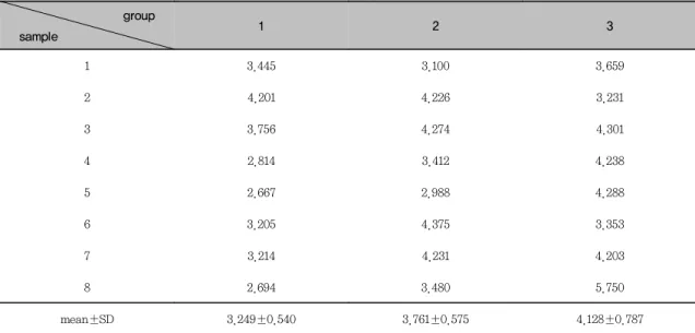
Table 1. MMP-2 intensity was normalized by β-actin intensity to quantify the expression levels of MMP-2 protein and the value were presented as a ratio of MMP-2/β-actin.
group
sample 1 2 3
1 3.445 3.100 3.659
2 4.201 4.226 3.231
3 3.756 4.274 4.301
4 2.814 3.412 4.238
5 2.667 2.988 4.288
6 3.205 4.375 3.353
7 3.214 4.231 4.203
8 2.694 3.480 5.750
mean±SD 3.249±0.540 3.761±0.575 4.128±0.787
Figure 1. MMP-2 Western analysis showing 4 representative samples in each group.
MMP-2 corresponding to molecular weight 72 kDa was shown to be expressed in all samples including healthy gingiva, and the expression levels of MMP-2 were increased in patients with type 2 diabetes mellitus than in control healthy subjects.
In order to quantify the MMP-2 levels, β-actin levels were also performed.
Group 1:healthy gingiva from systemically healthy person
Group 2:inflammed gingiva from patient with chronic periodontitis
Group 3:inflammed gingiva from patient with chronic periodontitis and type 2 DM
ured by anti-β-actin specific western blot analysis. Then MMP-2 levels were normalized by β-actin (ratio of MMP-2/β-actin). The expression levels of MMP-2 in each sample were measured by densitometer. The levels of gingival MMP-2 protein are given in Table 1.
The mean amount of MMP-2 (ratio of
MMP-2/β-actin) were 3.249 in group 1, 3.760 in group 2, 3.876 in group 3. The amount of MMP-2 was higher in group 3 than in group 1.
Also higher amount of MMP-2 was seen in group 3 compared to group 2. But, the differ- ences among three groups were not statistically significant (P>0.05).
Figure 2. Graphics showing the average amounts (Ratio of MMP-2/β-actin) and standard deviation of gelatinase MMP-2 in group 1, 2 and 3. In the inflamed gingiva (with or without diabetes, group 2 & 3), MMP-2 seemed to be increased compared to healthy gingiva. But the difference was not statistically significant (P>0.05).
Group 1:healthy gingiva from systemically healthy person
Group 2:inflammed gingiva from patient with chronic periodontitis
Group 3:inflammed gingiva from patient with chronic periodontitis and type 2 DM
Figure 3. MMP-9 Western analysis showing 4 representative samples in each group.
MMP-9 corresponding to molecular weight 92 kDa was shown to be expressed in all sam- ples including healthy gingiva and the expression levels of MMP-9 were increased in pa- tients with type 2 diabetes mellitus than in control healthy subjects.
In order to quantify detected MMP-9, β-actin levels were also measured.
Group 1:healthy gingiva from systemically healthy person
Group 2:inflammed gingiva from patient with chronic periodontitis
Group 3:inflammed gingiva from patient with chronic periodontitis and type 2 DM
The comparison of MMP-9 protein level western blot analysis using the antibodies against MMP-9 detected about 92 kDa molec-
ular weight of MMP-9 in all three groups (Figure 3).
Generally, the bands seemed to be thicker in
Figure 4. Graphics showing the average amounts (Ratio of MMP-9/β-actin) and standard deviation of gelatinase MMP-9 in group 1, 2 and 3. In the inflamed gingiva (with or without diabetes, group 2 & 3), the levels of MMP-9 was higher compared to healthy gingiva. The difference between group 3 and group 1 was statistically significant (P<0.05).
* significant difference between group 1 and group 3 (P<0.05) Group 1:healthy gingiva from systemically healthy person
Group 2:inflammed gingiva from patient with chronic periodontitis
Group 3:inflammed gingiva from patient with chronic periodontitis and type 2 DM Table 2. MMP-9 intensity was normalized by β-actin intensity to quantify the expression levels
of MMP-9 protein and the value were presented as a ratio of MMP-9/β-actin.
group
sample 1 2 3
1 0.430 0.628 1.478
2 0.793 1.157 1.153
3 1.426 1.740 1.474
4 0.778 0.711 1.614
5 1.337 1.437 1.709
6 1.435 1.038 2.650
7 0.794 3.033 3.262
8 1.097 1.851 1.839
mean±SD 1.011±0.369 1.449±0.777 1.897±0.703*
* significant difference between group 1 and group 3 (P<0.05)
*
group 3 compared to group 1 and 2. The levels of MMP-9 proteins were also quantified (Figure 3). The levels of individual gingival MMP-9 are given in Table 2. MMP-9 in each sample and mean amounts of MMP-9 in three different groups were compared (Table 2) and (Figure 4).
The mean amounts of MMP-9 (ratio of MMP-9/
β-actin) were 1.011 in group 1, 1.449 in group 2 and 1.897 in group 3. The MMP-9 levels in group 3 were thicker than in group 1 (P<0.05).
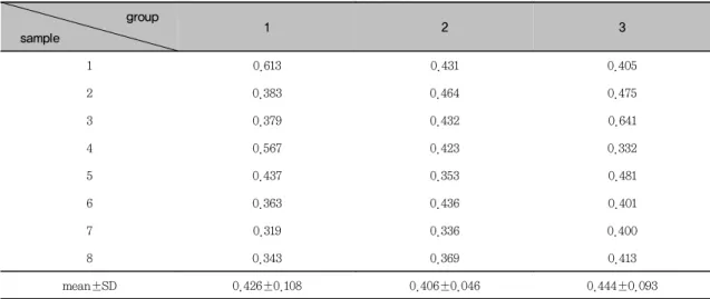
In the comparison of TNF-α protein levels using Western blot analysis, TNF-α of about
17 kDa molecular weight was identified and quantified (Table 3) and (Figure 3). The aver- age amounts of TNF-α (ratio of TNF-α/β -actin) were 0.425 in group 1, 0.405 in group 2 and 0.443 in group 3. There were no sig- nificant differences among the control group, the chronic periodontits group and the chron- icperiodontitis with type 2 DM group (P>0.05).
Both chronic periodontitis group & chronic periodontitis with type 2 DM group showed the expression of MMP-2, MMP-9 and TNF-α in all samples. The degrees of expression of
Table 3. TNF-α intensity was normalized by β-actin intensity to quantify the expression levels of TNF-α protein and the value were presented as a ratio of TNF-α/β-actin.
group
sample 1 2 3
1 0.613 0.431 0.405
2 0.383 0.464 0.475
3 0.379 0.432 0.641
4 0.567 0.423 0.332
5 0.437 0.353 0.481
6 0.363 0.436 0.401
7 0.319 0.336 0.400
8 0.343 0.369 0.413
mean±SD 0.426±0.108 0.406±0.046 0.444±0.093
Figure 5. TNF-α Western analysis showing 4 representative samples in each group. TNF-α corresponding to molecular weight 17 kDa was expressed in all samples including healthy gingiva and the expression levels of TNF-α were not increased significantly in patients with type 2 diabetes mellitus than in control subjects.
In order to quantify the TNF-α levels, β-actin levels were also measured.
Group 1:healthy gingiva from systemically healthy person
Group 2:inflammed gingiva from patient with chronic periodontitis
Group 3:inflammed gingiva from patient with chronic periodontitis and type 2 DM
MMP-2 showed only slight difference between the control group and the chronic periodontitis groups. The difference between the chronic periodontits groups and the chronic perio- dontitis with type 2 DM group was not statisti- cally significant either. In the case of MMP-9 more significant difference was observed be- tween the control group and the chronic perio- dontitis group. More prominent and significant difference was observed between the chronic periodontitis group and the chronic perio- dontitis with type 2 DM group compared to MMP-2. When the interrelationship of MMP-2, MMP-9 and TNF-α was analyzed. Although there were no proportional relationship between the interrelationship of MMP-2, MMP-9 and TNF-α,.as expression of TNF-α were in- creased, MMP-2, MMP-9 expressions showed increasing tendency in chronic periodontitis as- sociated to type 2 DM
Discussion
Multiple studies have demonstrated a link between diabetes and periodontal disease in human subjects. Although diabetes itself does not cause periodontitis, periodontal disease progresses more rapidly and leads to more tooth loss in patients whose diabetes is poorly controlled11-13). Various pathogenetic factors have been suggested to explain the increased prevalence and severity of periodontitis in dia- betes14,15). Mükelü and colleagues31) reported that during active phase of peridontitis, MMP-2 and MMP-9 levels in G ingival crev- icular fluid are pathologically elevated and Collin et al20) found that MMP-9 in salivary tissue is elevated and they suggested that MMPs may have a key role of periodontal de- struction2).
MMP-2 and MMP-9 is thought to play an Figure 6. Graphics showing the average amounts (Ratio of TNF-α/β-actin) and standard
deviation of TNF-α in group 1, 2 and 3. In the inflamed gingiva (with or without dia- betes, group 2 & 3), TNF-α seemed to be not increased compared to healthy gingiva.
And the difference was not statistically sig nificant (P>0.05).
Group 1:healthy gingiva from systemically healthy person
Group 2:inflammed gingiva from patient with chronic periodontitis
Group 3 : inflammed gingiva from patient with chronic periodontitis and type 2 DM
important role in the degradation of denatured collagens (gelatin), basement membrane (type IV collagen) and other matrix. The degenerative process causes the loss of attachment apparatus between tooth and epithelium, connective tissue which accelerates inflammatory process result- ing in deepening of periodontal pocket. In gen- eral, inducers such as Tumor Necrosis Factor- α(TNF-α) or EGF, IL-1β enhances MMP-9 without altering MMP-2 levels. TNF-α is a protein secreted by lipopolysaccharide- stimu- lated macrophages and causes tumor necrosis in vivo when injected into tumor bearing mice6,26). TNF-α appears directly toxic to vascular en- dothelial cells. Other actions of TNF-α include stimulating growth of human fibroblasts27) and other cell lines, activating polymorphonuclear neutrophils and osteoclasts, and the induction of Interleukin-1, Prostaglandin E2 and collage- nase production36). Several studies on the ex- pression and activities of these MMPs were executed in saliva, crevicular fluid20,30,32,33) but direct study on the expression of MMP-2, MMP-9, TNF-α in diabetic gingival tissue re- lated to periodontitis is limited. The purpose of this study was to quantify and compare the ex- pression of MMP-2, MMP-9 and TNF-α in the gingival tissues of the patients with chronic periodontitis associated to type 2 DM, in order to contribute to understand the mechanisms of periodontal destruction in type 2 diabetic pa- tients, especially MMP-mediated host response in type 2 diabetic patients.
In this study MMP-2 corresponding to mo- lecular weight 72 kDa was expressed in almost samples and the mean MMP-2 level was in- creased in chronic periodontitis groups than in control subject, although the difference was
statistically non-significant (P>0.05). Uitto et al2) demonstrated that active form of MMP-2 and MMP-9 in healthy gingiva increased sig- nificanlty as inflammation progressed. Several studies on the activity of MMP-2 and MMP-9 in oral fluid and cell culture were also the same with this results2,4,5). When the level of expression between non diabetic patients with chronic periodontitis and chronic periodontitis with type 2 DM was compared, chronic perio- dontitis with type 2 DM showed higher level than chronic periodontitis without DM. These findings seen in human diabetes are strongly supported by observation in rat models of in- sulin-deficient diabetes21,34). These animal model studies have shown reduced collagen sol- ubility and turnover resulting evidently from the formation and action of elevated levels of advanced glycation end-products (AGEs). AGEs can induce the expression of MMPs through the enhanced production of TNF-α and IL-β, vascular hardening and basement membrane disintegration, reduced solubility and decreased turnover rate4,35,36). Reactive oxygen species are produced to a significant extent by triggered degranulating neutrophils, to a lesser extent by other cells such as monocytes and macrophages.
Collin et al20) reported that the roll of oxidative stress in diabetes can exert multifactorial ac- tion regarding the development of periodontitis and it can be potent non-proteolytic pro-MMP activators.
The expression levels among our ex- perimental groups were prominent in MMP-9 than MMP-2. Since the intensity of MMP aciv- ity correlates with the activity of extracellular matrix breakdown and remodelling of the in- flammatory sites, the higher activity of MMP-9
compared to MMP-2 appears to be due to en- hanced elaboration of the PMNLs and mono- cytes in gingiva. Since MMP-2 is constitutively present in gingival tissue, leakage of proteases from the gingival suculs or stimulus by other pathogen could activate the latent form of MMP-2 and so affect gingival inflammation8).
The main sources of MMP-9 is PMNLs. In ad- dition keratinocytes of normal oral epithelium in vivo and in vitro and granulation tissue also strongly expreses MMP-930). These differences in cell lines could influence on inflammation resulting in higher expression level of MMP-9.
In the case of MMP-9 the expression of activ- ity showed elicit increasing tendency in chronic periodontitis group (group 2 and group 3) and a little difference between nondiabetic chronic periodontitis group and chronic perio- dontitis with type 2 DM group. Salvi et al have reported impaired PMN function in dia- betes regarding to chemotaxis, chemokinesis and degranulation 14,17,36-38). This might explain the increased susceptibility of diabetic patients to various infectious diseases including periodontitis. These results make it possible to consider that PMN dysfunction may be reflected in the gingival neutrophil degranulation product which is released from neutrophils recruited to inflammatory gingiva. In addition, it couldn't be excluded that age, blood glucose also and other unknown factors influence on the results although sex difference was not found among groups.
The increase of MMP in chronic periodontitis with type 2 DM was prominent in MMP-9 than in MMP-2. This might be due to the fact that MMP-9 is secreted from PMNLs, inflammatory cells that can react immediately to any stim-
ulus, and can be secreted easily by other vari- ous cytokines. It also might be due to that AGEs produced from diabetic environments en- hances production of MMPs especially MMP-940-44). Uemura et al24) also reported that the activity of MMP-9, but not MMP-2 is preferentially enhanced in endothelial cells by hyperglycemia. MMP-9 can be expected to be used as a factor for the parameter and the di- agnosis of peridontal diaease.
The mean level of TNF-α did not show sig- nificant differences among the groups (P>0.05).
Body Mass Index (BMI) and smoking did not show any difference among groups on the mean level of TNF-α. Based on the fact that TNF- α also expressed in control subjects, it is pre- sumed that cells included in sample may be in initial stage of inflammation, or that TNF-α was expressed to stimulate the growth of hu- man fibroblast in collagen remodelling during turnover, and also can be presumed that TNF- α constitutively secreted by adipose tissue ar- rived through circulation at gingival tissue to induce an effect antagonistic to insulin4).
In interrelationship between MMP-2, MMP-9 and TNF-α, although MMP-2 and MMP-9 ex- pressions showed increasing tendency in chronic periodontitis associated to type 2 DM as ex- pression of TNF-α were increased in Group 3 than G roup 2, but, there were no significant interrelationship among MMP-2, MMP-9 and TNF-α (P>0.05). This assumed that absolute concentration of TNF-α is not important enough to be a single parameter for severity of periodontal inflammation. Thus several possible reasons for the results are suggested. First, according to the effect of antimicrobial perio- dontal treatment on circulating TNF-α4) and
glycated Hb level in patient with type 2 DM and other studies by Katsuki et al45), the con- centration of circulating TNF-α itself was very low. So, it can be suggested that the differ- ence on the concentration of TNF-α between the groups was not significant enough to influ- ence solely on the expression of MMP-2 and MMP-9. Other cytokines such as EGF, IL-1β, TGF-β may influence on the expression.
Second, the changes in MMPs synthesis and activity might be time dependent. After TNF- α produced by monocytes or adipocytes acted on the target cells that were activated at the early stages of inflammation, the immune cells that arrived in the later stage or other resi- dent gingival cells (fibroblast or epithelial cells) activated by TNF-α could start to produce MMPs. Ejeil et al.3) reported that MMP-9 and MMP-13 showed significant increase in the later stage of inflammation. Third, though the concentration of TNF-α expressed was not enough to show the difference of MMP ex- pression between the groups, the target cells to TNF-α in the inflammatory areas are in- creased in periondontitis group compared to control group which might enhance the chance of activation10,23). Fourth, the activity of MMPs is tightly regulated at several levels including gene expression, secretion of pro-enzymes that require activation and inhibition by TIMPs46-50). Death et al46) found that high glucose may reg- ulate MMP gene expression via its effects on gene transcription or growth factors through the increased activation of its own response el- ements such as Activating Protein-2 enhance site (MMP-2) and Activating Protein-1, Nuclear Factor-κB (MMP-9).
Finally, it seemed that more studies are
needed to investigate the effect and inter- relationship between MMPs and other cytokines that affect the progression of periodontal dis- ease at a higher level.
Summary
The purpose of this study was to quantify and compare the level of MMP-2, MMP-9 and TNF-α in the healthy, inflammed gingival tis- sue and inflammed gingival tissue associated with type 2 DM. We tried to investigate whether expression of MMP-2, MMP-9 and TNF-α are increased by chronic periodontitis and those are more upregulated by type 2 DM.
But the results of quantification of MMP-2, MMP-9 and TNF-α were different.
Gingival tissue samples were obtained during periodontal surgery or tooth extraction.
According to the patient's systemic condition &
clinical criteria of gingiva, each gingival sam- ple was devided into three groups. Group 1 (n=8) is clinically healthy gingiva without bleeding and no evidence of bone resorption or periodontal pockets, obtained from systemically healthy 8 patients. Group 2 (n=8) is inflammed gingiva from patients with chronic periodontitis. Group 3 (n=8) is inflammed gin- giva from patients with chronic periodontitis associated to type 2 diabetes. Tissue samples were prepared and analyzed by Western blotting. The quantification of MMP-2, MMP-9, TNF-α were performed using a densitometer and statistically analyzed by one-way ANOVA followed by Scheffe test.
1. The expressions of MMP-2 and MMP-9 showed increased tendency in group 2 & 3
compared to group 1, but TNF-α showed slightly increased tendency in G roup 3 than G roup 1 and G roup 2. The level of expression of MMP-9 was more sig- nificantly increased in group 3 compared to group 2.
2. As MMP-2 levls were increasing, MMP-9 levels also were increased comparing to MMP-2 levels between group 2 and group 3. But there were no statistically sig- nificant difference between MMP-2 and MMP-9.
3. As expression of TNF-α were increased, MMP-2 and MMP-9 expressions showed increasing tendency in G roup 3 than Group 1 and Group 2, although there were no proportional relationship.
In conclusion, this study demonstrated that although there were no proportional relation- ship between the interrelationship of MMP-2, MMP-9 and TNF-α,. as expression of TNF-α were increased, MMP-2, MMP-9 expressions showed increasing tendency in chronic perio- dontitis associated to type 2 DM.
It can be assumed that TNF-α in part af- fect to expression of MMP-2 and MMP-9 in progression of periodontal inflammation asso- ciated to type 2 DM
References
1. Yoshihio l, Fusanori N, Masatsugu N et al.
The Effect of Antimicrobial Periodontal Treatment on Circulating Tumor Necrosis Factor-Alpha and G lycated Hemoglobin Level in Patients With Type 2 Diabetes. J Periodontol. 2001;72:774-778.
2. Uitto VJ, Overall CM and McCulloch C Proteolytic host cell enzymes in gingival- crevice fluid. Periodentology 2000 2003;31:
77-104.
3. Ejeil AL, Igondjo-Tchen S, Ghomrasseni S et al. Expression of Matrix Metalloproteinases (MMPs) and Tissue Inhibitors of Metalloproteinases (TIMPs) in Healthy and Diseased Human G ingiva. Periodontol.
2003;74:188-195.
4. Ryan EM, Golub LM Modulation of Matrix metalloproteinase activities in periodontitis as a treatment strategy. Periodontology 2000. 2000:24:226-238.
5. Souza AP, Gerlach RF and Line SRP Inhibition of human gingival gelatinases (MMP-2 and MMP-9) by metal salts.
Dental Materials 2000;16:103-108.
6. Masanori N, Yoshinori Y, Kazusada Y and Yukikazu S Effects of TNF-α and prosta- glandin E2 on the expression of MMPs in human periodontal ligament fibroblasts.
Periodont Res. 2002;37: 167-176.
7. Meikle MC, Hembry RM, Holley J et al.
Immunolocalization of matrix metal- loproteinases and TIMP-1 (tissue inhibitor of metalloproteinases) in human gingival tissues from periodontitis patients.
Periodont Res. 1994;29:118-126.
8. Chang Y-C, Yang S-F, Lai C-C, Liu J-Y and Hsieh Y-S Regulation of matrix met- alloproteinase production by cytokines, pharmacological agents and periodontal pathogens in human periodontal ligament fibroblast cultures. Periodont Res. 2002;
37:196-203.
9. Kiili M, Cox SW, Chen HW et al.
Collagenase-2 (MMP-8) and collage- nase-3(MMP-13) in adult periodontitis :
Molecular forms and levels in gingival crevicular fluid and immunolocalisation in gingival tissue. J Clin Periodontol. 2002;
29: 224-232.
10. Desfaits A-C, Serri O and Renier G Normalization of Plasma Lipid Peroxides, Monocyte Adhesion and Tumor Necrosis Factor-α Production in NIDDM Patients After Gliclazide Treatment. Diabetes Care.
1998;21 487-493.
11. Erwin van der Zee, Everts V and Beertsen W Cytokines modulate routes of collagen breakdown. J Clin Periodontol. 1997;24:
297-305.
12. Collin HL, Sorsa T, Meurman JH et al.
Salivary Matrix Metalloproteinase (MMP-8) levels and Gelatinase(MMP-9) activities in patients with type 2 diabetes mellitus. J Periodont Res. 2000;35:259-265.
13. Wall SJ, Sampson MJ, Levell N and Murphy G Cutaneous Biology. Elevated matrix met- alloproteinase-2 and -3 production from human diabetic dermal fibroblasts. British Journal of Dermatology. 2003;149:13-16.
14. G rayson R, Douglas CWI, Heath J et al Activation of human matrix metal- loproteinase 2 by gingival crevicular fluid and Porphyromonas gingivalis. J Clin Periodontol. 2003;30:542-550.
15. Birkendal-Hansen H Role of Matrix Metalloproteinases In Human Periodontal Diseases. J Periodontol. 1993;64:474-484.
16. Uemura S, Matsushita H, Li W et al.
Diabetes Mellitus Enhances Vascular Matrix Metalloproteinase Activity. Role of Oxidative Stress. Circulation Research 2001;88:1291-1298..
17. Portik-Dobos V, Anstadt MP, Hutchinson J, Bannan M and Ergul A Evidence for a
Matrix Metalloproteinase Induction/
Activation System in Arterial Vasculature and Decreased Synthesis and Activity in Diabetes. Diabetes 2002;51:3063-3068.
18. Emrich LJ, Shlosman M and G enco RJ : Periodontal disease in non-insulin depend- ent diabetes mellitus. J Periodontol.1991;
62:123-12.
19. Taylor GW, Burt BA, Becker MP et al.
Non-insulin dependent diabetes mellitus and alveolar bone loss progression over 2 years. J Periodontol. 1998;69:76-83.
20. Collin H-L, Uusitupa M and Niskanen L Periodontal findings in elderly patient with non-insulin depedent diabetes mellitus. J Periodontol. 1998;69:962-966.
21. Salvi G E, Yalda B, Collins JG et al.
Inflammatory Mediator Response as a Potential Risk Marker for Periodontal Diseases in Insulin-Dependent Diabetes mellitus patients. J Periodontol. 1997;68:
127-135.
22. AAP Position Paper : Diabetes and peri- dontal disease. J Periodontol. 1999;70:
935-949.
23. Yalda B, Offenbacher S and Collins JG Diabetes as a modifier of periodontal dis- ease expression. Periodontology 2000.
1994;6:37-49.
24. Bissada NF, Manoucher-Pour M, Haddow M and Spagnulo PJ Neutrophil functional ac- tivity in juvenile and adult onset diabetic patients with mild and severe periodontitis.
J Periodont Res. 1982;17:500-502.
25. Armitage GC Development of a classi- fication system for periodontal disease and conditions. Ann Periodontol 19994:1-6.
26. Fusanori N, Yoshihiro I, Junji M et al.Periodontal Disease and Diabetes Mellitus:
The Role of Tumor Necrosis Factor-α in a 2-way Relationship. Periodontol. 2003;74:
97-102.
27. Quintero JC, Piesco NP, Langkamp HH, Bowen L and Agarwal S : LPS responsive- ness in periodontal ligament cells is regu- lated by tumor necrosis factor-alpha. J Dent Res. 1995;74(11):1802-1811.
28. Persson L, Bergstrüm J and Gustafsson A Effect of Tobacco Smoking on Neutrophil Activity Following Periodontal Surgery. J Periodontol. 2003;74:1475-1482.
29. Mühlman HR and Son SH Gingival Bleeding-a leading symptom in initial gingivitis. Helvitica Odontologica Acta.
1971;15:107-113.
30. Cho J-Y, Xing S, Liu X et al. Expression and activity of human Na+/I- symporter in human glioma cells by adenovirus-mediate gene delivery. Gene Therapy. 2000;7:
740-749.
31. Mükelü M, Jalo T, Uitto V-J and Larjava H: Matrix Metalloproteinase (MMP-2 &
MMP-9) of the oral cavity. J Dent Res.
73:1397-1406. 1994.
32. Page RC and Schroeder HE Parhogenesis of inflammatory periodontal disease. A sum- mary of current work. Lab Invest.1976;
34:235-249.
33. Ingman T, sorsa T, Lindy O, Koski H and Konttinen YT Multiple forms of gelati- nases/type IV collagenases in saliva and gingival crevicular fluid of periodontitis patients. J Clin Periodontol. 1994;21:26-31.
34. G olub LM, Lee HM and Lehrer G Minocycline reduces gingival collagenolytic activity during diabetes. Preliminary ob- servations and a proposed new mechanism of action. J Periodont Res. 1983;18:516-526.
35. Lalla E, Lamster IB, Feit M et al. Blockade of RAGE suppresses peiodontitis-associated bone loss in diabetic mice. J. Clin. Invest.
2000;105:1117-1124.
36. Ide M, Jagdev D, Coward PY et al. The short-term effects of treatment of chronic periodontitis on circulating levels of endo- toxin, C-reactive protein, tumor necrosis factor-α and interleukin-6. J Peridontol.
2004;75:420-428.
37. Wahlgren J, Maisi P, Sorsa T et al.
Expression and induction of collage- nases(MMP-8 and -13) in plasma cells as- sociation with bone-destructive lesions.
Journal of Pathology 2001;194:217-224.
38. Manoucher-Pour M, Spagnuolo PJ and Bissada NF Impaired neutrophil chemotaxis in diabetic patients with severe periodontitis. J Dent Res. 1981;60:729-730.
39. Uematsu S, Mogi M and Deguchi T Interleukin(IL)-1β IL-6, Tumor Necrosis Factor-α, Epidermal Growth Factor, and β2-microglobulin levels are elevated in gingival crevicular fluid during human or- thodontic tooth movement. J Dent Res 1996;75:562-567.
40. Noda K, Ishida S, Inoue M et al.
Production and activation of matrix metal- loproteinase-2 in proliferative diabetic retinopathy. Invest Ophthalmol Vis Sci.
2003;44:2163-70.
41. Nagase H and Woessner JF Jr. Matrix metalloproteinases. J Biol Chem. 1994;274:
21491-21494.
42. McLennan SV, Martell SK and Yue DK Effects of mesangium glycation on matrix metalloproteinase activities : possible role in diabetic nephropathy. Diabetes. 2002;51:
2612-8.
43. Diamant M, Hanemaaijer R, Berheijen JH et al. Elevated matrix metalloproteinase-2 and -9 in urine, but not in serum, are markers of type 1 diabetic nephropathy.
Diabet Med. 2001;18:423-4.
44. Jin M, Kashiwagi K, lizuka Y, Tanaka Y, Imai M and Tsukahara S : Matrix metal- loproteinases in human diabetic and non- diabetic vitreous. Retina. 21(1) : 28-33.
2001.
45. Katsuki A, Sumida Y, Murashima S et al.
Serum Levels of Tumor Necrosis Factor-α Are Increased in Obese Patients with Noninsulin-Dependent Diabetes Mellitus. J Clinical Endoclinology and Metabolism.
1998;83:859-862.
46. Death AK, Fisher EJ, McG rath KCY and Yue DK High glucose alters matrix metal- loroteinase expression in two key vascular cells : potential impact on artherosclerosis in diabetes. Arthersclerosis. 2003;168:
263-269.
47. Sakuta T, Tokuda M, Tamura M et al. Dual regulatory effects of interferon-alpha, -beta and -gamma interleukin-8 gene ex- pression by human gingival fibroblasts in culture upon stimulation with lip- opolysaccharide from Prevotell intermedia, interleukin-1alpha, or tumor necrosis fac- tor-alpha. J Dent Res. 1998;77:1597-1605.
48. Ryan ME, Ramamurthy NS, Sorsa T and Golub LM MMP-mediated events in diabetes. Ann. NY Acad Sci. 1999;873:
311-314.
49. Choi DH, Moon IS, Choi BK et al. Effects of sub-antimic robial dose doxycycline therapy on crevicular fluid MMP-8 and gingival tissue MMP-9, TIMP-1 and IL-6 levels in chronic periodontitis. J Periodont Res. 2004;39:20-26.
50. Dahan M, Nawrocki B, Elkaїm R et al.
Expression of matrix metallopoteinases in healhty and diseases human gingiva. J.
Clin Periodontol 2001;28:128-136.
-Abstract-
단순 만성 치주염 환자 및 2형 당뇨병을 가진 만성 치주염 환자의 치은조직에서 Matrix Metalloproteinase
와 TNF-α의 발현 양상 비교
김도훈, 박의균, 신홍인, 조제열, 서조영,이재목 경북대학교 치의학대학원
치주질환의 병원균은 세포벽의 항원에 의하여 조직내 존재하는 mononuclear phagocytes가 활성화되어 cy- tokine들이 생성됨 으로써 치주 결체조직의 파괴를 진행시킨다. 이런 관련된 cytokine들은 순차적으로 상주하 는 치은세포 및 대식세포가 Matrix metalloproteinase 합성을 하도록 유도하여 조직파괴를 시작한다. 이들 Matrix metalloproteinase중 MMP-2, MMP-9 (Gelatinase A,B)는 type IV collagen 및 변성된 interstitial collagen을 파괴하며 치주환자의 치은 열구액, 치은조직, 타액 내에서 높게 보고 되어왔다. 당뇨병은 치주질 환의 위험요소중 하나로 당뇨 환자에서는 치주질환의 유병율이 일반인에 비해 높고 치주질환의 중증도도 더 심 하여 진행도 빠르다고 알려져 있다. 그 병리 기전 중 하나로는 당뇨 환자에서는 치은 열구액 내 중성구 유래의 Matrix metalloproteinase의 활성 증가 및 TNF-α 의 활성 증가가 추정되고 있다.
본 실험에서는 제2형 당뇨병 환자와 비당뇨 환자들에서 만성 치주염 부위의 치은 및 건강한 치은에서 염증 매개체 중 하나인 MMP-2, MMP-9 및 TNF-α 의 발현에 대해 상호 비교 분석함으로서 염증, 혈당이 미치는 영향을 밝히고 제 2형 당뇨병 환자에서 심한 치주조직 파괴의 기전을 연구하고자 하였다.
경북대학교병원 치주과 내원환자 중 제2형 당뇨병 환자와 비당뇨 환자들 및 치주질환이 없는 건강인 대조군 을 대상으로 여러 가지 환자요소, 임상 치주상태를 기록하고 , 전신적으로 건강한 환자의 건강한 부위(n=8, Group 1), 전신적으로 건강한 환자의 만성 치주염 부위(n=8, Group 2), 제2형 당뇨병 환자의 만성 치주염 부 위 (n=8, Group 3)에서 각각 변연치은을 채득하고 액화질소에 급속 동결하였다. Western blotting을 이용하 여 각 조직 내 MMP-2, MMP-9 및 TNF-α 의 발현을 관찰, densitometer를 이용하여 상대적 발현을 정량, 각 조직의 β-actin을 이용하여 표준화하여 실험군과 대조군들의 평균치를 비교하였다.
비당뇨 환자들의 만성 치주염 부위 및 제2형 당뇨병 환자의 만성 치주염 부위에서 모두 건강 대조군에 비해 MMP-2와 MMP-9 의 발현이 증가되었다. 또한 MMP-2 와 MMP-9는 2형 당뇨 환자의 만성 치주염 부위가 비당뇨 환자의 만성 치주염 부위보다 증가된 발현양상을 보였으며, TNF-α발현 비교시 각 군간 유의성 있는 변화는 없었으나 2형 당뇨환자군에서 MMP-2 및 MMP-9의 증가와 함께 다소 증가 양상을 보였다. 결론적으 로 본 실험에서 MMP-2 및 MMP-9의 증가가 만성 치주염 및 2형 당뇨 환자에서의 만성치주염에서 비당뇨환 자 보다 MMP-2, MMP-9의 증가양상을 보여 주었으며 TNF-α가 2형 당뇨환자의 만성치주염 진행과정에 기 여인자로써 역할을 하는 것으로 생각된다.2)
주요어:치주염, 당뇨병, MMP ,TNF-α