서론
만성 치주염은 치아 주위 조직의 부착 상실과 치조골 파 괴를 특징으로 하는 감염성 질환이다. 치주조직의 세포외기
질 구성성분 특히, 교원질은 이러한 치주염의 주요한 파괴 대상이 된다. 세포외기질을 파괴하는 숙주 단백분해효소 중 matrix metalloproteinases(MMPs)는 치주질환에서 조직 파괴와 개조에 많이 관련된 것으로 보고되고 있다1).
MMPs는 정상적으로 유전자 발현 수준에서부터 세포 외 로의 분비까지 잘 조절되지만, 이러한 조절이 붕괴되면 결 체 조직의 병적인 파괴를 일으킨다. 치주조직에서 MMPs의 높은 수준은 교원질의 생산과 파괴의 불균형을 초래하여 결 국 치아 부착 상실을 일으키는 것으로 알려져 있다2).
MMPs는 그것의 기질 특성과 배열 상동관계에 기초하여,
단순 만성 치주염 환자와 제 2형 당뇨병을 동반한 만성 치주염 환자에서 Stromelysin-1와 Membrane type-MMP-1 Expressions
류상호, 박진우, 서조영, 이재목
*경북대학교 치의학전문대학원 치주과학교실
Stromelysin-1 and Membrane type-MMP-1 Expressions in Human Chronic Periodontitis with Type 2 Diabetes Mellitus
Sang-Ho Ryu, Jin-Woo Park, Jo-Young Suh, Jae-Mok Lee
*Department of Periodontology, School of Dentistry, Kyungpook National University
ABSTRACT
Purpose: The purposes of this study were to compare and quantify the expression of Stromelysin-1 and MT-MMP-1 in the gingival tissues of patients with type 2 diabetes mellitus(DM) and healthy adults with chronic periodontitis.
Materials and Methods: Gingival tissue samples were obtained during periodontal surgery or tooth extraction. According to the patient's systemic condition & clinical criteria of gingiva, each gingival sample was devided into three groups. Group 1 (n=8) is clinically healthy gingiva without bleeding and no evidence of bone resorption or periodontal pockets, obtained from systemically healthy 8 patients. Group 2 (n=8) is inflammed gingiva from patients with chronic periodontitis. Group 3 (n=8) is inflammed gingiva from patients with chronic periodontitis associated with type 2 DM. Tissue samples were prepared and analyzed by Western blotting. The quantification of Stromelysin-1 and MT-MMP-1 were performed using a densitometer and statistically analyzed by one-way ANOVA followed by Tukey test.
Results: In the analysis of expression levels, Stromelysin-1 and MT-MMP-1 expressions were similar in group 1 and 2.
Stromelysin-1 and MT-MMP-1 expressions was more increased in group 3 than group 1, 2. The difference between group 3 and group 1, 2 was statistically significant. Also, in the interrelationship of Stromelysin-1 and MT-MMP-1 expressions, expressions of Stromelysin-1 and MT-MMP-1 showed increasing tendency in chronic periodontitis associated with type 2 DM and it seems that the MT-MMP-1 expressions were increasing in proportion to Stromelysin-1 expressions.
Conclusion: It is suggested that Stromelysin-1 and MT-MMP-1 may be partly involved in the progression of periodontal inflammation associated with type 2 DM, as related to a metabolism of other factors, such as AGE, plasmin and other inflammatory mediators. Therefore, the expression levels of Stromelysin-1 and MT-MMP-1 can be inflammatory markers of periodontal inflammed tissue with type 2 DM. (J Korean Acad Periodontol 2008;38:629-638)
KEY WORDS: Stromelysin-1; MT-MMP-1; DM; chronic periodontitis.
Correspondence : Jae-Mok Lee
Department of Periodontology, School of Dentistry, Kyungpook National University, 188-1 Samduk-Dong 2ga, Jung-Gu, Daegu, 700-412, Korea.
E-mail : [email protected], Tel: 82-53-600-7511, Fax: 82-53-427-3263
Received: Oct. 13, 2008; Accepted: Nov. 28, 2008
크게 4가지로 구분된다3): (1) collagenase 1, 2, 3, 4 (2) gelastinases (3) stromelysins-1(MMP-3),-2, matrilysin (4) MT-MMP-1, MMP-15, MMP-16, MMP-17과 같이 최 근 발견된 membrane type matrix metalloproteinases (MT-MMPs). MMP-12(Metalloelastase), MMP-19와 MMP- 20(enamelysin) 또한 MMPs 군에 속한다.
이 중 Stromelysin-1(MMP-3)는 치은 섬유아세포 뿐 아 니라, 단핵구, 내피세포, 연골세포, 활막세포를 포함한 다양 한 세포에서 합성된다4). Stromelysin-1는 gelatin, pro- teoglycan, laminin, fibronectin과 type I, type IX col- lagen을 포함한 많은 세포외기질의 분해에 효과적이다5). 또 한 이러한 다양한 결체조직의 구성성분을 파괴하는 능력 외 에도 Stromelysin-1는 잠재적인 pro-MMP-1, -8, -9의 단백질 분해 활성에 역할을 하는 것으로 알려져 있다6). Stromelysin-1에 의해 매개되는 교원질 분해는 치주염에서 결체조직의 파괴와 개조에 주요 경로가 될 수 있다.
1994년 Sato 등7)은 membrane type-MMP(MT-MMP-1, MMP-14)를 처음 발견하여, 이것이 pro-MMP-2를 활성화 시킨다고 설명하였다. 이후, MT-MMP-1는 type I, II, III collagen, fibronectin, laminins 그리고 proteoglycans을 포함한 다양한 세포외 기질의 구성성분을 파괴함이 관찰되 었다8). MT-MMP-1는 분비되지 않고 세포막에서 역할을 하 며, pro-MMP-8과 -13을 활성화 시킨다. Achong 등9)은 치주 병소의 개조와 치유 단계 동안, 세포막에서 활성화된 MMP-2와 함께 MT-MMP-1가 세포 이주와 세포외기질의 재구성을 일으킬 것이라 설명하였다.
당뇨병은 전신적 영향을 받을 수 있는 치주질환과 연관되 어져 왔다. 또한 당뇨병을 동반한 심한 치주염은 신장, 심혈 관계 그리고 감염성 합병증을 일으킬 가능성이 높은 것으로 보고 되고 있다10). 당뇨병 환자의 감염에 대한 감수성과 느 린 상처 치유는 잘 알려져 있다11). 당뇨병은 그 자체가 치주 염을 일으키지는 않지만, 대사 조절 정도와 관련하여 치주 질환의 높은 이환율에 부분적으로 관여하는 것으로 알려져 왔다12). Bissada 등13)은 경도의 치주염을 가진 당뇨병 환자 와 경도 또는 중등도의 치주염을 가진 건강한 환자와 비교 하여 당뇨병을 동반한 치주염 환자에서 다형핵 백혈구의 화 학 주성이 더욱 부족함을 관찰하고, 손상된 다형핵 백혈구 기능이 치주염과 당뇨병에 관련됨을 보고하였다.
염증 반응에서 Stromelysin-1과 MT-MMP-1의 역할과 상호작용은 아직 명백하지 않다. 치주염의 병인에서
Stromelysin-1과 MT-MMP-1의 상대적인 기여도 또한 아 직 잘 알려져 있지 않다. 또한, 당뇨를 동반하거나 동반하지 않은 만성 치주염 환자에서 각 Stromelysin-1과 MT-MMP-1 그리고 그것들의 상호관계를 동시에 분석한 연 구는 아직 미흡한 실정이다. 이번 연구의 목적은 제2형 당 뇨병 환자와 전신적으로 건강한 성인의 만성 치주염 부위 치은 조직에서 Stromelysin-1과 MT-MMP-1의 발현을 정 량하고 상호 비교 분석하고자 하였다.
재료 및 방법
1. 환자 선정 및 조직 표본 채득
이번 연구에 참여한 환자는 만성 치주염 치료를 위해 XX 대학교 병원을 내원한 환자들 중 제2형 당뇨병을 동반한 만 성 치주염 환자 8명, 전신적으로 건강한 만성 치주염 환자 8명 그리고 전신적으로 건강한 성인 8명으로 구성되었다.
치주조직 표본은 모든 환자에게 동의를 얻은 후, 치주 수술 (치관연장술 포함) 또는 발치 시 내사면절개에 의해 얻어졌다.
환자의 전신적 상태(나이, 성별, 혈당 수치, 흡연 유무), 치은의 임상적 기준(치은 열구 출혈 지수, 치주낭 깊이) 그 리고 골 흡수의 방사선학적 증거에 따라, 각 치은 조직 표본 은 3가지 군(Group 1, 2, 3)으로 나누어졌다.
첫 번째 군(Group 1, 대조군)은 전신적으로 건강한 8명 의 성인으로부터 얻어진, 출혈이 없고 골 흡수 또는 치주낭 이 없는 임상적으로 건강한 치은 조직으로 하였고, 두 번째 군(Group 2)은 만성 치주염 환자로부터 얻어진 염증성 치은 조직으로 하였다. 만성 치주염은 임상적 그리고 방사선학적 (골 흡수) 기준을 기초로 치주 질환과 상태에 대한 분류에 따라 진단되었다14). 두 번째 군의 모든 환자는 전신적으로 건강하고, 치주낭 깊이가 5 mm 이상인 부위를 한 부위 이 상 가지며, 4 mm 이상의 부착 소실을 보이는 치아를 적어 도 한 개 이상 가지고 있었다. 모든 치은 조직 표본은 5 mm 이상의 치주낭 깊이, 변연 치은의 부종 그리고 Mühlman &
Son15)에 따른 치은 열구 출혈 지수 3을 보이는 치아로부터 채득되었다.
세 번째 군(Group 3)은 제2형 당뇨병을 동반한 만성 치 주염 환자로부터 얻어진 염증성 치은 조직으로 하였다. 세 번째 군은 적어도 6개월 이전에 제2형 당뇨병 진단을 받고, 식 후 2시간째 혈당 수치가 200mg/dl 이상 보이는 환자를
대상으로 하였다. 두 번째 군과 세 번째 군의 환자는 유사한 치주 상태를 보이지만, 두 번째 군은 전신적으로 건강한 환 자를 대상으로 하였고, 세 번째 군은 약물치료 중인 제2형 당뇨병 환자를 대상으로 하였다.
수술 시, 채득된 조직 표본은 즉시 액화 질소에 급속 동 결되었다(-70゚C).
2. 단백질 분리와 Western blotting
Western blotting은 이전에 Park 등16)의 연구에서 시행 된 방법과 동일하게, 동결된 조직을 Cho 등17)의 방법을 따 라 1 : 30으로 희석된 protease inhibitor cocktail(Roche, Germany)을 함유한 RIPA 용해완충액(10 mM EDTA, 0.15M NaCl)에서 균질화 시키고, 그 용해물을 10초 동안 세 번 초음파 분쇄하였으며, 4゚C, 12000rpm에서 15분 동 안 원심 분리하였다. 상층액의 단백질 농도는 BSA를 표준 용액으로 이용하여 Braford protein assay(Quick Start, BIO-RAD, USA) 통해 결정되었다.
그 용해물의 상층액을 SDS samples buffer(1M Tris- HCl(pH 6.8), 40% glycerol, 8% SDS, 2% mercapto-ethanol, 0.002% Bromophenol blue)에서 끓인 후, 15% sodium dodecyl sulfate(SDS)-polyacrylamide gels에서 분리하고, polyvinylidene difluride(PVDF) 막으로 전이하였다.
막은 비특이적인 반응을 제거하기 위해 5% powdered milk와 1% BSA를 함유한 Tris-buffered saline(TBS)으로 1시간동안 처리하고, 실온에서 polyclonal anti- Stromelysin- 1과 anti-MT-MMP-1(prepared in rabbit, 각각 TBS에서 1 : 1,000, 1 : 2,000으로 희석, Sigma- Aldrich, Inc. USA) 항체와 1.5시간 동안 반응시켰다.
이러한 막은 Tween 20으로 5분씩 5회 세척되고, an- ti-Stromelysin-1과 anti-MT-MMP-1에 대하여 horse- radish peroxidase(HRP)-conjugated goat anti-rabbit 이차항체를 TBS에서 1 : 2,000으로 희석하여, 실온에서 1시 간동안 반응시켰다. 다시 막은 Tween 20으로 5분씩 5회 세 척된 후 ECL Plus development kit(Amsterdam, Beckinghamshire, UK)으로 밴드를 확인하였다.
Stromelysin-1과 MT-MMP-1 발현에 대한 정량 분석은 densitometer(Scion Image β 4.02, Scion Corporation, USA)을 이용하여 시행되었다. 각 표본에서 β-actin (Abcam, UK)에 대한 표준화를 시행하고, Stromelysin-1과, MT-MMP
-1의 발현은 Stromelysin-1 또는 MT-MMP-1/β-actin의 비율로 나타내었으며, 세 군 사이의 차이가 결정되었다.
3. 통계
모든 수치는 평균±표준편차로 나타내었고, 결과는 통계 학적으로 분석되었다. 각 3군 사이의 Stromelysin-1과 MT-MMP-1 수준은 one way ANOVA를 이용하여 비교되 었고, Tukey test에 의해 사후검증 되었다(P<0.05).
결과
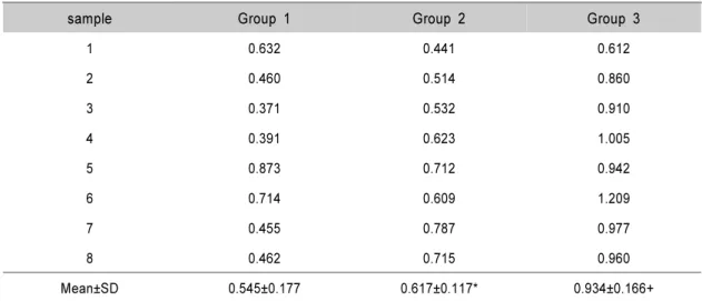
제 2형 당뇨병을 동반한 만성 치주염 환자와 전신적으로 건강한 만성 치주염 환자의 채득된 모든 조직 표본에서 Stromelysin-1(59kDa)과 MT-MMP-1(63kDa)의 발현이 관찰되었다. 세 군 모두에서 Stromelysin-1가 검출되었으 며 Stromelysin-1 발현 수준은 β-actin(ratio of Stromelysin-1/ β-actin)으로 표준화되었다(Fig. 1A). 표 준화된 Stromelysin-1의 발현 수준은 Table 1에 제시하였 고, Fig. 1B에 도표로 요약하였다.
Stromelysin-1의 평균 발현 양(ratio of Stromelysin-1/
β-actin)에서 1군은 0.545±0.177, 2군은 0.617±0.117, 3 군은 0.934±0.166로 나타났다. 1군과 2군 사이에는 유의한 차이가 없었지만 1군과 3군, 2군과 3군 사이의 차이는 통계 학적으로 유의하였다(P<0.05).
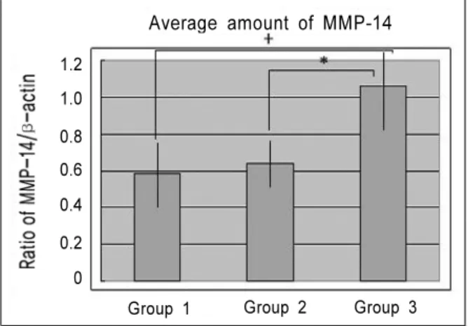
MT-MMP-1 발현도 세 군에서 MT-MMP-1가 검출되었 다(Fig. 2A). MT-MMP-1 발현 수준은 역시 β-actin(ratio of MT-MMP-1/ β-actin) 표준화로 정량되었으며(Fig.
2B), 표준화된 MT-MMP-1의 발현 수준은 Table 2에 제시 되었다. MT-MMP-1의 평균 발현 양(ratio of PGE2/ β -actin)은 1군은 0.582±0.178, 2군은 0.637±0.123, 3군 은 1.060±0.217로 나타났다. 이 경우 Stromelysin-1과 마 찬가지로, 1군과 2군 사이에는 유의한 차이가 없었지만, 1군 과 3군, 2군과 3군 사이의 차이는 통계학적으로 유의하였다 (P<0.05).
또한, Stromelysin-1과 MT-MMP-1 발현의 상관관계에 서, Stromelysin-1과 MT-MMP-1의 발현 모두 제 2형 당 뇨병 환자의 염증 조직에서 증가되는 경향을 나타내었고, MT-MMP-1의 발현이 Stromelysin-1의 발현에 비례하여 증가하는 것처럼 보였다.
Table 1. Normalized Stromelysin-1 expressions by Stromelysin-1/β-actin
sample Group 1 Group 2 Group 3
1 0.632 0.441 0.612
2 0.460 0.514 0.860
3 0.371 0.532 0.910
4 0.391 0.623 1.005
5 0.873 0.712 0.942
6 0.714 0.609 1.209
7 0.455 0.787 0.977
8 0.462 0.715 0.960
Mean±SD 0.545±0.177 0.617±0.117* 0.934±0.166+
Group 1 : healthy gingiva from systemically healthy person Group 2 : inflammed gingiva from patient with chronic periodontitis
Group 3 : inflammed gingiva from patient with chronic periodontitis and type 2 DM + significant difference between group 1 and group 3 (P<0.05)
* significant difference between group 2 and group 3 (P<0.05)
Figure 1A. Stromelysin-1(MMP-3) Western analysis showing 4 representative samples in each group.
Stromelysin-1 corresponding to molecular weight 59kDa was shown to be expressed in all samples including healthy gingiva. The expression levels of Stromelysin-1 were increased in order of group 1, group 2, group 3.
In order to quantify detected Stromelysin-1, β-actin levels were also measured.
Group 1 : healthy gingiva from systemically healthy person Group 2 : inflammed gingiva from patient with chronic periodontitis
Group 3 : inflammed gingiva from patient with chronic periodontitis and type DM MMP-3
β-actin
1 2 3 4 1 2 3 4 1 2 3 4
Group 1 Group 2 Group 3
Figure 1B. Graphics showing the average amounts (Ratio of Stromelysin-1(MMP-3)/β-actin) and standard deviation of Stromelysin-1 in group 1, 2 and 3. In the inflammed gingiva with diabetes (group 3), Stromelysin-1 seemed to be increased compared to group 1 and group 2.
Group 1 : healthy gingiva from systemically healthy person Group 2 : inflammed gingiva from patient with chronic periodontitis
Group 3 : inflammed gingiva from patient with chronic periodontitis and type 2 DM + significant difference between group 1 and group 3 (P<0.05)
* significant difference between group 2 and group 3 (P<0.05)
Average amount of MMP-3
1.2 1.0 0.8 0.6 0.4 0.2 0
Group 1 Group 2 Group 3
Figure 2A. MT-MMP-1(MMP-14) Western analysis showing 4 representative samples in each group.
MT-MMP-1 corresponding to molecular weight 63kDa was shown to be expressed in all samples including healthy gingiva and the expression levels of MT-MMP-1 were increased in order of group 1, group 2, group 3. In order to quantify detected MT-MMP-1, β-actin levels were also measured.
Group 1 : healthy gingiva from systemically healthy person Group 2 : inflammed gingiva from patient with chronic periodontitis
Group 3 : inflammed gingiva from patient with chronic periodontitis and type DM MMP-14
β-actin
1 2 3 4 1 2 3 4 1 2 3 4
Group 1 Group 2 Group 3
Table 2. Normalized MT-MMP-1 expressions by MT-MMP-1/β-actin
sample Group 1 Group 2 Group 3
1 0.461 0.437 0.999
2 0.480 0.578 0.920
3 0.578 0.812 1.170
4 0.602 0.601 0.928
5 0.981 0.709 1.176
6 0.615 0.780 1.512
7 0.396 0.569 0.907
8 0.545 0.607 0.870
Mean±SD 0.582±0.178 0.637±0.123* 1.060±0.217+
Group 1 : healthy gingiva from systemically healthy person Group 2 : inflammed gingiva from patient with chronic periodontitis
Group 3 : inflammed gingiva from patient with chronic periodontitis and type 2 DM + significant difference between group 1 and group 3 (P<0.05)
* significant difference between group 2 and group 3 (P<0.05)
Figure 2B. Graphics showing the average amounts (Ratio of MT-MMP-1(MMP-14)/β-actin) and standard deviation of MT-MMP-1 in group 1, 2 and 3. In the inflammed gingiva with dia- betes (group 3), MT-MMP-1 seemed to be increased compared to group 1 and group 2.
Group 1 : healthy gingiva from systemically healthy person Group 2 : inflammed gingiva from patient with chronic periodontitis
Group 3 : inflammed gingiva from patient with chronic periodontitis and type 2 DM + significant difference between group 1 and group 3 (P<0.05)
* significant difference between group 2 and group 3 (P<0.05)
Average amount of MMP-14
1.20 0.2 0.4 0.6 0.8 1.0
Group 1 Group 2 Group 3
고찰
인간을 대상으로 한 당뇨와 치주 질환 사이의 상호관계에 대해 많은 연구가 보고되고 있다. 잘 조절되지 않는 당뇨병 환자의 경우 치주 질환이 더욱 급속히 진행되고, 더 많은 치 아 상실이 일어나는 것으로 알려져 있다18). 당뇨를 가진 경 우 치주염의 증가된 이환율과 심도를 설명하는 다양한 병인 적 요소들이 제안되어 오고 있다19).
상처 치유에 있어 중심적 구성 성분인 교원질은 우리 몸 의 가장 많은 양을 차지하는 단백질이고 치은의 가장 중요 한 구성성분이라 할 수 있다. MMPs는 조직의 개조 그리고 치주질환과 같은 병적인 과정을 포함한 많은 생리적인 과정 에 역할을 하는데, 치주조직이 파괴되는데 있어 교원질 파 괴에 주요한 역할을 하는 것으로 알려져 있다.
이 연구의 목적은 제 2형 당뇨병 환자에서 치주 파괴 특 히, MMP에 의한 숙주 반응에 대한 Stromelysin-1과 MT-MMP-1의 기여도를 이해하기 위해서 제 2형 당뇨병을 동반한 만성 치주염을 가진 환자의 치은 조직에서 Stromelysin-1과 MT-MMP-1의 발현을 정량화하여 비교하 는 것이다.
이번 연구에서, 전신적으로 건강한 환자의 정상 치은 조 직과 염증성 치은 조직에서 Stromelysin-1과 MT-MMP-1 의 발현은 유사한 양상을 나타내었다. 이것은 조직 표본의 다양한 염증 정도에 기인한 것으로 여겨진다.
지금까지 제 2형 당뇨병을 동반한 만성 치주염 환자에서 Stromelysin-1의 발현 양상에 관한 연구는 미미한 실정이 다. 이번 연구에서 Stromelysin-1 발현의 정량적 분석은 제 2형 당뇨병 환자의 염증성 치은 조직에서 전신적으로 건 강한 환자의 정상 치은 조직과 염증성 치은 조직과 비교하 여 Stromelysin-1의 발현이 다소 증가된 양상을 보여 주었 고, 그 차이가 통계학적으로 유의하였다. 이러한 결과는 Stromelysin-1이 제 2형 당뇨병 환자에 있어 질환의 진행 과정에 다른 반응 경향을 나타냄을 보여 주고, 이러한 전신 적 질환을 가진 환자의 증가된 염증 반응에 있어 부분적인 역할을 하는 것을 나타낸다.
당뇨병은 고혈당을 특징으로 하고, 고혈당에 지속된 노출 은 다양한 당뇨 합병증이 발생하는데 일차적인 요인이 된다
20). 당뇨병에 있어 일반적인 생화학적 기전은 고혈당에 의 해 매개되는 비효소적 advanced glycation end products
(AGEs)의 형성이다. 당에 의해 매개되는 AGE의 축적은 단 핵구와 탐식세포의 이주와 탐식 능에 영향을 미쳐서 결과적 으로 더욱 병적인 치은연하 미생물총을 형성하게 한다.
이것은 cytokine upregulation의 infection-mediated pathway를 자극하고, 특히 tumor necrosis factor(TNF)- α와 interleukine(IL)-1의 분비를 자극한다21). Wassenaar 등과 Tewari 등은 각각 TNF-α와 IL-1에 의해 치은 섬유모 세포에서 Stromelysin-1의 발현이 자극된다고 설명하였다22). 치주염 환자의 치은 열구액에는 호중구에 의해 유도된 cathepsin G와 elastase가 증가되어 있는 것으로 보고되고 있으며23), 이러한 효소들은 pro-Stromelysin-1을 활성화 시킬 수 있는 것으로 알려져 있다24). 그러므로, 이것은 치은 열구액 내에서 치은 섬유모세포에서 유래된 pro- Stromelysin-1을 활성화시킬 수 있는 것으로 생각된다. 또 한, pro-MMP-8과 pro-MMP-9가 활성화되고 탈과립된 호중구로부터 분비되면, 활성화된 Stromelysin-1은 pro-MMP-8과 pro-MMP-9의 활성화 부위를 막고 있는 10 kDa의 peptide를 잘라낸다25). 활성화된 pro-collage- nase-8(MMP-8)과 pro-gelatinase B(MMP-9)는 연속적 으로 각각 type I collagen과 gelatin을 파괴하게 된다26).
또한 MT-MMP-1의 발현에서도 전신적으로 건강한 환자 의 정상 치은 조직과 염증성 치은 조직과 비교하여 제 2형 당뇨병을 동반한 만성 치주염 환자의 치은 조직에서 더욱 높게 나타났고, 그 차이가 통계학적으로 유의하였다.
섬유모세포는 만성적인 병소에서 관찰되는 MMPs의 증가 된 수준에 주요 기여 인자가 될 수 있다. 염증성 치은 조직 에서 상피하 섬유모세포(subepithelial fibroblasts)는 MT-MMP-1를 생산하는 주요 세포로 알려져 있다27). 당뇨 상태에서 관찰되는 고혈당은 다양한 염증매개체를 유도하고 이것은 치은 섬유모세포를 자극할 수도 있다고 생각할 수 있으나 정확한 신호전달 기전을 밝히기는 이번 실험으로는 어려우며, 치은 조직 내에서 다른 매개체의 염증성 반응에 대한 좀 더 많은 연구가 필요할 것으로 생각된다.
Ohuchi 등28)은 MT-MMP-1를 간질성 collagen 분해 효 소(interstitial collagenases)와 그 기질 특성을 공유하고, 세포외기질의 분해 효소로 설명하였다. 그리고, 세포외기질 분해에서 직접 작용하거나 병적인 상태 하에 pro-MMP-2 의 활성화를 통해 그 역할을 할 것이라고 보고하였다. 또한, 열구 상피의 기저 세포 또는 치은 결합 조직 내에서 발현되 는 MT-MMP-1는 procollagenase(MMP-13)의 활성 인자
로서 역할을 하는 것으로 보고되고 있다29).
Stromelysin-1과 MT-MMP-1 사이의 관계에서, pro-Stromelysin-1과 Stromelysin-1은 각각 tissue-type 과 urokinase-type의 plasminogen activator에 의해 plasminogen을 활성화시키는 것으로 알려져 있는데30), 이 러한 plasmingen activator들은 Arg561-Val562 peptide bond를 끊으면서 plasminogen을 활성화된 plasmin으로 전 환시키고 활성화된 plasmin은 MT-MMP-1의 전구체를 활 성화시키며31), MT-MMP-1-dependent mechanism에 따라 pro-MMP-2를 활성화 시키는 것으로 보고되고 있다32).
고혈당은 당을 매개로 한 AGE 축적을 증가시키고20), AGE는 cytokine upregulation에 의해 치은 섬유모세포에 서 Stromelysin-1의 발현을 유도할 수 있다21-22). 그러므로, 당뇨병을 동반한 환자에서 Stromelysin-1이 plasmin에 의 해 MT-MMP-1의 활성화를 자극할 수 있다고 생각할 수 있 으나 좀 더 많은 연구가 필요하리라 생각된다.
이번 연구에서, Stromelysin-1과 MT-MMP-1의 발현이 제 2형 당뇨병을 동반한 만성 치주염 환자의 치은 조직에서 다소 증가되는 양상을 관찰하였고, MT-MMP-1의 발현이 Stromelysin-1의 발현에 비례하여 증가되는 것처럼 보였 다. 이것은 Stromelysin-1이 제 2형 당뇨병을 동반한 만성 치주염 환자의 염증성 조직에서 MT-MMP-1의 활성화에 관 여할 것으로 생각되나 다른 염증성 인자나 tissue degra- dation enzymes이 MT-MMP-1의 발현에 복잡하게 관여됨 을 배제할 수 없다.
결론적으로, 제 2형 당뇨병 환자의 염증성 조직에서 Stromelysin-1과 MT-MMP-1이 전신적으로 건강한 환자의 염증성 조직과 정상 조직보다 유의하게 증가하는 양상을 보 여 Stromelysin-1과 MT-MMP-1가 AGE, plasmin 그리고 다른 염증인자와 연관하여 부분적인 기여 인자로서 역할을 한 것으로 생각되며, 제 2형 당뇨병 환자의 만성 치주염 부 위에서 향후 염증상태에 대한 지표개발에 응용될 수 있으리 라 사료된다.
마지막으로, 치주 질환의 진행과정에 영향을 미치는 MMPs와 다른 염증매개체 사이의 영향과 관계에 대해 높은 수준에서의 더 많은 연구가 필요하고, 이러한 연구들은 질 환의 진단과 치료방법의 발달에 기여할 수 있을 것으로 생 각된다.
참고문헌
1. Reynolds, J. J. Collagenases and tissue inhibitors of metal- loproteinases : a functional balance in tissue degradation.
Oral Diseases 1995;2:70-76.
2. M. S. Kumar, G. Vamsi, R. Sripriya, and P. K. Sehgal.
Expression of matrix metalloproteinases(MMP-8 and -9) in chronic periodontitis patients with and without diabetes mellitus. J Periodontol 2006;77:1803-1808.
3. Werb, Z. ECM and cell surface proteolysis : regulating cel- lular ecology. Cell 1997;91:439-442.
4. Domeij, H., Yucel-Lindberg, T. & Modeer, T. Signal path- ways involved in the production of MMP-1 and Stromelysin-1 in human gingival fibroblasts. European Journal of Oral Science 2002;110:302-306.
5. Nakaya H., Oates, T. W., Hoang, A. M., Kamoi, K. &
Cochran, D. L. Effects of IL-1 beta on matrix metal- loproteinase 3 levels in human periodontal ligament cells. J Periodontol 1997;68:517-523.
6. Sorsa, T., Suomalainen, K. & Uitto, V. J. The role of gin- gival crevicular fluid and salivary interstitial collagenases in human periodontal diseases. Archives of Oral Biology 1990;35:193-196.
7. H. Sato, T. Takino, Y. Okada et al. A matrix metal- loproteinase expressed on the surface of invasive tumour cells. Nature 1994;370:61-65.
8. M. Seiki. The cell surface: the stage for matrix metal- loproteinase regulation of migration. Curr. Opin. Cell Biol.
2002;14:624-632.
9. R. Achong, I Nishimura, H. Ramachandran et al.
Membrane Type(MT)1-matrix metalloproteinase(MMP) and MMP-2 expression in ligature-induced periodontitis in the rat. J Periodontol 2003;74:494-500.
10. Thorstensson H, Kuylenstiern J, Hugoson A. Medical status and complications in relation to periodontal disease experi- ence in insulin-dependent diabetics. J Clin Periodontol 1996;23:194-202.
11. Terranova A. The effects of diabetes mellitus on wound healing. Plast Surg Nurs 1991;11:20-25.
12. Grossi SG, Genco RJ. Periodontal disease and diabetes mellitus: A two-way relationship. Ann periodontol 1998;3:
51-61.
13. Bissada NF, Manouchehr-Pour M, Haddow M, Spagnuolo
PJ. Neutrophil functional activity in juvenile and adult on- set diabetic patients with mild and severe periodontitis. J Periodont Res 1982;17:500-502.
14. Amitage GC. Development of a classification system for periodontal diseases and conditions. Ann Periodontol 1999;4:1-6.
15. Muhlemann HR and Son S. Gingival sulcus bleeding a leading symptom in initial gingivitis. Helv Odontol Acta 1971;15:107.
16. Park HK and Lee JM. Interrelationship of matrix metal- loproteinase-13 and elastase expression in human gingiva with chronic periodontitis associated to type 2 diabetes mellitus. Journal of Korean Academy of Periodontology.
2006;36:397-408.
17. Cho JY, Xing S, Liu X et al. Expression and activity of human Na+/I-symporter in human glioma cells by ad- enovirus-mediateed gene delivery. Gene Therapy 2000;7:
740-749.
18. Portik-Dobos V, Anstadt MP, Hutchinson J, Bannan M and Ergul A. Evidence for a matrix metalloproteinase in- duction/activation system in arterial vasculature and de- creased synthesis and activity in diabetes. Diabetes 2002;51:3063-3068.
19. Taylor GW, Burt BA, Becker MP et al. Non-insulin de- pendent diabetes mellitus and alveolar bone loss pro- gression over 2 years. J Periodontol 1998;69:76-83.
20. The Diabetes Control and Complications Trial Research Group. The effect of intensive treatment of diabetes on the development and progression of long-term complication in insulin-dependent diabetes mellitus. N Engl J Med 1993;329:977-986.
21. Sara G., Grossi, Robert J. Genco, Periodontal disease and diabetes mellitus: A two-way relationship. Ann Periodontol 1998;3:51-61.
22. Wassenaar A., Verschoor T., Kievits F. et al. CD40 en- gagement modulates the production of matrix metal- loproteinases by gingival fibroblasts. Clin Exp Immunol
1999;115:161-167.
23. Tervahartiala T, Konttinen YT, Ingman T. et al. Cathepsin G in gingival tissue and crevicular fluid in adult periodontitis. J Clin Periodontol 1996;23:68-75.
24. Jenne DE. Structure of the azurocidin, proteinase 3, and neutrophil elastase genes. Implications for inflammation and vasculitis. Am J Respir Crit Care Med 1994;150(6 Pt 2):s147-s154.
25. Weiss SJ. Tissue destruction by neutrophils. N Engl J Med 1989;320:365-376.
26. A. Beklen, G. Tuter, T. Sorsa et al. Gingival tissue and crevicular fluid co-operation in adult periodontitis. J Dent Res 2006;85(1):59-63.
27. Trengove NJ. Stacey MC. MacAuley S et al. Analysis of the acute and chronic wound environments: the role of pro- teases and the inhibitors. Wound Repair Regeneration 1999;7:442-52.
28. Ohuchi E, Imai K, Fujii Y. et al. Membrane type-1 matrix metalloproteinase digests interstitial collagens and other ex- tracellular matrix macromolecules. J Biol Chem 1997;272:
2446-2451.
29. von Bredow DC, Cress AE, Howard EW, Bowden GT, Nagle RB. Activation of gelatinase-tissue-inhibitors-of- metalloproteinase complexes by matrilysin. Biochem J 1998;331(Pt 3):965-972.
30. B. Arza, M.F. Hoylaerts, J. Felez, D. Collen, H. R. Lijnen.
Prostromelysin-1 (proStromelysin-1) stimulates plasminogen activation by tissue-type plasminogen activator. Eur J Biochem 2000;267:6378-6384.
31. Y. Okumura, H. Sato, M. Seiki, H. Kido. Proteolytic acti- vation of the precursor of membrane type 1 matrix metal- loproteinase by human plasmin: A possible cell surface activator. FEBS Letters 1997;402:181-184.
32. S. Monea, K. Lehti, J. Keski-oja, P. Mignatti. Plasmin acti- vates pro-matrix metalloproteinase-2 with a membrane-type 1 matrix metalloproteinase-dependent mechanism. J cell physiol 2002;192:160-170.