대한치주과학회지 : Vol. 37, No. 2(Suppl.), 2007
The comparison of IL-6, elastase and α1-PI expressions in human chronic periodontitis with type
2 diabetes mellitus
Jae-Wan Park, Jae-Mok Lee*
Department of Periodontology, School of Dentistry, Kyungpook National University
I. INTRODUCTION
Diabetes mellitus is an important risk fac- tor of periodontal diseases. It is one of the main contributing factors for periodontal dis- ease and also limiting factor for periodontal treatments such as implant therapy. Although diabetes itself does not cause periodontitis, periodontal disease progresses more rapidly and leads to more tooth losses in patients with poorly controlled blood glucose1-5). Severe periodontitis has been associated with an increased risk of poor glycemic control and, in turn untreated advanced periodontal disease can deteriorate the metabolic control of diabetes6). Various pathogenetic factors have been suggested to explain the increased prevalence and severity of periodontitis in diabetes7,8).1)
Reduced polymorphonuclear leukocyte(PMN) function has been found in patients with diabetes. This impairment of function was noted in assays of PMN chemotaxis, adher- ence and phagocytosis9-11). Studies of PMN defects suggest that this dysfunction could lead to impaired host resistance to in- fection12). Gingival fibroblasts from diabetic patients synthesize less collagen compared to non-diabetic subjects13). In addition to de- creased collagen production, crevicular fluid collagenolytic activity also was increased in diabetic patients14). This increased crevicular fluid collagenase activity appears to be pri- marily of neutrophil origin. Non-enzymatic glycosylation process results in increased cross-linking between collagen molecules.
This cross-linking of collagen significantly contributes to reduced solubility and de-
* Correspondence: Jae-Mok Lee, Department of Periodontology, School of Dentistry, Kyungpook National University, 50, Samduk 2Ga, Jung-Gu, Daegu, 700-422, Korea (E-mail: [email protected])
creases turnover rate15-17). Vascular changes are common in patients with diabetes. Basement membrane proteins become glycosylated in a hyperglycemic environment, with thickening and changes in the physical properties18,19).
Chronic Periodontitis is an inflammatory disease initiated and maintained by bacterial plaque and its metabolic products that trig- ger the local infiltration of inflammatory cells associated with the breakdown of col- lagenous extracellular matrices (ECM)20). The degradation of gingival connective tissue during periodontitis could be a disturbance of cell-cell and cell-matrix interactions in- volving the production of enzymes, activa- tors, inhibitors, and regulatory molecules such as cytokines and growth factors21,22).
Interleukin-6(IL-6) is recognized factors for progression of gingivitis to periodontitis.
It has been suggested that IL-6 accumu- lation within inflamed gingiva is also a sig- nificant factor in progression of periodontal disease. IL-6 concentrations become elevated later in the pathogenesis of periodontal dis- ease23,24) or at refractory sites25,26). IL-6 in- crease the potential for periodontitis and al- veolar bone loss, as this cytokine has been implicated in the tissue destruction character- istic of this disease27,28).
Elastase is secreted upon release of azur- ophilic granules during neutrophil phag- ocytosis, stimulation, and cell lysis. Parts of these proteinase is bound to the external sur- face of the cell membrane after their transport from the granules. Membrane-bound leukocyte elastase has a strong activity against fi-
bronectin and type IV(basement membrane) collagen29). Consequently elastase may be an important enzyme facilitating neutrophil transmigration through subendothelial and subepithelial basement membranes. The se- creted elastase has potential for extensive deg- radation of extracellular matrix30,31). Elastase activity in gingival crevicular fluid(GCF) of humans is elevated during adult periodontitis and this analyte has been used as an in- dicator to predict gingival attachment loss in longitudinal studies on humans with perio- dontal disease32,33).
α1-Proteinase Inhibitor(α1-PI), has been implicated in the modulation of periodontal inflammation and destruction34). This plasma protein, produced by liver cells and mono- cytes, is an acute phase reactant and ele- vated during inflammation and other disease conditions35). It was reported that the α1-PI rapidly forms a 1:1 inactive complex with the enzyme and the α1-PI/elastase complex is al- so increased during gingival inflammation in regulating elastase activity36). Conversely, de- ficiency of α1- PI levels in serum can result in several disease processes associated with excess degradation of elastase substrates(e.g.
elastic fibers, fibronectin, proteoglycan)37). Several groups have reported about relation of elastase-complex with α1-PI in GCF33,38).
In inflammatory response, the roles and interactions of IL-6, elstase and α1-PI are not clear. The relative contribution of IL-6, elstase and α1-PI in the pathogenesis of pe- riodontitis is still not entirely established.
Moreover none of the in vivo studies simul-
taneously analysed each IL-6, elstase and α1- PI and it's interrelationship for the diabetic and nondiabetic patients with chronic periodontitis. The purposes of this study were to compare and quantify the expression of IL-6, elstase, α1-PI and α1-PI/elstase rela- tion in the gingival tissues of patients with type 2 diabetes mellitus and healthy adults with chronic periodontitis.
II. MATERIALS AND METHODS
1. Study population and Tissue sampling
Study population consisted of 8 patients with type 2 diabetes and chronic perio- dontitis, 8 patients with chronic periodontitis, and 8 healthy individuals. Marginal gingival tissue samples were obtained by intenal bev- el incision at the time of periodontal surgery (including surgical crown lengthening) or tooth extraction and informed consent was obtained from all of the participants before the surgery.
Clinical criteria of gingiva (Sulcus bleed- ing index value, probing depths) and radio- graphic evidences of bone resorption, each gingival sample was divided into the three groups. Group 1 (normal, n=8) is clinically healthy gingiva without bleeding and no evi- dence of bone resorption or periodontal pockets, obtained from systemically healthy 8 patients. Group 2 (chronic periodontitis, n=8) is inflamed gingiva from patients with chronic periodontitis. The diagnosis of
chronic periodontitis was established on the basis of clinical and radiographic criteria (bone resorption) according to the classi- fication system for periodontal disease and condition. All patients of group 2 were sys- temically healthy and had more than one pe- riodontal pockets ≥5 mm and at least one pocket with ≥4mm loss of attachment. All gingival samples were obtained from the teeth with probing depth ≥5 mm, swelling of the marginal gingiva, and bleeding corre- sponding to gingival sulcus bleeding indexes 3 according to Mühlman and Son39). Group 3 (chronic periodontitis & type 2 DM, n=8) is inflamed gingiva from patients with chronic periodontitis associated with type 2 diabetes. Patients in group 3 were diagnosed type 2 diabetes mellitus since 6 months and showed above 200 mg/dl blood glucose level in postprandial 2 hours. Patients in group 2
& 3 have similar periodontal condition, but systemically patients in group 2 were healthy and patients in group 3 had type 2 diabetes with treatment. Gingival sample were obtained by similar way described above.
Following surgery, excised tissue speci- mens were immediately placed on liquid ni- trogen and subsequently frozen (-70℃).
2. Protein Isolation and Western blotting
For Western blotting, as previously de- scribed technique by Kim et al40) frozen tissues were homogenized in RIPA lysis buffer (10 mM EDTA, 0.15M NaCl) with
1:30 diluted protease inhibitor cocktail (Roche, Germany)41). The lysates were soni- cated 3 times for 10 seconds and centrifuge at 12,000g for 15 minutes. Protein concen- trations of supernant were routinely determined by a Braford protein asssay (Quick Start, BIO-RAD, USA) using BSA as standard.
Lysates were boiled in SDS samples buf- fer (1M Tris-Cl (pH6.8), 40% glycerol, 8%
SDS, 2% mercapto-ethanol, 0.002%
Bromophenole blue). Prepared samples were separated by 15 % sodium dodecyl sulfate (SDS)- polyacrylamide gels and transferred to a polyvinylidene difluride membrane.
The membranes were subsequently blocked in Tris-buffered saline (TBS) containing 5%
powdered milk and 1% BSA for 1 hour, and then incubated with polyclonal anti-IL-6, anti-elastase, and anti-α1-PI (Santa Cruz Biotechnology, Inc. USA) antibody for 1.5 hours at room temperature.
The membranes were washed (five times for 5 minutes with Tween 20) and incubated with a horseradish peroxidase(HRP)- con- jugated goat anti-rabbit secondary antibody for anti-IL-6 antibody and donkey anti-goat secondary antibody for anti-elastase, anti-α1- PI antibody (diluted 1: 2000 in TBS) for 1 hour at room temperature. After additional washing (five times for 5 minutes with Tween 20) the Western blot procedure was completed with an ECL Plus development kit (Amsterdam, Beckinghamshire, U.K.)
The quantification analysis of IL-6, el- stase, α1-PI expression was performed using a densitometer (Image Gauge V 3.46,
Koshin Graphic Systems, FUJI PHOTO FILM CO, Japan). After normalization to β -actin (Abcam® U.K.) in each sample, level of IL-6, elstase and α1-PI were expressed as a ratio of IL-6 or elstase or α1-PI/β-actin and the differences of density between 3 groups were determined. α1- PI/elastase ratio was also compared.
3. Statistical Analysis of the Western blot results
All data were presented as means ± standard deviation and results were statisti- cally analyzed. The IL-6, elstase, α1-PI lev- els and α1-PI/elastase ratio among each 3 groups were compared using one way ANOVA followed by Tukey test. P value <
0.05 was considered to statistically significant.
III. RESULTS
Both chronic periodontitis group & chron- ic periodontitis with type 2 DM group showed the expression of IL-6, elastase and α1-PI in all samples. To compare IL-6 ex- pression levels in human gingiva with chron- ic periodontitis with or without associated to Type 2 diabetes mellitus, IL-6 specific anti- bodies were used to detect the cytokine in the tissues (Figure 1A, B). Representative Western blot data were presented in Figure 1A. The expression levels of β-actin were also measured by anti-β-actin specific west- ern blot analysis. In order to quantify the level of IL-6 expression in the groups, the
Figure 1A. IL-6 Western analysis showing 4 representative samples in each group. IL-6 corresponding to molecular weight 21kDa was shown to be expressed in all samples including healthy gingiva, and the expression levels of IL-6 were increased in order of group1, group2, group3. In order to quantify the IL-6 levels, β-actin levels were also performed.
Group 1 : healthy gingiva from systemically healthy person
Group 2 : inflammed gingiva from patient with chronic periodontitis
Group 3 : inflammed gingiva from patient with chronic periodontitis and type 2 DM
Figure 1B. Graphics showing the average amounts (Ratio of IL-6/β-actin) and standard deviation of IL-6 in group 1, 2 and 3. In the inflamed gingiva with diabetes (group 3), IL-6 was highest in to group 3.
Group 1 : healthy gingiva from systemically healthy person
Group 2 : inflammed gingiva from patient with chronic periodontitis
Group 3 : inflammed gingiva from patient with chronic periodontitis and type 2 DM +significant difference between group 1 and group 2 (P<0.05)
* significant difference between group 2 and group 3 (P<0.05)
** significant difference between group 1 and group 3 (P<0.05) expression levels of IL-6 in each sample
were measured by densitometer. Then IL-6 expression levels were normalized by β-actin (ratio of IL-6/β-actin). The levels of gingival normalized IL-6 expression were summarized as a graph in Figure 1B
The mean amount of IL-6 expression (ratio of IL-6/β-actin) were 0.793±0.135 in
group 1, 0.962±0.113 in group 2, 1.148±
0.142 in group 3. The expression levels of IL-6 among group 1, group 2 and group 3 were significantly different and were highest in group 3. (P<0.05)
The comparison of elastase expression lev- els were also studied by Western blot analy- sis using elastase specific antibody which
Figure 2A. Elastase Western analysis showing 4 representative samples in each group. Elastase corresponding to molecular weight 30kDa was shown to be expressed in all samples including healthy gingiva. The expression levels of elastase were increased in order of group1, group2, group3. In order to quantify detected elastase, β-actin levels were also measured.
Group 1 : healthy gingiva from systemically healthy person
Group 2 : inflammed gingiva from patient with chronic periodontitis
Group 3 : inflammed gingiva from patient with chronic periodontitis and type DM
Figure 2B. Graphics showing the average amounts (Ratio of elastase/β-actin) and standard deviation of elastase in group 1, 2 and 3. In the inflammed gingiva with diabetes (group 3), elastase seemed to be increased compared to group 1 and group 2.
Group 1 : healthy gingiva from systemically healthy person
Group 2 : inflammed gingiva from patient with chronic periodontitis
Group 3 : inflammed gingiva from patient with chronic periodontitis and type 2 DM + significant difference between group 1 and group 3 (P<0.05)
* significant difference between group 2 and group 3 (P<0.05) detected about 30kDa molecular weight of
elastase in all three groups (Figure 2A). The levels of elastase expression were also quan- tified with β-actin normalization (Figure 2B). The mean amounts of elastase expression (ratio of elastase/β-actin) were 0.818± 0.112 in group 1, 0.977±0.174 in group 2 and 1.500±0.231 in group 3. There was no sig- nificant difference between group 1 and
group 2, but the differences between group 1 and group 3 and between group 2 and group 3 were statistically significant (P<0.05).
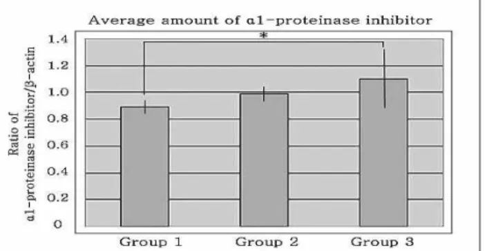
In the study of α1-PI expression levels us- ing Western blot analysis, molecular weight of α1-PI was identified as 42kDa size(Figure 3A). The mean amounts of α1-PI expression (ratio of α1-PI/β-actin) were 0.892±0.053 in group 1, 0.989±0.054 in group 2 and 1.107±
Figure 3A. α1-PI Western analysis showing 4 representative samples in each group. α1-PI corresponding to molecular weight 42kDa was shown to be expressed in all samples including healthy gingiva and the expression level of α1- PI was increased in patients with type 2 diabetes mellitus than in control healthy subjects. In order to quantify detected elastase, β-actin levels were also measured.
Group 1 : healthy gingiva from systemically healthy person
Group 2 : inflammed gingiva from patient with chronic periodontitis
Group 3 : inflammed gingiva from patient with chronic periodontitis and type DM
Figure 3B. Graphics showing the average amounts (Ratio of α1-PI/β-actin) and standard deviation of α1-PI in group 1, 2 and 3. In the inflamed gingiva (with or without diabetes, group 2 & 3), the levels of α1-PI was higher compared to healthy gingiva.
Group 1 : healthy gingiva from systemically healthy person
Group 2 : inflammed gingiva from patient with chronic periodontitis
Group 3 : inflammed gingiva from patient with chronic periodontitis and type 2 DM
* significant difference between group 1 and group 3 (P<0.05) 0.226 in group 3. The significant difference
was observed only in between group 1 and group 3.(P<0.05)
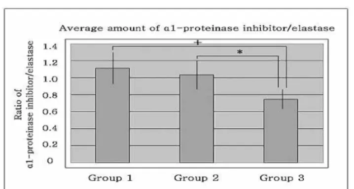
The ratios of α1-PI/elastase were also cal- culated, and compared between groups. The mean amounts of α1-PI/elastase ratio were 1.113±0.192 in group 1, 1.038±0.177 in group 2 and 0.742±0.114 in group 3. The ratio was decreased in the order of group 1, 2, and 3. The differences between group 1 and group 2 and between group 1 and group
3 were statistically significant. (P<0.05) In the interrelationship of IL-6, elastase and α1-PI expressions, as expression of IL-6 was increased, elastase expressions showed increasing tendency in chronic periodontitis associated to type 2 DM. Although α1-PI levels were increased in DM according to increase of IL-6 and elastase, α1-PI/elastase ratios were decreased in chronic periodontitis with type 2 DM.
Figure 4. Graphics showing the average amounts and standard deviation of α1-PI/elastase ratio. The inflammed gingiva with DM is lower than group 2 and 3.
Group 1 : healthy gingiva from systemically healthy person
Group 2 : inflammed gingiva from patient with chronic periodontitis
Group 3 : inflammed gingiva from patient with chronic periodontitis and type 2 DM +significant difference between group 1 and group 3 (P<0.05)
* significant difference between group 2 and group 3 (P<0.05)
IV. DISCUSSION
Multiple studies have demonstrated the link between diabetes and periodontal dis- ease in human subjects. Although diabetes itself does not cause periodontitis, perio- dontal disease progresses more rapidly and leads to more tooth loss in poorly controlled patientsㅃ. Various pathogenetic factors have been suggested to explain the increased prevalence and severity of periodontitis in diabetes3,4).
The purpose of this study was to quantify and compare the expression of IL-6, elastase and α1-PI in the gingival tissues of the pa- tients with chronic periodontitis associated to type 2 DM, in order to understand the con- tribution of these proteins to periodontal de- struction in type 2 diabetic patients, espe- cially elastase-mediated host response in type 2 diabetic patients.
The amount of IL-6 expression was higher
in inflammed gingiva with chronic perio- dontitis associated to type 2 DM than in healthy gingiva from systemically healthy person and inflammed gingiva from patients with chronic periodontitis. The differences among three groups were statistically significant between group 1, 2 and group 3. (P<0.05).
(Figure 1B)
When the expression levels of IL-6 be- tween chronic periodontitis without type 2 DM and chronic periodontitis with type 2 DM were compared, chronic periodontitis with type 2 DM showed higher level than chronic periodontitis without DM. These re- sults were similar with previous reports seen in inflammatory conditions23,24,26). It was considered that IL-6 levels were overex- pressed in inflammed ginginva with or with- out type 2 DM and chronic periodontitis pa- tient with type 2 DM expressed higher cyto- kine activity and inflammatory response.
It was reported the impaired PMN func-
tion in diabetes regarding to chemotaxis, chemokinesis and degranulation41,42). This might explain the increased susceptibility of diabetic patients to various infectious dis- eases including periodontitis. These results make it possible to consider that PMN dys- function may be reflected in the gingival neutrophil degranulation product, which is released from neutrophils recruited to in- flammatory gingiva.
The amounts of elastase expression were higher in chronic periodontitis with type 2 DM compared to chronic periodontitis group of systemic healthy person and healthy gin- giva from systemically healthy person.
The levels in inflammed gingiva with type 2 DM were higher than chronic periodontitis in systemic healthy person and there was statistically significant difference between in- flammed gingiva from patients with chronic periodontitis and inflammed gingiva with chronic periodontitis associated to type 2 DM. These results were similar with the re- sults reported in GCF of chronic perio- dontitis condition of various report43-45). It is assumed that IL-6 stimulated elastase activ- ity in chronic periodontitis associated with type 2 DM. But, it is also considered that other cytokines or other tissue degradation enzymes are involved complexly in elasatse expression.
α1-PI has been studied in this study. The level of α1-PI was increased in inflammed gingiva. In inflammatory response, as the level of elastase increased, α1-PI level was increased, but in the case of inflammed gin-
giva with type 2 DM, the elastase value was not proportional to α1-PI. There was sig- nificant difference between group 1 and group 3.
The comparison of α1-PI/elastase ratio was also studied in some previous reports as an important factor in inflammatory condition.
Some studies had investigated balalnce be- tween α1-PI and elastase in GCF43,46) and a few reports in whole mouth saliva47). It was considered that the ratio was decreased in inflammed gingiva from patients with chron- ic periodontitis and inflammed gingiva with chronic periodontitis associated to type 2 DM. There was significant difference be- tween group 1, 2 and 3. It was considered that α1-PI activity was suppressed because the relative amount of α1-PI was decreased due to the relative increase of elastase in chronic periodontitis group without DM. The ratio in DM group was further decreased.
Neutrophils and monocytes migrating into the gingival crevice were probably the main source of elastase while α1-PI would have derived from extravasated serum, possibly from some local production by macro- phage48,49). It was considered that α1-PI ac- tivity of playing a role in controlling elas- tase activity was decreased in DM and it is suggested that defensive factors are asso- ciated with immune response related to neu- trophils, monocytes, and macrophage were further decreased in DM group, but relation- ship with other immune factors should also be considered.
In interrelationship between IL-6, elastase
and α1-PI, α1-PI level of Group 3 showed increasing pattern according to increasing tendency of IL-6 and elastase expressions in chronic periodontitis associated to type 2 DM. It was shown positive relation between IL-6 and elastase expression, although they were shown reverse effect to α1-PI ratio in inflammed tissue with DM
In inflammed gingiva from patient with chronic periodontitis and type 2 DM, despite of increasing IL-6 and elastase, α1-PI values were not significantly different from the gin- giva with chronic periodontitis.
In conclusion, this study demonstrated that the expression levels of IL-6 and elastase had increasing tendency and positive relation in inflammed tissue and inflammed tissue associated with DM. It is suggested that IL-6 and elastase may be partly involved in the progression of periodontal inflammation associated with type 2 DM. The α1-PI/elas- tase ratio also was futrher decreased in DM group. It is considered that as the quantity and activity of defensive factors associated with immune response is depressed in DM group, the inflammatory response in DM is higher, so the IL-6 and elastase levels are higher.
This ratio of α1-PI/elastase also may be important measuring inflmmatory factors in the progression of periodontal inflammation associated to type 2 DM.
Finally, it seemed that more studies are needed to investigate the effect and inter- relationship between inflammatory enzymes and other immune factors that affect the
progression of periodontal disease in in- flammatory tissue with DM.
V. SUMMARY
The purposes of this study were to com- pare and quantify the expression of IL-6, el- stase and α1-PI in the gingival tissues of patients with type 2 diabetes mellitus and healthy adults with chronic periodontitis.
Gingival tissue samples were obtained dur- ing periodontal surgery or tooth extraction.
According to the patient's systemic condition
& clinical criteria of gingiva, each gingival sample was devided into three groups.
Group 1 (n=8) is clinically healthy gingiva without bleeding and no evidence of bone resorption or periodontal pockets, obtained from systemically healthy 8 patients. Group 2 (n=8) is inflammed gingiva from patients with chronic periodontitis. Group 3 (n=8) is inflammed gingiva from patients with chron- ic periodontitis associated with type 2 diabetes. Tissue samples were prepared and analyzed by Western blotting. The quantifi- cation of IL-6, elastase and α1-PI were per- formed using a densitometer and statistically analyzed by one-way ANOVA followed by Tukey test.
1. The expression levels of IL-6 showed in- creasing tendency in group 2 and 3, and It was highest in group 3.
2. The expression of elastase showed in- creasing tendency in group 2 and 3, and It was highest in group 3.
3. The expression of α1-PI showed increas-
ing tendency in group 3 compared to group 1.
4. The α1-PI/elastase ratio was decreased in group 2 and 3 compared to group 1, es- pecially most decreased in group 3.
5. As IL-6 levels were increasing, elastase showed increasing tendency in group 3, and although IL-6 and elastase levels were increasing, α1-PI level in group 3 showed slightly increasing pattern com- paring to group 1.
In conclusion, this study demonstrated that the expression levels of IL-6 and elastase will be inflammatory markers of periodontal inflammed tissue and DM. The α1-PI/elas- tase ratio also may be important measuring inflmmatory factors in the progression of pe- riodontal inflammation associated to type 2 DM.
VI. REFERENCES
1. Uemura S, Matsushita H, Li W et al.
Diabetes Mellitus Enhances Vascular Matrix Metalloproteinase Activity. Role of Oxidative Stress. Circulation Research 2001;
88:1291-1298
2. Portik-Dobos V, Anstadt MP, Hutchinson J, Bannan M and Ergul A. Evidence for a Matrix Metalloproteinase Induction/
Activation System in Arterial Vasculature and Decreased Synthesis and Activity in Diabetes. Diabetes 2002;51:3063-3068.
3. Emrich LJ, Shlosman M and Genco RJ.
Periodontal disease in non- insulin de- pendent diabetes mellitus. J Periodontol 1991;62:123-129.
4. Taylor GW, Burt BA, Becker MP, Genco
RJ, Shlossman M, Knowler WC and Pettitt DJ. Non-insulin dependent diabetes mellitus and alveolar bone loss progression over 2 years. J Periodontol 1998;69:76-83.
5. Collin H-L, Uusitupa M and Niskanen L.
Periodontal findings in elderly patient with non-insulin depedent diabetes mellitus. J Periodontol 1998;69:962-966.
6. Salvi GE, Yalda B, Collins JG, et al.
Inflammatory Mediator Response as a Potential Risk Marker for Periodontal Diseases in Insulin-Dependent Diabetes mellitus patients. J Periodontol 1997;68:
127-135.
7. AAP Position Paper. Diabetes and peri- dontal disease. J Periodontol 1999;70:935 -949.
8. Yalda B, Offenbacher S and Collins JG.
Diabetes as a modifier of periodontal dis- ease expression. Periodontology 2000 1994;
6:37-49.
9. Mowat AG, Baum J. Chemotaxis of poly- morphonuclear leucocytes from patients with diabetes mellitus. N Engl J Med 1971;284:621-627.
10. Bagdade JD, Stewart M, Walters E.
Impaired granulocyte adherence. A rever- sible defect in host defense in patients with poorly controlled diabetes. Diabetes 1978;27:677-681.
11. Wilson RM, Reeves WG. Neutrophil phagocytosis and killing in insulin-depend- ent diabetes. Clin Exp Immunol 1986;63:
478-484.
12. Iacono VJ, Singh S, Golub LM, Ramamurthy NS, Kaslick R. In vivo as- say of crevicular leukocyte migration. Its development and potential applications. J Periodontol 1985;56:56-62.
13. el-Kishky M, Mahfouz SA, el-Habbak SM. An in vitro study of hydroxyproline synthesis by gingival fibroblasts in pa-
tients wity juvenile diabetes. Egypt Dent J 1986;32:15-27.
14. Sorsa T, Ingman T, Suomaainen K, et al.
Cellular source and tetracycline inhibition of gingival crevicular fluid collagenase of patients with labile diabetes mellitus. J Clin Periodontol 1992;19:146-149.
15. Seibold JR, Uitto J, Dorwart BB.
Collagen synthesis and collagenase activ- ity in dermal fibroblasts from patients with diabetes mellitus and digital sclerosis.
J Lab Clin Med 1985;105:664-667.
16. Vlassara H. Non-enzymatic glycosylation.
Diabetes Annual 1991;6:371-389.
17. Salmela PI, Oikarinen A, Pirttiaho H.
Increased non-enzymatic glycosylation and reduced solubility of xkin collagen in in- sulin-dependent diabetic patients. Diabetes Res 1989;11:115-120.
18. Brownlee M, Cerami A, Vlassara H.
Advanced products of nonenzymatic gly- cosylation and the pathogenesis of dia- betic vascular disease. Diabetes Metab Rev 1988;4:437-451.
19. Sastrowijoto SH, van der Velden U, et al.
Improved metabolic control, clinical perio- dontal status and subgingival microbiology of healthy diseased periodontal pockets in Type I diabetes mellitus patients. J Clin Periodontol 1989;16:233-242.
20. Listgarten MA. Pathogenesis of periodontitis.
J Clin Periodontol 1986;13:418-425.
21. Birkedal-Hansen H. Role of matrix metal- loproteinases in human periodontal diseases.
J Periodontol 1993;64:474-484.
22. Reynolds JJ and Meikle MC. Mechanisms of connective tissue matrix destruction in periodontitis. Periodontol 2000 1997;14:
144-157.
23. Johnson RB, Serio FG, Dai X. Vascular endothelial growth factors and progression of periodontal diseases. J Periodontol
1999;70:848-852.
24. Geivelis M, Turner DW, Pederson ED, Lamberts B. Measurements of inter- leukin-6 in gingival crevicular fluid from adults with destructive periodontal disease.
J Periodontol 1993;64:980-983.
25. Reinhardt RA, Masada MP, Kaldahl WB et al. Gingival fluid IL-1 and IL-6 levels in refractory periodontitis. J Clin Periodontol 1993;20:225-231.
26. Guillot JL, Pollock SM, Johnson RB.
Gingival IL-6 concenturation following phase I therapy. J Periodontol 1995;66:
667-672.
27. Yamazaki K, Nakajima T, Gemmell E, et al. IL-4 and IL-6 producing cells in hu- man periodontal disease tissue. J Oral Pathol Med 1994;23:347-353.
28. Irwin CR, Myrillas TT. The role of IL-6 in the pathogenesis of periodontal disease.
Oral Dis 1998;4:43-47.
29. Owen CA, Campbell EJ. The cell biology of leukocyte-mediated proteolysis. J Leukoc Biol 1999;65:137-150.
30. Dallegri F, Ottonello L. Tissue injury in neutrophilic inflammation. Inflamm Res 1997;46:382-391.
31. Watanabe H, Hattori S. Human neutrophil elastase:degradation of basement mem- brane components and immunolocalization in the tissue. J Biochem 1990;108:753-759.
32. Armitage CG, Jeffcoat MK, Chadwick DE. Longitudinal evaluation of elastase as a marker for the progression of perio- dontitis. J Periodontol 1994;65:120-128.
33. Mansson SA, Soder B, KAri K, Meurman JH. Influence of combinations of bacteria on the levels of Prostaglandin E2, IL-1beta, and granulocyte Elastase in gingival crev- icular fluid and on the sevirity of peri- dontal disease. J Periodontol 2006;77:1025 -1031.
34. Sandholm L. Proteases and their inhibitors in chronic inflammatory periodontal disease.
J Clin Periodontol 1986;13:19-26.
35. Rinehart AR, Mallya SK, Simon SR.
Human α1-proteinase inhibitor binds to extracellular matrix in vitro with retention of inhibitory activity. Am J Respir Cell Mol Biol 1993;9:666-679.
36. Giannopoulou C, Andersen E, Demeurisse C, Cimasoni G. Neutrophil elastase and its inhibitors in human gingival crevicular fluid during experimental gingivitis. J Dent Res 1992;71:359-363.
37. Janoff A. Elastases and emphysema: cur- rent assesment of the proteinase-anti- proteinase hypothesis. Am Rev Respir Dis 1985;132:417-433.
38. Adonogianaki E, Mooney J, Kinane DF.
The ability of gingival crevicular fluid acute phase proteins to distinguish health, gingivitis and periodontitis sites. J Clin Periodont 1992;19:98-102.
39. Mühlman HR and Son SH. Gingival Bleeding - a leading symptom in initial gingivitis. Helvitica Odontologica Acta 1971;15:107-113.
40. Kim DH, Park EK, Shin HI et al. The comparison of MMP-2, MMP-9 and tu- mor necrosis factor-α expressions in hu- man gingiva with chronic periodontitis with or without associated to Type 2 Diabetes Mellitus. Journal of Korean Academy of Periodontology 2006;36:409-426.
41. Cho JY, Xing S, Liu X et al. Expression and activity of human Na+/I- symporter in human glioma cells by adenovirus -mediated gene delivary. Gene Therapy 2000;7:740-749.
42. Golub LM, Stiegel K and Ramamurty NS. Some characteristics of collagenase activity in gingival crevicular fluid and its relationship to gingival diseases in human.
J Dent Res 1976;55:1049-1057.
43. R. Buchmann, A. Hasilik, T.E. Van Dyke, and D.E. Lange. Amplified crevicular leu- kocyte activity in aggresive periodontal disease. J Dent Res 2002;81:716-712.
44. Figueredo CMS and Gustafsson A.
Activity and inhibition of elastase in GCF. J Clin Periodontol 1998;25:531-535.
45. Uitto V-J, Nieminen A, Coli J, Hurttia H, Larjava H. Oral fluid elastase as an in- dicator of periodontal health. J Clin Periodontol 1996;23:30-37.
46. Cox SW, Rodriguez-Gonzalez EM, Booth V and Eley BM. Secretory leukocyte pro- tease inhibitor and its potential inter- actions with elastase and cathepsin B in gingival crevicular fluid and saliva from patients with chronic periodontitis. J Periodont Res 2006;41:477-485.
47. Pederson ED, Stanke SR, Whitener SJ et al. Saliva levels of α2-macroglobulin, α1- antitrypsin, C-reactive protein, cathepsin G and elastase in humans with or without destructive periodontal disease. Arch Oral Biol 1995;40:1151-1155.
48. Kennett CN, Cox SW, Eley BM.
Investigations into the cellular contribution to host tissue proteases and inhibitors in gingival crevicular fluid. J Clin Periodontol 1997;24:424-431.
49. Kennett CN, Cox SW, Eley BM.
Localisation of active and inactive elas- tase, alpha-1-proteinase inhibitor and al- pha-2-macroglobulin in human gingiva. J Dent Res 1995;74:667-674.
-국문초록-
단순 만성 치주염 환자 및 2형 당뇨병환자의 만성 치주염 치은조직에서 IL-6, elastase 및 α
1-PI의 발현
양상 비교
박재완, 이재목*
경북대학교 치과대학 치주과학교실
본 실험에서는 제2형 당뇨병 환자와 비당뇨 환자들에서 만성 치주염 부위의 치은 및 건강한 치은에서 염증 매개체 중 하나인 IL-6, elastase 및 α1-PI의 발현에 대해 상호 비교 분석함으로서 염증, 혈당이 미치는 영향을 밝히고 제 2형 당뇨병 환자에서 심한 치주조 직 파괴의 기전을 연구하고자 하였다.
경북대학교병원 치주과 내원환자 중 제2형 당뇨병 환자와 비당뇨 환자들 및 치주질환 이 없는 건강인 대조군을 대상으로 여러 가지 환자요소, 임상 치주상태를 기록하고, 전신 적으로 건강한 환자의 건강한 부위(n=8, Group 1), 전신적으로 건강한 환자의 만성 치주 염 부위(n=8, Group 2), 제2형 당뇨병 환자의 만성 치주염 부위 (n=8, Group 3)에서 각각 변연치은을 채득하고 액화질소에 급속 동결하였다. Western blotting을 이용하여 각 조직 내 IL-6, elastase 및 α1-PI의 발현을 관찰, densitometer를 이용하여 상대적 발현을 정량, 각 조직의 β-actin을 이용하여 표준화하여 실험군과 대조군들의 평균치를 비교하여 다음과 같은 결과를 얻었다.
1. IL-6의 발현은 2군과 3군에서 증가되는 경향을 보였으며 3군에서 가장 높게 나타났다.
2. Elastase의 발현도 2군과 3군에서 증가양상을 보였으며 3군에서 가장 높게 나타났다.
3. α1-PI의 발현정도는 1군에 비해 3군에서 증가되는 경향을 보였다.
4. α1-PI/elastase 비율은 1군에 비해 2군과 3군에서 감소되었으며, 특히 3군에서 가장 감 소되었다.
5. 3군에서 IL-6 발현이 증가됨에 따라 Elastase 발현이 증가되는 경향을 보였으나, IL-6 와 Elastase 수치가 증가함에도 불구하고 α1-PI 수치는 1군에 비해 약간 증가되는 경 향을 보였다.
결론적으로 IL-6 와 elastase는 만성 치주염과 제2형 당뇨병 환자의 만성 치주염 부위에 서 염증상태에 따른 지표로 응용할 수 있으리라 생각되며, α1-PI/elastase 비율 또한 2형 당뇨환자의 만성치주염 부위에서 중요한 측정인자가 될 수 있으리라 생각된다.2)
Key worlds: inflammation, inflammatory mediator, chronic periodontitis, diabetes mellitus