: Vol. 35, No. 3, 2005
Expression of Matrix metalloproteinase-1 between Simple Chronic Periodontitis and Type 2 Diabetes
associated Chronic Periodontitis on Protein level
Jae-Mok Lee
Department of Periodontology, College of Dentistry, Kyungpook National University
Ⅰ. Introduction
1)Chronic Periodontitis is an inflammatory disease initiated and maintained by bac- terial plaque and its metabolic products that trigger the local infiltration of inflammatory cells associated with the breakdown of collagenous extracellular matrices(ECMs)1). The degradation of gingival connective tissue during periodontitis could be a disturbance of cell-cell and cell-matrix interactions involving the production of enzymes, acti- vators, inhibitors, and regulatory molecules such as cytokines and growth factors2,3).
In periodontitis, although some tissue- destructive enzyme activities may derive from specific bacteria, it is more likely that plaque bacteria inaddition to their other
pro-inflammatory effects, generate prote- inases that activate latent forms of mam- malian collagenase, or even stimulate the release of collagenase and other matrix metalloproteinases(MMPs) from host cells4). MMPs are a family of zinc-dependent endo- peptidases that collectively degrade all ex- tracellular matrix proteins and basement membrane at neutral pH and are essential for cellular migration and tissue remodeling under physiological and pathological condi- tions5,6). Most of them are secreted as inac- tive form(proenzyme) and are activated in the extracellular compartment or in the vicinity of the cell membranes by other MMPs or serine proteinases7). Interstitial collagenase, including matrix metallopro- teinase-1(MMP-1) and MMP-8, serve as ini-
*This work was supported by BioMedical Research Institute grant, Kyungpook National University Hospital (2004)
Correspondence author: Jae-Mok Lee, Department of Periodontology, College of Dentistry, Kyungpook National University, 50 Samduk-Dong, Jung-Gu, Daegu, 700-422, South Korea
tiators of extracellular matrix destruction in periodontal disease. Fibroblast-type inter- stitial collagenase(MMP-1) is distributed widely in tissues and expressed by fibro- blasts, keratinocytes, endothelial cells, osteoblasts, chondrocytes and monocytes/
macrophages4,8). MMP-1 is a regulator of connective tissue remodeling and is present in high concentrations especially in inflamed gingival regions, including in periodontal disease8). Enhanced protein levels and m- RNA expression of MMP-1 have been de- monstrated in inflammatory disease, inclu- ding periodontitis9-11). The increased levels of MMP-1 observed in periodontitis may be a result of an alteration in the regulation of MMP-1 by gingival fibroblast, which is the most predominant cell in the gingivae. MMP -1 production in human gingival fibroblasts is stimulated by cytokines such as inter- leukin-1β and tumor necrosis factor-α12).
Periodontal disease is frequently men- tioned among the oral problems encountered in patients with diabetes. Although diabetes in itself does not cause periodontitis, perio- dontal disease progresses more rapidly and leads to more tooth loss in patients whose diabetes is poorly controlled13-17). Severe Periodontitis has been associated with an increased risk of poor glycemic control and, in turn, untreated advanced periodontal disease can deteriorate the metabolic control of diabetes18-22). Collagen undergoes non- enzymatic glycosylation when subjected to a hyperglycemic environment and the glucose- derived cross-links between the molecules contribute to reduced collagen solubility and turn-over rate23). Advanced glycosylation
end-products(AGEs) play a central role in diabetic complications. Several studies have linked an increased development of perio- dontal disease in diabetes to an decreased collagen production and exaggerated colla- genase activities in periodontium. In dia- betic patients, collagenolytic activity of gin- gival crevicular fluid is increased and gingival fibroblasts from diabetic patients synthesize less collagen than those form non-diabetic subjects24,25).
It is well known that MMP-1 regulates ECM turnover, however, whether diabetes affects the expression and activity of the MMP-1 in patients with diabetes remains unknown. Therefore, the aim of this study is to investigate the MMP-1 levels in human gingival tissue and to compare MMP-1 levels of diabetic and nondiabetic periodontal pa- tients, using systemically healthy indivi- duals as the control group.
Ⅱ. Materials and Methods
1. Study population and Tissue sampling
Study population comprised 8 patients with type 2 diabetes and chronic perio- dontitis, 8 patients with chronic perio- dontitis, and 8 healthy individuals. Marginal gingival tissue samples were obtained during periodontal surgery(including surgical crown lengthening) or tooth extraction. Before sur- gery, patient's systemic condition(age, sex, blood glucose level, smoking, obesity) and clinical criteria of gingiva were recorded.
According to the patient's systemic con- dition & clinical criteria of gingiva(gingival
color, gingival bleeding, probing depths, and radiographic evidence of bone resorption), each gingival sample was devided into the three group. Group 1(control, n=8, aged 38.1 [20-48], 5 males and 3 females) is clinically healthy gingiva without bleeding and no evidence of bone resorption or periodontal pockets, obtained from syste- mically healthy 8 patients. Group 2(perio- dontitis, n=8, aged 42.8 [38-48], 5 males and 3 females) is inflamed gingiva from patients with chronic periodontitis. The diagnosis of chronic periodontitis was esta- blished on the basis of clinical and radio- graphic criteria(bone resorption) according to the classification system for periodontal diseases and conditions26). All patient in group 2 was systemically healthy and had more than one pocket ≥5 mm and at least on pocket with ≥4 mm loss of attachment.
All gingival samples were obtained from teeth with probing depth ≥5 mm, swelling of the marginal gingiva, and bleeding corre- sponding to sulcus bleeding index 3 accor- ding to Muhlemann and Son27). Group 3 (periodontitis & type 2 DM, n=8, aged 58.3 [44-71], 6 males and 2 females) is inflamed gingiva from patients with chronic perio- dontitis and type 2 diabetes. Patients in group 2 & 3 has similar periodontal con- dition, but systemically, patients in group 2 was healthy and patients in group 3 was type 2 diabetics. Mean fasting glucose level in group 3 was 142.88 mg/dl and mean 2 hour postprandial glucose level was 212.5 mg/dl. Diabetic control was performed by insulin medication in all 8 patients and, additionally, diet control was also performed
in 4 of them. Gingival samples were obta- ined by similar way described above.
The sample cohort consisted of 8 clinically healthy, 8 inflamed and 8 diabetic patients' inflamed samples from total of 24 subjects.
Following surgery, excised tissue specimens were immediately placed on liquid nitrogen and subsequently frozen(-70℃).
2. Protein Isolation and Western blotting
The visualization of Protein levels of MMP-1 in tissue samples was performed by immunoblotting using antibody specific for the species.
For Western blotting, as previously des- cribed technique by Cho28), frozen tissues were homogenized in RIPA lysis buffer(10 mM phosphate buffer, pH 7.4, 10% glycerol, 1% NP-40, 0.1% SDS, 4 mM EDTA, 0.15 M NaCl) with 1:30 diluted protease inhibitor cocktail(Roche). The lysates were sonicated 3 times for 10 seconds and centrifuged at 12,000g for 15 minutes. Protein concen- tration of supernatant were routinely deter- mined by a Bradford protein assay(Bio-Rad) using BSA as a standard.
Lysates were boiled in SDS sample buffer (1M Tris-Cl (pH6.8), 40% glycerol, 8% SDS, 2%-mercaptoethanol, 0.002% Bromophenole blue). Prepared sample were separated by 10% sodium dodecyl sulfate(SDS)-polyacry- lamide gels and transferred to a polyvin- ylidene difluride membrane.
The membranes were subsequently block- ed in Tris-buffered saline(TBS) containing 5% powdered milk and 1% BSA for 1 hours, and then incubated with anti-MMP-1 anti-
Group 1 Group 2 Group 3 MMP-1
β-actin
Figure 1. Western analysis of samples for MMP-1. MMP-1 corresponding to molecular weight 53 kDa was expressed in all samples including healthy gingiva, and expression of MMP-1 was increased in Group 2 than Group 1, 3. In order to quantify detected MMP-1, normalization to β-actin was performed.
body(Sigma, ST. Luis, U. S. A., prepared in rabbits, diluted 1:1000 in TBS/1% BSA) for 1.5 hours at room temperature. The mem- brane was washed(five times for 5 minutes with 0.2% Tween 20) and incubated with a horseradish peroxidase(HRP)-coupled goat anti-rabbit secondary antibody(diluted 1:
2000 in TBS) for 1 hour at room tempe- rature. After additional washing(five times for 5 minutes with Tween 20) the Western blot procedure was completed with an ECL Plus development kit(Amersham, Becking- hamshire, U. K.)
The quantitative analysis(the ratio of MMP-1/β-actin) of MMP-1 was performed using a densitometer(Image Gauge V 3.46, Altura Software, Koshin Graphic Systems, FUJI PHOTO FILM CO). After normali- zation to β-actin(AbcamⓇ, Cambridge Science Park, U. K.) in each sample, level of MMP -1 was expressed as a ratio and the differ- ence of density among 3 group was determined.
3. Statistical Analysis of the Western blot results
All data were presented as means and standard deviation and results were stati- stically analyzed. The MMP-1 levels among each 3 groups were compared using one way ANOVA followed by Scheffe test.
It was considered to be statistically signi- ficant when P<0.05. Statistical analysis in this study was performed using appro- priate SPSS 12.0 K software(SPSS, Korea).
Ⅲ. Results
The purpose of this study was to compare the relative amounts of MMP-1 in healthy versus diseased gingiva with or without type 2 diabetes mellitus. Antibodies to MMP-1 cross-reacted with 53 kDa of MMP-1 in all 3 groups were observed(Figure 1). In order to quantify detected MMP-1, normalization to β -actin was performed.
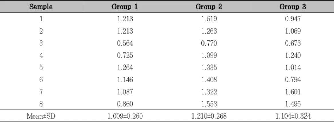
Using a densitometer, the quantitative analysis(the ratio of MMP-1/β-actin) of MMP-1 of each sample was performed. The identified MMP-1 levels of gingival tissue samples are presented in Table 1. Average amounts of MMP-1 between 3 groups were compared(Figure 2).
MMP-1 levels of gingival tissue samples were varied within the same group(Table 1).
The mean amount of MMP-1 in group 1, 2 and group 3 was 1.009, 1.209 and 1.104, respectively. But, the differences among 3 groups were not statistically significant (P>0.05). It is because sample size was too small and various factor of each individuals was not excluded.
Table 1. The quantitative analysis(the ratio of MMP-1/β-actin) of MMP-1 of each sample was performed using a densitometer and mean amount of MMP-1 in 3 groups were identified
Sample Group 1 Group 2 Group 3
1 1.213 1.619 0.947
2 1.213 1.263 1.069
3 0.564 0.770 0.673
4 0.725 1.099 1.240
5 1.264 1.335 1.014
6 1.146 1.408 0.794
7 1.087 1.322 1.601
8 0.860 1.553 1.495
Mean±SD 1.009±0.260 1.210±0.268 1.104±0.324
Group 1: healthy gingiva from systemically healthy patient
Group 2: inflamed gingiva from systemically healthy patient with chronic periodontitis Group 3: inflamed gingiva from patients with diabetic patient with chronic periodontitis
Figure 2. Mean amount(the ratio of MMP-1/β-actin) and standard deviation of MMP-1 in group 1, 2, and 3. In group 2, MMP-1 increased compared to group 1. But the difference was not statistically significant(P>0.05).
Ⅳ. Discussion
Diabetic subjects are 2.3 times more likely to afflicted with periodontal disease than non-diabetic subjects30) and several cross- sectional studies suggested that the bidirec- tional relationship between DM and perio-
dontal diseases14,21,29). Considering the in- creasing number of diabetics in the aging population, assessment of MMP system in periodontal tissue and diabetic patients may help to provide appropriate health/oral care to this population.
In present study, MMP-1 levels of in-
flamed gingiva of systemically healthy pa- tient with chronic periodontitis were higher than normal gingiva of healthy patient.
Periodontal disease is characterized by loss of collagen fiber and other extracellular matrix constituents in periodontal tissues.
Most likely, periodontal tissue destruction is mediated to a significant extent by the host cell-derived MMPs. The neutrophil-type col- lagenase(MMP-8) secreted by PMN leuko- cytes may have a more important role than the fibroblast-type collagenase(MMP-1) in the pathological destruction of periodontal connective tissues24,35). However, MMP-1 is an important regulator of CT remodelling and is present in high concentration in inflamed regions, including in periodontal disease. Aiba et al.8) indicated that MMP-1 mRNA increased in inflammatory lesions of adult periodontitis. It was in agreement with the findings obtained by Nomura et al36). Meikle et al.37) immunohistochemically de- tected the presence of MMP-1, -2, and -3 and TIMP-1 in inflamed gingiva of patients with chronic inflammatory periodontal disea- se. Moreover the protein amounts and acti- vities of MMP-1 in gingival crevicular fluid (GCF) and gingival tissue were reported to be higher in periodontitis affected sites than in healthy site11). Thus, MMP-1 may parti- cipate in collagen degradation in advanced periodontal disease. These are consistent with this result.
In this study, although the severity of gingival inflammation in group 2 and 3 are similar, MMP-1 expression is decreased in diabetic patients than systemically healthy periodontal patients. It is assumed that
AGE formation of collagen might reduced MMP-1 production and decreased remo- delling capacity of fibroblast. Uncontrolled diabetes with peridontal disease frequently exhibit an altered inflammatory cell func- tions and impaired neutrophil and monocyte /macrophage functions23,31-33). Increasing glu- cose concentrations can reduce the synthesis of collagens and glycosaminoglycans23). Both the function of proteins and cells involved in the host defence can be modified by non- enzymatic glycosylation23,32). And prolonged exposure to hyperglycemia may result in vascular dysfunction and cellular changes33). Most histological studies have demonstrated that small blood vessels of the gingiva in long-term diabetic patients frequently show microangiopathic changes with occlusion and increased vascular thickness. Chappey et al.32) have indicated that microvascular complications due to long term hypergly- cemia may occur due to modified proteins, the so called advanced glycosylation end- products(AGEs). AGEs can induce diabetic collagen cross-links and expansion of ECM, such as hardening of arteries and narrowing of vascular lumina. Potentially AGEs may also induce oxidant stress in the gingiva, resulting in accelerated periodontal tissue destruction34).
There is little data on MMP expression in human type 2 diabetes, despite of the great interest in the MMP system and periodontal disease. Death et al.38) showed that high glucose exposure could promote increased MMP-1 expression from two key vascular cell, endothelial cell and macrophage. But Portik-Dobos et al.39) demonstrated that
MMP induction and activation system exists in human arterial vasculature and that is downregulated in diabetes. They commented that decreased MMP activity(MMP-1, -2 and -9) may contribute to increased collagen deposition and pathological remodeling in diabetes. Rittie et al.40) studied the influ- ence of collagen glycation on matrix metal- loproteinase production by dermal fibro- blasts using the model of lattice culture.
Contraction of glycated collagen lattices was strongly reduced and fibroblast synthesized lower amount of interstitial collagenase (MMP-1). These results demonstrate that nonenzymatic glycation of type I collagen could greatly impair healing by decreasing the collagen remodeling capacity of fibro- blast.
In diabetic patient, further evaluation is needed to investigate the relationship bet- ween MMP-1 and other MMP system and the influence of inflammatory mediator (cytokine) on MMP-1 expression. Under- standing expression patterns and levels of MMPs in periodontal tissues is essential for monitoring the course of periodontitis as well as the effects of various treatment modalities.
Ⅴ. Summary
The purpose of this study was to quantify and compare the level of MMP-1 in the healthy or inflamed gingival tissue of patients with or without type 2 diabetic mellitus. We investigated whether mean
amount of MMP-1 was changed by chronic periodontitis and type 2 DM.
Gingival tissue samples were obtained during periodontal surgery or tooth extrac- tion. According to the patient's systemic condition & clinical criteria of gingiva, each gingival sample was divided into the three group. Group 1(n=8) was clinically healthy gingiva without bleeding and no evidence of bone resorption or periodontal pockets, obtained from systemically healthy 8 patients. Group 2(n=8) was inflamed gin- giva from patients with chronic periodon- titis. Group 3(n=8) was inflamed gingiva from patients with chronic periodontitis and type 2 diabetes. Tissue samples were pre- pared and analyzed by Western blotting.
The quantitative analysis of MMP-1 was performed using a densitometer and sta- tistically analyzed by ANOVA.
MMP-1 was expressed in all samples and an increased MMP-1 level was observed in group 2 compared to group 1 and decreased MMP-1 level was found group 3 compared to group 2, but the differences among 3 groups were not statistically significant.
In conclusion, this study demonstrated that MMP-1 levels of inflamed gingiva of systemically healthy patient(group 2) were higher than normal gingiva of systemically health patients and although the severity of gingival inflammation in group 2 and 3 were similar, MMP-1 expression was decreased in diabetic patients than systemically healthy periodontal patients.
Ⅵ. References
1. Listgarten MA. : Pathogenesis of perio- dontitis. J Clin Periodontol 13:418-425.
1986.
2. Birkedal-Hansen H. : Role of matrix me- talloproteinases in human periodontal diseases. J Periodontol 64:474-484. 1993.
3. Reynolds JJ and Meikle MC. : Mecha- nisms of connective tissue matrix de- struction in periodontitis. Periodontol 2000 14:144-157. 1997.
4. Westerlund U, Ingman T and Lukin- mmaa P-L : Human neutrophil gelati- nase and associated lipokalin in adult and localized juvenile periodontitis. J Dent Res 75:1553-1563. 1996.
5. Nagase H and Woessner JF Jr. : Ma- trix metalloproteinases. J Biol Chem 274:21491-21494. 1999.
6. Wincenti MP. : The matrix metal- lopro- teinase(MMP) and tissue inhibitor of metalloproteinase genes. Methods Mol Biol 151:121-148. 2001.
7. Nagase H : Activation mechanisms of matrix metalloproteinases. J Biol Chem 378:151-160. 1997.
8. Aiba T, Akeno N, Kawane T, Okamoto H and Horiuchi N. : Matrix metallopro- teinases-1 and -8 and TIMP-1 mRNA levels in normal and diseased human gingivae. Eur J Oral Sci 104:562-569.
1996
9. Kubota T, Nomura T, Takahashi T and Kara K. : Expression of mRNA for matrix metalloproteinases and tissue inhibitors of metalloproteinases in periodontitisaf- fected human gingival tissue. Arch Oral
Biol 41:253-262. 1994.
10. Ingman T, Tervahartiala T and Ding Y : Matrix metalloproteinases and their inhibitors in gingival crevicular fluid and saliva of periodontitis patients. J Clin Periodontol 23:1127-1132. 1996.
11. Soell M, Elkaim R and Tenenbaum H. : Cathepsin C, matrix metalloproteinases, and their tissue inhibitors in gingiva and gingival crevicular fluid from periodon- titis affected patients. J Dent Res 81:
174-178. 2002.
12. Zee E, Everts V and Beertsen W. Cyto- kines modulate routes of collagen break- down. Review with special emphasis on mechanism of collagen degradation in the periodontium and the burst hypothesis of periodontal disease progression. J Clin Periodontol 24:297-305. 1997.
13. Nelson RG, Shlossman M and Budding LM. : Periodontal disease and NIDDM in Pima Indians. Diabetes Care 13:836-840.
1990.
14. Emrich LJ, Shlossman M and Genco RJ.
: Periodontal disease in non-insulinde- pendent diabetes mellitus. J Perio- dontol 62:123-13. 1991.
15. Thorstensson H and Hugoson A. : Perio- dontal disease experience in adult long duration insulin-dependent diabetics. J Clin Periodontol 20:352-358. 1993.
16. Taylor GW, Burt BA, Becker MP, Genco RJ, Shlossman M, Knowler WC and Pettitt DJ. : Non-insulin dependent dia- betes mellitus and alveolar bone loss progression over 2 years. J Periodontol 69:76-83. 1998.
17. Collin HL, Uusitupa M and Niskanen L.
: Periodontal findings in elderly patient with non-insulin dependent diabetes mellitus. J Periodontol. 69:962-966.
1998.
18. Wolf J. : Dental and periodontal condi- tions in diabetes mellitus. A clinical and radiographic study. Proc Finn Dent Soc 73(4-6 Suppl.):1-56. 1977.
19. Seppala B, Seppala M and Ainamo J. : A longitudinal study on insulin-depen- dent diabetes mellitus and periodontal disease. J Clin Periodontol 20:161-165.
1993.
20. Seppala B and Ainamo J. : A site- by-site follow-up study on the effect of controlled versus poorly controlled insulin-dependent diabetes mellitus. J Clin Periodontol 21:161-165. 1994.
21. Taylor GW, Burt BA and Becker MP : Severe periodontitis and risk for poor glycemic control in patient with non- insulin-dependent diabetes mellitus. J Periodontol. 67:1085-1093. 1996.
22. Taylor GW. : Bidirectional interrelation- ships between diabetes and periodontal diseases : An Epidomiologic Perspective.
Ann Periodontol 6:99-112. 2001.
23. Cohen MP. : Non-enzymatic glycosyla- tion. Diabetes Annual 4:469-484. 1988.
24. Golub LM, Lee HM and Lehrer G. : Minocycline reduces gingival collageno- lytic activity during diabetes. J Perio Res 18:516-526. 1983.
25. AAP. Position Paper : Diabetes and periodontal disease. J Periodontol 70:
935-949. 1999.
26. Armitage GC. : Development of a classi- fication system for periodontal diseases
and conditions. Ann Periodontol 4:1-6.
1999.
27. Muhlemann HR. and Son S. : Gingival sulcus bleeding a leading symptom in initial gingivitis. Helv Odontol Acta 15:107. 1971
28. Cho J-Y, Xing S, Liu X, Buckwalter TLF , Hwa L, Sferra TJ, Chiu IM and Jhiang SM. : Expression and activity of human Na+/I- symporter in human glioma cells by adenovirus-mediated gene delivery.
Gene Therapy 7:740-749. 2000.
29. Tervonen T. and Oliver RC. : Long- term control of diabetes mellitus and periodontitis. J Clin Periodontol 20:431 -435. 1993.
30. Grossi SG, Zambon JJ, Ho AW, Koch G, Dunford RG, Machtei EE, Norderyd OM and Genco RJ. : Assessment for risk of periodontal disease. I. Risk indicators for attachment loss J Periodontol 65:
260-267. 1994.
31. Ueta E, Osaki T, Yoneda K and Yama- moto T. : Prevalence of diabetes mel- litus in odontogenic infections and oral candidacies: an analysis of neutrophil suppression. J Oral Patho Med 22:168- 194. 1993.
32. Chappey O, Dosquet C, Wautier MP and Wautier JL. : Advanced glycation end products, oxidant stress and vascular lesions. Eur J Clin Invest 27:97-108.
1997.
33. Feener EP and King GL. : Vascular dys- function in diabetes mellitus. Lnacet 350:S19-S113. 1997.
34. Schmidt AM, Weidman E, Lalla E, Yan SD, Hori O, Cao R, Brett JG and
Lamster IB. : Advanced glycation end products induce oxidant stress in gin- giva: a potential mechanism underlying accelerated periodontal disease associat- ed with diabetes. J Periodont Res 31:
508-515. 1996.
35. Sorsa T, Ding Y and Ingman T. : Cel- lular source, activation and inhibition of dental plaque collagenase. J Clin Perio- dontol 22;709-717. 1995.
36. Nomura T, Takahashi T and Hara K. : Expression of TIMP-1, TIMP-2 and colla- genase mRNA in periodontitis-affected human gingival tissue. J Perio Res 28:354-362. 1993.
37. Meikle MC, Hembry RM, Holley J, Horton C, McFarlane CG and Reynolds JJ. : Immunolocalization of matrix metalloproteinases and TIMP-1 in hu- man gingival tissues from periodontal patients. J Periodont Res 29:118-126.
1994.
38. Death AK. Fisher EJ, McGrath KCY and Yue DK. : High glucose alters matrix metalloproteinase expression in two key vascular cells: potential impact on atherosclerosis in diabetes. Athero- scle- rosis 168:263-269. 2003.
39. Portik-Dobos V, Anstadt MP, Hutch- inson J, Bannan M and Ergul A. : Evi- dence for a Matrix Metalloproteinase induction/activation system in arterial vasculature and decreased synthesis and activity in diabetes. Diabetes 51: 3063 -3068. 2002.
40. Rittie L, Berton A, Monboisse JC, Hor- nebeck W and Gillery P. : Decreased contraction of glycated collagen lattices coincides with impaired matrix metal- loproteinase production. Biochemical and Biophysical Research Communications 264:488-492. 1999.
-Abstract-
단순만성치주염환자와 2형 당뇨환자의 만성치주염에서 Matrix metalloproteinase-1의 발현양상
이 재 목
경북대학교 치과대학 치주과학교실
본 연구의 목적은 전신적으로 건강한 치주질환자를 대조군으로 하여 제 2형 당뇨병을 동반한 치주질환자의 치은조직에서 MMP-1의 발현양상을 관찰, 비교하는 것으로 당뇨병을 동반한 경우 MMP-1의 발현양상이 변화 되는지의 여부를 연구하였다.
경북대학교 병원 치주과에 내원한 환자 중 검사 및 수술에 동의한 환자로 전신 질환이 없고 부착 소실이 없 거나 안정되어 있으며 치은 염증 소견이 없는 환자를 정상조직군, 임상적 치주낭 깊이가 5 mm 이상이고 방사 선 사진상 치조골 소실이 분명한 환자를 만성 치주염 환자군, 심각한 전신적 합병증, 감염등의 위험요인이 없 고 2형 당뇨병으로 진단받은 환자로서 만성 치주염으로 진단된 환자군 각 8명을 대상으로 하였다. 만성 치주염 환자와 당뇨병을 가진 만성 치주염 환자에서 치은 염증 소견을 보이는 치은조직을 채득하고 액화질소에 넣어 급속 동결고정시킨 후 MMP-1의 발현 양상을 western blot analysis를 통해 관찰하였고, densitometer를 이용하여 상대적 발현을 정량, 각 조직의 β-actin을 이용하여 표준화하여 각 군의 평균치를 비교하였다. 각 군 간의 차이를 one way ANOVA test로 분석하였다.
모든 군에서 분자량 53 kDa의 MMP-1에 상응하는 띠가 나타났으며 정량결과 전신적으로 건강한 치주염 환자군에서 MMP-1의 발현이 당뇨병을 동반한 치주염 환자군과 정상조직군의 치은조직에서보다 높게 나타났 으나 통계적으로 유의한 차이는 나타나지 않았다.
치은염증의 존재시 MMP-1의 발현이 다소 증가됨을 관찰하였으나 통계적으로 유의한 수준은 아니였으며, 당뇨병을 동반한 치주염 환자군에서 전신적으로 건강한 치주염 환자군에서 보다 MMP-1의 발현이 감소되는 경향을 보였다.2)
주요어 : 치주염, 당뇨병, Matrix metalloproteinase, MMP-1, 치은조직