www.jpis.org
pISSN 2093-2278 eISSN 2093-2286 Copyright © 2010 Korean Academy of PeriodontologyThis is an Open Access article distributed under the terms of the Creative Commons Attribution Non-Commercial License (http://creativecommons.org/licenses/by-nc/3.0/).
The expressions of inflammatory factors and tissue inhibitor of matrix metalloproteinase-2 in human chronic periodontitis with type 2 diabetes mellitus
Dong-Seok Shin, Jin-Woo Park, Jo-Young Suh, Jae-Mok Lee* Department of Periodontology, Kyungpook National University School of Dentistry, Daegu, Korea
Purpose: The purpose of this study was to observe and quantify the expression of interleukin-4 (IL-4), interferon-g (IFN-g), and tissue inhibitor of matrix metalloproteinase-2 (TIMP-2) in the gingival tissue of patients with type 2 diabetes mellitus (DM) and healthy adults with chronic periodontitis.
Methods: Twelve patients with type 2 DM and chronic periodontitis (Group 3), twelve patients with chronic periodontitis (Group 2), and twelve healthy individuals (Group 1) were included in the study. Clinical criteria of gingival (sulcus bleeding in- dex value, probing depths) and radiographic evidences of bone resorption were divided into three groups. The concentrations of cytokines were determined by a western blot analysis and compared using one-way ANOVA followed by Tukey’s test.
Results: The expression levels of IFN-g and TIMP-2 showed an increasing tendency in Groups 2 and 3 when compared to Group 1. On the other hand, the expression of IL-4 was highest in Group 1.
Conclusions: The findings suggest that IFN-g and TIMP-2 may be involved in the periodontal inflammation associated with type 2 DM. IL-4 may be involved in the retrogression of the periodontal inflammation associated with type 2 DM.
Keywords: Chronic periodontitis, Tissue inhibitor of metalloproteinases, Type 2 diabetes mellitus.
INTRODUCTION
Chronic periodontal diseases are bacterial infections affect- ing the periodontium resulting in the loss of tooth support and are associated with bacteremia, inflammation, and a strong immune response. They represent primarily anaerobic Gram- negative oral infection that leads to gingival inflammation, destruction of periodontal tissues and loss of alveolar bone [1,2].
Diabetes mellitus (DM) is a highly prevalent metabolic dis- order. The more common form, type 2 diabetes, results from a combination of impaired insulin production and insulin re- sistance [3].
The mechanisms responsible for these outcomes in patients
with diabetes are mainly related to the increased risk of in- fections, impairment of the synthesis of collagen and glycos- aminoglycan by gingival fibroblasts, and increased collagenoly- tic activity in crevicular fluid [4,5]. Patients with diabetes and periodontitis have enhanced production of inflammatory mediators in the gingival tissues compared to non-diabetics.
These changes can contribute to the pathogenesis of periodon- tal diseases and to alterations in wound healing because col- lagen is the major structural protein in the periodontium [6,7].
The immune response against periodontopathic bacteria is regulated by the balance between cytokines produced by T helper 1 (Th1) and T helper 2 (Th2) cells. The typical secretory products of Th1 cells are interleukin (IL)-2, IL-12, tumor necro-
Received: Dec. 15, 2009; Accepted: Feb. 3, 2010
*Correspondence: Jae-Mok Lee
Department of Periodontology, Kyungpook National University School of Dentistry, Samduk-dong 2-ga, Jung-gu, Daegu 700-412, Korea E-mail: [email protected], Tel: +82-53-600-7511, Fax: +82-53-427-3263
are IL-4, IL-5, IL-6, IL-10, and IL-13 [8].
IL-4 is a glycosylated cytokine secreted by activated T lym- phocyte, basophils and mast cells. It is a potent down-regula- tor of macrophage function [9]. Furthermore IL-4 can down- regulate the CD14 receptor and is also found to induce apop- tosis in monocytes. IL-4 also inhibits the IL-1-induced expres- sion of matrix metalloproteinase (MMP)-3 mRNA and pro- tein in human gingival fibroblasts isolated from patients with periodontitis [10].
IFN-g is an antiviral and antiparasitic agent produced by CD4+/CD8+ lymphocytes and natural killer cells that under- go activation by antigens or mitogens. IFN-g production mod- ulates T cell growth and differentiation and inhibits the growth of B cells. Synthesis of IFN-g is inducible by IL-2, fibroblast growth factor, and epidermal growth factor. During the gen- eration of a primary Th1 response, IFN-g acts as a positive reg- ulator by selectively inducing Th1 differentiation through the increased transcription of T-bet, which results in enhanced IL-12 responsiveness and suppressed Th2 lineage commit- ment [11]. In some studies [12,13], IFN-g seemed to be the pre- dominant cytokine produced by T cells in periodontal dis- eases, and an enhancement of IFN-g-producing cells was cor- related with the progression of disease.
MMPs belong to the matrixin family, which is composed of at least 23 related zinc-dependent endopeptidases that are able to degrade extracellular matrix proteins [14].
Tissue inhibitor of matrix metalloproteinases (TIMPs), which consist of four members, TIMP-1, 2, 3, and 4, have many basic similarities, but they exhibit structural and biochemical dif- ferences. These molecules inhibit the proteolytic activity of activated MMPs by forming 1:1 stochiometric inhibitory com- plex with the enzyme [15]. The balance between activated MMPs and TIMPs controls the extent of extracellular matrix remodeling [16], and a disruption of the MMP-TIMP balance can result in pathological processes such as arthritis, athero- sclerosis and periodontitis, in which the loss of extracellular matrix (ECM) is a major feature. TIMP-2 is also able to bind noncovalently to the latent proform of MMP-2 away from its active sites, thereby preventing its activation and inhibiting enzyme activity [17].
Cytokines are considered to play a key role in the inflam- mation process [18]. In inflammatory response with bone re- sorption, the role and interactions of IL-4, IFN-g and TIMP-2 are not clear, and their relative contribution to the pathogen- esis of periodontitis and alveolar bone resorption is not en- tirely established yet. The purpose of this study was to observe and quantify the expression of IL-4, IFN-g, and TIMP-2 in the gingival tissue of patients with type 2 DM and systemically healthy adults with chronic periodontitis.
Study population and tissue sampling
The study population consisted of 12 patients with type 2 diabetes and chronic periodontitis (Group 3), 12 patients with chronic periodontitis (Group 2), and 12 healthy individuals (Group 1). Marginal gingival tissue samples were obtained by internal bevel incision at the time of periodontal surgery (in- cluding surgical crown lengthening) or tooth extraction and informed consent was obtained from all of the participants before the surgery. This study was approved by the Ethical Committee of Clinical Experiments, Kyungpook National Uni- versity (74005-1119).
Clinical criteria of gingiva (sulcus bleeding index value, prob- ing depths) and radiographic evidences of bone resorption were divided into three groups according to Joo and Lee’s study [19].
Following surgery, excised tissue specimens were immedi- ately placed on liquid nitrogen and subsequently frozen (-70°C).
Protein isolation and western blotting
For western blotting, as previously described by Kim et al. [20]
frozen tissues were homogenized in RIPA lysis buffer (10 mM EDTA, 0.15 M NaCl) with 1:30 diluted protease inhibitor cock- tail (Roche, Mannheim, Germany) according to Cho et al.’s method [21]. Protein concentrations of supernatant were rou- tinely determined by a Bradford protein assay (Quick StartTM, BIO-RAD, Hercules, USA) using bovine serum albumin as stan- dard.
The quantification analysis of IL-4, IFN-g and TIMP-2 ex- pression was performed using a densitometer (Scion Image b 4.02, Scion Corporation, Frederick, USA). After normaliza- tion to b-actin (Abcam, Edinburgh, UK) in each sample, level of IL-4, IFN-g and TIMP-2 were expressed as a ratio of IL-4, IFN-g, or TIMP-2/b-actin and the differences of density be- tween the three groups were determined.
Statistical analysis of the western blot results
All data were presented as means ± SD and results were sta- tistically analyzed. The IL-4, IFN-g and TIMP-2 levels among the 3 groups were compared using one-way ANOVA followed by Tukey’s test. A P-value < 0.05 was considered to be statisti- cally significant.
RESULTS
Both the chronic periodontitis group and the chronic peri- odontitis with type 2 DM group showed the expression of IL- 4, IFN-g and TIMP-2 in all samples.
IL-4 specific antibodies were used to detect the cytokine in
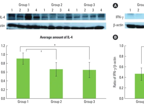
the tissues (Figs. 1A and B). Representative western blot data (Fig. 1A) detected about an 18 kDa molecular weight of IL-4 in all three groups. The expression levels of b-actin were also measured by anti-b-actin specific western blot analysis. In order to quantify the level of IL-4 expression in the groups, the expression levels of IL-4 in each sample were measured by densitometer. Then IL-4 expression levels were normal- ized by b-actin (ratio of IL-4/b-actin).
The mean amount of IL-4 expression (ratio of IL-4/b-actin) were 0.911 ± 0.131 in Group 1, 0.664 ± 0.155 in Group 2, and 0.648 ± 0.177 in Group 3. There was a significant difference between Groups 1 and 3, and between Groups 2 and 3, but there was no statistically significant difference (P < 0.05) be- tween Groups 1 and 2.
The comparison of IFN-g expression levels were also made by western blot analysis using IFN-g specific antibody (Fig.
2A). The levels of IFN-g expression which detected a molecu- lar weight within the range of 20-25 kDa were also quantified with b-actin normalization (Fig. 2B). The mean amounts of IFN-g expression (ratio of IFN-g/b-actin) were 0.465 ± 0.120 in
Group 1, 0.553 ± 0.161 in Group 2 and 0.664 ± 0.160 in Group 3.
There was a significant difference between Groups 1 and 3 (P < 0.05).
In this study, the molecular weight of TIMP-2 was identi- fied as 21 kDa size in western blot analysis (Fig. 3A). The mean value of TIMP-2 expression (ratio of TIMP-2/b-actin) was 0.407 ± 0.080 for Group 1, 0.559 ± 0.158 for Group 2 and 0.649 ± 0.165 for Group 3. There was a significant difference between Groups 1 and 3, and between Groups 2 and 3 (P < 0.05).
DISCUSSION
The association between DM and periodontitis has long been discussed with conflicting conclusions. Current studies tend to support a higher incidence and severity of periodon- titis in patients with DM. It has been shown that diabetes up- regulates the production of inflammatory cytokines and chemokines [22], leading to increased inflammation, tissue damage, and apoptosis in patients who have periodontitis [23].
The immune response against periodontopathic bacteria is
IL-4 b-actin
1 2 3 4 1 2 3 4 1 2 3 4
Group 1 Group 2
*
†
Group 3
Ratio of IL-4
/ b-actin
1.2 1.0 0.8 0.6 0.4 0.2 0.0
B Average amount of IL-4
Figure 1. (A) Interleukin (IL)-4 western blot analysis showing 4 rep- resentative samples in each group. IL-4 levels were quantified on the basis of b-actin levels. IL-4 corresponding to molecular weight 18 kDa was shown to be expressed in all samples including healthy gingiva, and the expression levels of IL-4 were decreased in order of Groups 1-3. (B) Graphics showing the average amounts (ratio of IL-4/b-actin) and standard deviation of IL-4 level in Groups 1-3. In the healthy gin- gival tissues (Group 1), the levels of IL-4 were significantly decreased as compared to Groups 1 and 2 (P < 0.05). * Significant difference be- tween Groups 1 and 3 (P < 0.05). † Significant difference between Groups 1 and 2 (P < 0.05). Group 1: healthy gingiva from systemically healthy individuals, Group 2: inflamed gingiva from patients with chronic periodontitis, Group 3: inflamed gingiva from patients with chronic periodontitis and type 2 diabetes mellitus.
IFN-g b-actin
1 2 3 4 1 2 3 4 1 2 3 4
Group 1
*
Group 2 Group 3
Ratio of IFN-g /
b-actin
1.0 0.8 0.6 0.4 0.2 0.0
B Average amount of IFN-g
Figure 2. (A) Interferon (IFN)-g western blot analysis showing 4 rep- resentative samples in each group. IFN-g levels were quantified on the basis of b-actin levels. IFN-g corresponding to molecular weight 20-25 kDa was shown to be expressed in all samples including healthy gingiva. The expression levels of IFN-g increased in order of Groups 1-3. (B) Graphics showing the average amounts (ratio of IFN-g/b-ac- tin) and standard deviation of IFN-g level in Groups 1-3. In the in- flamed gingiva (with or without diabetes, Groups 2 and 3), the levels of IFN-g were higher than those in healthy gingiva. * Significant difference between Groups 1 and 3 (P < 0.05). Group 1: healthy gin- giva from systemically healthy individuals, Group 2: inflamed gin- giva from patients with chronic periodontitis, Group 3: inflamed gingiva from patients with chronic periodontitis and type 2 diabetes mellitus.
regulated by the balance between cytokines produced by Th1 and Th2 cells. Th1 cells regulate a cell-mediated-type immune response, and Th2 cells regulate a humoral-type immune re- sponse. In addition, each subset can regulate the function of the other. The typical secretory products of Th1 cells are IL-2, IL-12, TNF-b, and IFN-g; those of Th2 cells are IL-4, IL-5, IL-6, IL-10, and IL-13 [8].
IL-4 is a pleiotropic cytokine that inhibits Th1 cells while stimulating a Th2-type of immune response. IL-4 has been shown to inhibit the IL-1 induction of MMP-3 expression in human skin fibroblasts [10]. IL-4 also inhibits the IL-1-induced expression of MMP-3 mRNA and protein in human gingival fibroblast isolated from patients with periodontitis. In a re- cent study, Salmon-Ehr et al. [24] demonstrated that IL-4, a pleiotropic cytokine, was able to activate connective tissue cells and stimulate accumulation of the extracellular matrix macromolecules. They concluded that IL-4 was implicated in wound healing.
in the periodontitis patient to be associated with type 2 DM.
In this study the quantitative analysis of the IL-4 level showed that IL-4 expression was rather decreased in inflamed gingi- va associated with type 2 DM as compared to healthy gingiva and inflamed gingiva of the systemically healthy patient, and the difference was statistically significant (P < 0.05). IL-4 in- hibits the secretion of PGE2 and cytokines by macrophage, and suppresses the synthesis of proinflammatory cytokines which induces inflammation, and suggests that the absence of IL-4 induces periodontal disease. This result indicates that IL-4 inhibits inflammatory response in disease progression in chronic periodontitis in type 2 DM patients and plays a role in decreased inflammatory response with bone resorp- tion in patients with this systemic disease.
IFN-g, released during the early and late stages of the im- mune response by natural killer cells and activated T cells, respectively, regulates several aspects of the immune response [25]. In addition, it mediates the host defense against infection and is a potent activator of mononuclear phagocytes. In our data, the level of IFN-g was increased in inflamed gingiva. The amounts of IFN-g expression were higher in chronic perio- dontitis patients with type 2 DM as compared to healthy gin- giva from a systemically healthy subjects, and the difference was statistically significant (P < 0.05). In some studies, IFN-g seemed to be the predominant cytokine produced by T cells in periodontal diseases, and an enhancement of IFN-g-pro- ducing cells was correlated with the progression of disease [12]. Gorska et al. [26] reported that the concentration of IFN- g was significantly higher in serum samples and gingival tis- sue biopsies from periodontitis patients than from healthy controls. Our data also demonstrated that the total amount of cytokine IFN-g in active sites in patients with the progres- sion of periodontitis is significantly higher than in inactive sites. IFN-g is a inflammatory cytokine associated with inflam- mation, tissue destruction, bone resorption and the produc- tion of matrix metalloproteinases and PGE2. The high expres- sion of these cytokines in chronic periodontitis patients may be a marker of continuous Th1 response against bacterial pathogens colonized in gingival tissue, suggesting that the Th1 response plays a destructive role in the periodontium.
The pro-inflammatory cytokines stimulate cells of the host to produce a number of MMPs, which are eventually respon- sible for degradation of periodontal connective tissues in the pathogenesis of periodontitis. TIMPs, which consist of four members, TIMP-1, 2, 3, and 4, have many basic similarities, but they exhibit structural and biochemical differences. TIMP-2 is able to bind noncovalently to the latent proform of MMP- 2 away from its active sites, thereby preventing its activation and inhibiting enzyme activity [17]. An imbalance between
TIMP-2 b-actin
1 2 3 4 1 2 3 4 1 2 3 4
Group 1 Group 2 Group 3
Ratio of TIMP-2
/ b-actin
1.0 0.8 0.6 0.4 0.2 0.0
*
†
B Average amount of TIMP-2
Figure 3. (A) Tissue inhibitor of matrix metalloproteinases-2 (TIMP- 2) western blot analysis showing 4 representative samples in each group. TIMP-2 levels were quantified on the basis of b-actin levels.
TIMP-2 corresponding to molecular weight 21 kDa was shown to be expressed in all samples including healthy gingiva. The expression levels of TIMP-2 were higher in patients with type 2 diabetes melli- tus (DM) than in control healthy subjects. (B) Graphics showing the average amounts (ratio of TIMP-2/b-actin) and standard deviation of TIMP-2 level in Groups 1-3. In the inflamed gingiva (with or with- out diabetes, Groups 2 and 3), the levels of TIMP-2 were higher than those in healthy gingiva (P < 0.05). * Significant difference between Groups 1 and 3 (P < 0.05). † Significant difference between Groups 1 and 2 (P < 0.05). Group 1: healthy gingiva from systemically healthy individuals, Group 2: inflamed gingiva from patients with chronic periodontitis, Group 3: inflamed gingiva from patients with chronic periodontitis and type 2 DM.
ECM components [16]. The balance between MMP-2 and TIMP-2 expression changes, and several studies have found that during periodontal disease, there is an imbalance of pro- teinases/inhibitors in favor of proteinases.
Larivee et al. [27] found the concentration of collagenase in- hibitors to be higher in healthy gingiva than in periodontitis- affected sites. In this study, the quantitative analysis of TIMP- 2 levels showed that TIMP-2 expression was rather increased in inflamed gingiva with or without type 2 DM as compared to healthy gingiva, and the difference was statistically signifi- cant. This might suggest that the host is producing TIMP-2 as the anti-proteolytic shield to overcome and regulate the tissue-destructive effects of MMPs in gingival tissues.
In conclusions, this study demonstrated that IFN-g and TIMP-2 expression levels in human gingival tissue have a positive correlation with the bone resorption process in in- flamed tissue and inflamed tissue associated with type 2 DM.
This suggests that IFN-g and TIMP-2 may be involved in the alveolar bone resorptive process of periodontal inflammation associated with type 2 DM. On the other hand, tissue with chronic periodontitis associated with type 2 DM showed sig- nificantly decreased IL-4 levels compared to healthy gingiva and non-diabetic inflamed gingiva. This suggests that IL-4 may be involved in the retrogression of periodontal inflam- mation associated with type 2 DM.
Finally, it seems that more studies are needed to investigate the effect and interrelationship between IL-4, IFN-g, TIMP-2, and other cytokines that affect the progression of periodon- tal disease at a higher level. Further studies will contribute to the development of disease diagnosis methods and treat- ment modalities.
CONFLICT OF INTEREST
No potential conflict of interest relevant to this article was reported.
ACKNOWLEDGEMENTS
This study supported by KNU Industry-Academic Research System (200808880000).
REFERENCES
Socransky SS, Haffajee AD. The bacterial etiology of de- 1.
structive periodontal disease: current concepts. J Perio- dontol 1992;63:322-31.
Liljenberg B, Lindhe J, Berglundh T, Dahlen G, Jonsson R.
2.
Some microbiological, histopathological and immunohis-
ease. J Clin Periodontol 1994;21:720-7.
Mealey BL, Ocampo GL. Diabetes mellitus and periodon- 3.
tal disease. Periodontol 2000 2007;44:127-53.
Diabetes and periodontal diseases. Committee on Research, 4.
Science and Therapy. American Academy of Periodontol- ogy. J Periodontol 2000;71:664-78.
Mealey B. Diabetes and periodontal diseases. J Periodon- 5.
tol 1999;70:935-49.
Nishimura F, Takahashi K, Kurihara M, Takashiba S, Mu- 6.
rayama Y. Periodontal disease as a complication of diabe- tes mellitus. Ann Periodontol 1998;3:20-9.
Stewart JE, Wager KA, Friedlander AH, Zadeh HH. The ef- 7.
fect of periodontal treatment on glycemic control in pa- tients with type 2 diabetes mellitus. J Clin Periodontol 2001;
28:306-10.
Gemmell E, Seymour GJ. Immunoregulatory control of 8.
Th1/Th2 cytokine profiles in periodontal disease. Perio- dontol 2000 2004;35:21-41.
Shapira L, van Dyke TE, Hart TC. A localized absence of in- 9.
terleukin-4 triggers periodontal disease activity: a novel hy- pothesis. Med Hypotheses 1992;39:319-22.
Prontera C, Crescenzi G, Rotilio D. Inhibition by Interleu- 10.
kin-4 of stromelysin expression in human skin fibroblasts:
role of PKC. Exp Cell Res 1996;224:183-8.
Mullen AC, High FA, Hutchins AS, Lee HW, Villarino AV, 11.
Livingston DM, et al. Role of T-bet in commitment of TH1 cells before IL-12-dependent selection. Science 2001;292:
1907-10.
Ukai T, Mori Y, Onoyama M, Hara Y. Immunohistological 12.
study of interferon-gamma- and interleukin-4-bearing cells in human periodontitis gingiva. Arch Oral Biol 2001;
46:901-8.
Roberts FA, McCaffery KA, Michalek SM. Profile of cytokine 13.
mRNA expression in chronic adult periodontitis. J Dent Res 1997;76:1833-9.
Nagase H. Activation mechanisms of matrix metallopro- 14.
teinases. Biol Chem 1997;378:151-60.
Hammani K, Blakis A, Morsette D, Bowcock AM, Schmutte 15.
C, Henriet P, et al. Structure and characterization of the hu- man tissue inhibitor of metalloproteinases-2 gene. J Biol Chem 1996;271:25498-505.
Ryan ME, Ramamurthy S, Golub LM. Matrix metallopro- 16.
teinases and their inhibition in periodontal treatment. Curr Opin Periodontol 1996;3:85-96.
Goldberg GI, Marmer BL, Grant GA, Eisen AZ, Wilhelm S, 17.
He CS. Human 72-kilodalton type IV collagenase forms a complex with a tissue inhibitor of metalloproteases desig- nated TIMP-2. Proc Natl Acad Sci U S A 1989;86:8207-11.
Reynolds JJ, Hembry RM, Meikle MC. Connective tissue 18.
roles of matrix metalloproteinases and their natural in- hibitors. Adv Dent Res 1994;8:312-9.
Joo SD, Lee JM. The comparison of inflammatory media- 19.
tor expression in gingival tissues from human chronic peri- odontitis patients with and without type 2 diabetes melli- tus. J Korean Acad Periodontol 2007;37(2 Suppl):353-69.
Kim DH, Park EK, Shin HI, Cho JY, Suh JY, Lee JM. Inter- 20.
relationship of matrix metalloproteinase and TNF-g in human gingiva with chronic periodontitis associated to type 2 diabetes mellitus. J Korean Acad Periodontol 2006;
36:409-5.
Cho JY, Xing S, Liu X, Buckwalter TL, Hwa L, Sferra TJ, et 21.
al. Expression and activity of human Na+/I- symporter in human glioma cells by adenovirus-mediated gene deliv- ery. Gene Ther 2000;7:740-9.
Park JW, Lee JM. The comparison of IL-6, elastase and
22. a1-
PI expressions in human chronic periodontitis with type 2 diabetes mellitus. J Korean Acad Periodontol 2007;37(2 Sup- pl):325-38.
tosis in chronic adult periodontitis analyzed by in situ DNA breaks, electron microscopy, and immunohistochemistry.
J Periodontol 2001;72:517-25.
Salmon-Ehr V, Ramont L, Godeau G, Birembaut P, Gue- 24.
nounou M, Bernard P, et al. Implication of interleukin-4 in wound healing. Lab Invest 2000;80:1337-43.
Boehm U, Klamp T, Groot M, Howard JC. Cellular respons- 25.
es to interferon-gamma. Annu Rev Immunol 1997;15:749- 95.
Gorska R, Gregorek H, Kowalski J, Laskus-Perendyk A, Sy- 26.
czewska M, Madalin@ski K, et al. Relationship between clin- ical parameters and cytokine profiles in inflamed gingival tissue and serum samples from patients with chronic peri- odontitis. J Clin Periodontol 2003;30:1046-52.
Larivee J, Sodek J, Ferrier JM. Collagenase and collagenase 27.
inhibitor activities in crevicular fluid of patients receiving treatment for localized juvenile periodontitis. J Periodon- tal Res 1986;21:702-15.