www.jpis.org
pISSN 2093-2278 eISSN 2093-2286 Copyright © 2011 Korean Academy of PeriodontologyThis is an Open Access article distributed under the terms of the Creative Commons Attribution Non-Commercial License (http://creativecommons.org/licenses/by-nc/3.0/).
The influence of type 2 diabetes mellitus on the expression of inflammatory mediators and tissue inhibitor of metalloproteinases-2 in human chronic periodontitis
Jae-Bung Kim1, Mi-Hwa Jung1, Je-Yeol Cho2, Jin-Woo Park1, Jo-Young Suh1, Jae-Mok Lee1,*
Departments of 1Periodontology and 2Oral Biochemistry, Kyungpook National University School of Dentistry, Daegu, Korea
Purpose: The purpose of this study was to compare and quantify the expression of C-reactive protein (CRP), matrix metallo- proteinase (MMP)-14, and tissue inhibitor of metalloproteinases (TIMP)-2 in gingival tissues of patients with chronic periodon- titis accompanied with inflammatory reaction related to alveolar bone resorption with or without type 2 diabetes mellitus (DM).
Methods: Twelve patients with type 2 DM and chronic periodontitis (group 3), twelve patients with chronic periodontitis (group 2), and twelve healthy individuals (group 1) were included in the study. Gingival tissue biopsies were collected from each pa- tient and from healthy individuals at the time of periodontal surgery (including surgical crown lengthening) or tooth extrac- tion. The concentrations of cytokines were determined by a western blot analysis.
Results: The expression levels of CRP and MMP-14 increased in group 2 and 3, and they were highest in group 3. The expres- sions of TIMP-2 also increased in group 2 and 3.
Conclusions: This study demonstrated that the expression levels of CRP, MMP-14, and TIMP-2 might be inflammatory mark- ers in periodontal inflamed tissue. It can be assumed that CRP, MMP-14, and TIMP-2 may be partly involved in the progres- sion of periodontal inflammation associated to type 2 DM.
Keywords: Chronic periodontitis, Type 2 diabetes mellitus, C-reactive protein, Matrix metalloproteinase 14, Tissue inhibitor of metalloproteinase-2.
INTRODUCTION
Diabetes mellitus (DM) is the most common metabolic dis- ease worldwide. More than 90% of DM patients have type 2 diabetes [1]. DM is the leading cause of blindness [2], renal failure, and lower limb amputations. DM is a major risk fac- tor for cardiovascular disease, stroke, neuropathy, and peri- odontitis [1,3].
Chronic periodontitis is the most common type of periodon-
tal disease, and results from extension of the inflammatory process initiated by bacteria in the gingiva to the supporting periodontal tissues. A reciprocal relationship exists between DM and periodontal disease [4]. Periodontal infections, like other infections, have a significant impact on diabetic con- trol.
Conversely, DM is a significant risk factor for the develop- ment of periodontal disease and aggravates the severity of periodontal infections [5]. The comorbidity of these two in-
Received: Mar. 7, 2011; Accepted: May 26, 2011
*Correspondence: Jae-Mok Lee
Department of Periodontology, Kyungpook National University School of Dentistry, 50 Samdeok-dong 2-ga, Jung-gu, Daegu 700-412, Korea E-mail: [email protected], Tel: +82-53-600-7522, Fax: +82-53-427-3263
ments of pathogenesis related to the risks for both conditions.
In patients with periodontal disease, chronic low-level sys- temic exposure to periodontal microorganisms may occur, leading to significant changes in the plasma levels of cyto- kines and hormones. Due to the dynamic nature of the in- flamed periodontium, the tissue may serve as an endocrine- like source of inflammatory mediators. Among the inflam- matory biomarkers examined, C-reactive protein (CRP), and Interleukin (IL)-6 appear to be involved, according to plausi- ble biological mechanisms through which they function, ex- amined in studies of the links between periodontal disease and cardiovascular disease [5]. Recently, Blüher et al. [6] in- vestigated whether the plasma concentrations of inflamma- tory markers were associated with measures of obesity, insu- lin sensitivity, and hyperglycemia, and discovered significant correlations between the plasma concentrations of all of the inflammatory markers examined and percent body fat, insu- lin sensitivity, and fasting plasma glucose. Fasting plasma glucose was a significant determinant of adiponectin, CRP, and IL-6 plasma concentrations, whereas body fat content was a significant predictor only of CRP plasma concentration [6]. In a similar study, the authors concluded that type 2 DM was highest among subjects with elevated levels of IL-18 and CRP or IL-18 and IL-6.
CRP is an acute-phase reactant synthesized by the liver in response to inflammatory cytokines, IL-6, IL-1, and tumor necrosis factor-alpha (TNF-α). Circulating CRP levels are a marker of systemic inflammation and are associated with periodontal disease [7], a chronic bacterial infection associat- ed with the elevation of proinflammatory cytokines and pros- taglandin [8]. Elevated immunoglobulin G induced by bacte- rial species associated with destructive periodontal diseases is associated with the increase in CRP [9]. Standard non-sur- gical periodontal therapy resulted in a decrease in serum CRP levels [10] in a previous study.
Microbial components, especially lipopolysaccharide, acti- vate macrophages to synthesize and secrete a variety of pro- inflammatory molecules, including the cytokines IL-1 and TNF-α; prostaglandins, especially prostaglandin E2; and hy- drolytic enzymes. Similarly, bacterial substances activate T lymphocytes to produce IL-1 and lymphotoxin, a molecule with similar properties to TNF-α. These cytokines manifest potent proinflammatory and catabolic activities, and play key roles in periodontal tissue breakdown through collagenolytic enzymes such as matrix metalloproteinases (MMPs) [11].
The MMPs are a family of structurally and functionally re- lated enzymes that are responsible for the proteolytic degra- dation of extracellular matrix components. More than 20 dif- ferent MMPs have been identified. These proteins can be
strate specificities and structural homologies: collagenase (MMP-1, -8, and -13), gelatinase (MMP-2, and -9), stromelysin (MMP-3, -10, and -11), membrane-type MMPs (MT-MMPs- : MMP-14, 15, -16, -17, -23, -24, and -25) and other MMPs, inclu- ding matrilysin (MMP-7) and metalloelastase (MMP-12) [12].
Expression of MMPs is low in normal cells, and these low levels allow for healthy connective tissue remodeling. In pa- thologic conditions, however, the level of MMP expression increases considerably, resulting in aberrant connective tis- sue destruction. Excess MMP production is associated with the pathology of many diseases, including periodontitis [13], atherosclerosis [14], tumor invasion/metastasis, and arthritic disease [15].
MT-MMPs are a unique class of MMPs anchored to the cell surface by transmembrane domains. MT-MMPs display a broad spectrum of activities, and because of their localiza- tion to the cell surface, they are thought to play a major role in controlling proteolytic events within the pericellular mi- croenvironment. MT1-MMP (MMP-14), can degrade a vari- ety of extracellular matrix proteins and, is capable of activat- ing both pro-MMP-2 and pro-MMP-13 [16].
MMP-17 and 25 are glycosylphosphatidylinositol-anchored whereas the other four MT-MMPs are type 1 transmembrane proteins with short cytoplasmic domains of about 20 amino acids. Like secreted MMPs, MT-MMPs can cleave extracellu- lar matrix molecules, as well as chemokines, cytokines, and growth factors [17]. MT-MMPs are generally thought to play important regulatory roles because of their ability to cleave substrates in the immediate vicinity of the cell membrane, where the cleaved products can interact with cell-surface re- ceptors. In addition, MT-MMPs are known to cleave and ac- tivate secreted MMPs, which were first described in the case of activation of MMP-2 by MMP-14 through interaction with TIMP-2.
MMPs are regulated at several levels including transcrip- tion, secretion, activation, and inhibition. Regulation by the latest of these mechanisms is via endogenous inhibitors, known as TIMPs. The balance between the levels of active enzymes and free TIMPs is thought to determine overall MMP activity. The TIMPs are small proteins (-23kDa) that in- hibit MMP activity by binding to them in a 1:1 stoichiomet- ric ratio [18]. The four members of the TIMP family (TIMP-1 through TIMP-4) are cysteine-rich proteins stabilized by di- sulphide bonds [19]. They are composed of a large N-termi- nal domain responsible for MMP inhibition and a smaller C- terminal domain. The TIMPs in general do not demonstrate specificity for any particular MMP [18], although TIMP-2 shows some degree of preference for MMP-2 and TIMP-1 for MMP- 9 [20].
MMP activation. The relative levels of TIMP-2 and MMP-14 are critical, since low TIMP-2 levels are associated with MMP- 14 mediated activation of pro-MMP-2 and at higher TIMP-2 levels MT1-MMP function is blocked, preventing pro-MMP-2 activation [21].
The particular roles and substrate specificities of MT-MMPs and TIMP-2 have not been described in detail, and the role of MT-MMPs and TIMP-2 in DM is unknown.
In inflammatory responses with bone resorption, the roles and interactions of CRP, MMP-14, and TIMP-2 are not clear.
Their contribution to the pathogenesis of periodontitis and alveolar bone resorption has not yet been established. More- over, no in vivo studies have simultaneously analyzed CRP, MMP-14, and TIMP-2, and their interrelationship in diabetic and nondiabetic patients with chronic periodontitis. The pur- pose of this study was to compare and quantify the expres- sions of CRP, MMP-14, and TIMP-2, in gingival tissues in or- der to reveal diagnostic factors in chronic periodontitis pa- tients with or without type 2 DM.
MATERIALS AND METHODS
Study population and tissue sampling
The study population consisted of 12 patients with type 2 DM and chronic periodontitis, 12 patients with chronic peri- odontitis, and 12 healthy individuals. Marginal gingival tissue samples were obtained in the Department of Periodontology, Kyungpook National University Hospital, Korea, by internal bevel incision at the time of periodontal surgery (including surgical crown lengthening) or tooth extraction. All partici- pants signed the Institutional Review Board-approved (No.
74 005-418) consent form prior to surgery.
According to the patient’s systemic condition (age, sex, blood glucose level, obesity, and smoking), the clinical criteria of the gingiva (sulcus bleeding index value and probing depths), and radiographic evidence of bone resorption, each gingival sample was assigned to one of three groups. Group 1 (normal, n=12) consisted of clinically healthy gingiva without bleed- ing, evidence of bone resorption, or periodontal pockets, ob- tained from 12 systemically healthy patients. Group 2 (chronic periodontitis, n=12) consisted of the inflamed gingiva of pa- tients with chronic periodontitis. The diagnosis of chronic periodontitis was established on the basis of clinical and ra- diographic criteria (bone resorption) according to the classi- fication system for periodontal disease and conditions. All patients in group 2 were systemically healthy and had more than one periodontal pockets ≥5 mm and at least one pocket with ≥5 mm loss of attachment. All gingival samples were obtained from teeth with a probing depth ≥5 mm, swelling
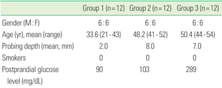
gival sulcus bleeding index 3 according to Mühlemann and Son [22]. Group 3 (chronic periodontitis and type 2 DM, n=12) consisted of the inflamed gingiva of patients with chronic periodontitis associated with type 2 DM. Patients in group 3 were diagnosed with type 2 DM at least 6 months prior and showed blood glucose levels of 200 mg/dL and above in the first postprandial 2 hours. Patients in groups 2 and 3 had sim- ilar periodontal conditions, but patients in group 2 were sys- temically healthy and patients in group 3 had type 2 DM. Gin- gival samples were obtained as described above. Patient char- acteristics are presented in Table 1.
Following surgery, excised tissue specimens were immedi- ately frozen in liquid nitrogen at -70°C.
Protein isolation and western blotting
The western blotting, technique has been as previously de- scribed by Park and Lee [13]. Frozen tissues were homo genized in radio-immunoprecipitation assay lysis buffer (10 mM eth- ylenediaminetetraacetic acid, 0.15 M NaCl) with 1:30 diluted protease inhibitor cocktail (Roche, Mannheim, Germany). The lysates were sonicated three times for 10 seconds and centri- fuged at 12,000 g for 20 minutes. Protein concentrations of the supernatant were routinely determined by a Braford pro- tein asssay (Quick Start, BIO-rad Laboratories Inc., Hercules, CA, USA) using bovine serum albumin (BSA) as standard.
Lysates were boiled in a sodium dodecyl sulfate (SDS) sam- ple buffer (1 M Tris-HCl [pH 6.8], 40% glycerol, 8% SDS, 2%
mercapto-ethanol, 0.002% Bromophenol blue). Prepared samples were separated by 15% SDS-polyacrylamide gels and transferred to a polyvinylidene difluoride membrane.
The membranes were subsequently blocked in tris-buff- ered saline (TBS) containing 5% powdered milk and 1% BSA for 1 hour, and then incubated with polyclonal anti-CRP anti- body, anti-MMP-14 antibody, and anti-TIMP-2 antibody (San- ta Cruz Biotechnology Inc., Santa Cruz, CA, USA) for 1.5 hours at room temperature.
The membranes were washed (five times for 5 minutes with Tween 20) and incubated with a horseradish peroxidase-con- jugated goat anti-rabbit secondary antibody for anti-CRP an-
Table 1. Patient characteristics.
Group 1 (n=12) Group 2 (n=12) Group 3 (n=12)
Gender (M:F) 6:6 6:6 6:6
Age (yr), mean (range) 33.6 (21-43) 48.2 (41-52) 50.4 (44-54)
Probing depth (mean, mm) 2.0 8.0 7.0
Smokers 0 0 0
Postprandial glucose 90 103 289
level (mg/dL)
(diluted 1:2,000 in TBS) for 1 hour at room temperature. Af- ter additional washing (five times for 5 minutes with Tween 20) the western blot procedure was completed with an ECL Plus development kit (Amsterdam, Beckinghamshire, UK).
The relative quantification analysis of CRP, MMP-14, and TIMP-2 expression was performed using a densitometer (Ima- ge Gauge V 3.46, Fuji Photo Film Co., Tokyo, Japan). After nor- malization to β-actin (Abcam plc, Cambri dge, UK) in each sample, levels of CRP, MMP-14, and TIMP-2 were expressed as a ratio of CRP, MMP-14, or TIMP-2 to β- actin and the dif- ferences in density between the 3 groups were determined.
Statistical analysis of the western blot results
All data were presented as means±standard deviation and results were statistically analyzed. The CRP, MMP-14, and TIMP-2 levels were compared using a one-way analysis of vaiance followed by Tukey’s test. P-value <0.05 was consid- ered to statistically significant.
RESULTS
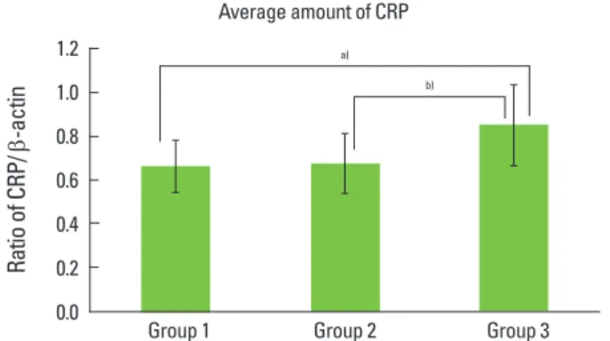
Representative western blot data comparing CRP expres- sion levels in human gingiva with chronic periodontitis with or without type 2 DM are presented in Fig. 1. The compari- son of CRP expression levels were also studied by detecting a CRP band with a molecular weight of about 27 kDa and mea- suring their density and areas in all three groups (Fig. 1). The comparative levels of normalized CRP expression are given in Fig. 2. The mean value of CRP expression (ratio of CRP/
β-actin) was 0.669±0.119 for group 1, 0.679±0.134 for group 2, and 0.859±0.186 for group 3. There were statistically signifi- cant differences between group 1 and group 3 and between group 2 and group 3, but there was no statistically significant difference (P<0.05) between group 1 and group 2. The com-
β-actin
Group 1
CRP
Group 2 Group 3
1 2 3 4 1 2 3 4 1 2 3 4
Figure 1. C-reactive protein (CRP) western analysis showing 4 rep- resentative blots in each group. C-reactive protein levels were quan- tified on the basis of β-actin levels. CRP corresponding to a molec- ular weight of 27 kDa was expressed in all groups, including healthy gingiva, and the expression levels of C-reactive protein increased in order from group 1 to group 2 to group 3. Group 1, healthy gingiva from systemically healthy persons; Group 2, inflamed gingiva from patients with chronic periodontitis; Group 3, inflamed gingiva from patients with chronic periodontitis and type 2 diabetes mellitus.
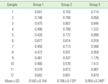
western blot analysis using MMP-14 specific antibody which detected MMP-14 in all three groups (Fig. 3). The compara- tive levels of MMP-14 expression were also quantified with β-actin normalization (Fig. 4). The mean value of MMP-14 expression (ratio of MMP-14/β-actin) was 0.542±0.104 for group 1, 0.706±0.133 for group 2, and 0.954±0.248 for group 3.
There were statistically significant differences between group 1 and group 2 (P<0.05) and between group 1 and group 3 (P<
0.05). In this study, the molecular weight of TIMP-2 was iden- tified to be 28 kDa in western blot analysis (Fig. 5). The com- parative levels of TIMP-2 expression were also quantified with β-actin normalization and presented in Fig. 6. The mean val- ue of TIMP-2 expression (ratio of TIMP-2/β-actin) was 0.485±
0.098 for group 1, 0.605±0.084 for group 2, and 0.746±0.181 for group 3. There were statistically significant differences between group 1 and group 3 and between group 2 and group 3 (P<0.05).
Figure 2. C-reactive protein (CRP) levels (ratio of CRP/β-actin) in groups 1, 2, and 3. In inflamed gingiva with diabetes (group 3), the levels of CRP were significantly increased as compared to group 1 and group 2 (P<0.05). a)Significant difference between group 1 and group 3 (P<0.05). b)Significant difference between group 2 and group 3 (P<0.05).
1.2 1.0 0.8 0.6 0.4 0.2 0.0
Ratio of CRP/ β-actin
Group 1 Group 2 Group 3
Average amount of CRP
a) b)
Group 1 Group 2 Group 3
1 2 3 4 1 2 3 4 1 2 3 4
MMP-14 β-actin
Figure 3. Matrix metalloproteinase (MMP)-14 western analysis show- ing 4 representative blots in each group. MMP-14 levels were quan- tified on the basis of β-actin levels. MMP-14 corresponding to a mol- ecular weight of 60 kDa was shown to be expressed in all groups in- cluding healthy gingiva. The expression levels of Matrix metallo- proteinase 14 increased in order from group 1 to group 2 to group 3.
Group 1, healthy gingiva from systemically healthy persons; Group 2, inflamed gingiva from patients with chronic periodontitis; Group 3, inflamed gingiva from patients with chronic periodontitis and type 2 diabetes mellitus.
DISCUSSION
Among the many oral problems that can occur because of diabetes, the prevalence of severe periodontitis is significant- ly higher among people with poorly controlled DM [23], to the extent that periodontitis has been called the “sixth com- plication of diabetes” [24]. The reason for the higher rates of periodontal disease in people with diabetes is not completely understood, but studies have reported that there is little dif- ference in the periodontal flora of people with and without DM, and suggest that the increased destruction of tissue among those with diabetes may be due to an altered host susceptibility to periodontal pathogens mediated by the ac- cumulation of advanced glycation end products in the tis- sues, microvascular changes, and perhaps impaired lipid me- tabolism [25]. Conversely, these data also suggest that the presence of periodontal infection can adversely affect glyce- mic control in people with diabetes and that there appears to be a bi-directional relationship between the two conditions.
The purpose of this study was to quantify and compare the expression of CRP, MMP-14 and TIMP-2 in the gingival tis- sues of patients with chronic periodontitis associated to type 2 DM, in order to understand the diagnostic contribution of these proteins to periodontal destruction accompanied with alveolar bone resorption in type 2 diabetic patients.
CRP is an acute-phase protein suggesting a central role in immunological response [26]. It is synthesized in the liver mainly in response to IL-6 and binds to the polysaccharides of pathogens promoting phagocytosis [27]. Several studies have shown that CRP could be useful in infection diagnosis [28], as well as in monitoring the response to antibiotic thera- py [29].
In this study, the quantitative analysis of CRP levels showed that CRP expression increased significantly in inflamed gin- giva with or without type 2 DM compared to healthy gingiva.
Differences were statistically significant between group 1 and group 3 and between group 2 and group 3 (P<0.05) (Table 2, Fig. 2).
Our results are similar to previous studies in which Noack et al. [7] and Craig et al. [9] found that subjects with periodon- tal disease demonstrated higher levels of CRP than subjects without periodontitis.
We found a statistically significant difference between peri- odontitis subjects with DM (group 3) and subjects with peri- odontitis only (group 2). The ongoing acute phase response (seen in insulin-resistant subjects and type II DM patients) is induced by cytokines, and is reflected in elevated circulating inflammatory markers, such as CRP, IL-1, IL-6, TNF-α, leptin, plasminogen activator inhibitor-1, angiotensinogen, and fi- brinogen, as described by Hsueh and Bruemmer [30]. It seems Figure 4. Matrix metalloproteinase (MMP)-14 levels (ratio of MMP-
14 to β-actin) in groups 1, 2, and 3. In the inflamed gingiva (with or without diabetes, group 3 and group 2, respectively), the levels of MMP-14 were higher than those of healthy gingiva. a)Significant difference between group 1 and group 2 (P<0.05). b)Significant dif- ference between group 1 and group 3 (P<0.05).
1.4 1.2 1.0 0.8 0.6 0.4 0.2 Ratio of MMP-14/ β-actin 0.0
Group 1 Group 2 Group 3
a) b)
Figure 5. Tissue inhibitor of metalloproteinase (TIMP)-2 western analysis showing 4 representative blots in each group. TIMP-2 levels were quantified on the basis of β-actin levels. TIMP-2 correspond- ing to a molecular weight of 28 kDa was expressed in all samples in- cluding healthy gingiva. The expression levels of TIMP-2 were in- creased in patients with type 2 diabetes mellitus compared to healthy control subjects. Group 1, healthy gingiva from systemically healthy persons; Group 2, inflamed gingiva from patients with chronic peri- odontitis; Group 3, inflamed gingiva from patients with chronic periodontitis and type 2 diabetes mellitus.
Group 1 Group 2 Group 3
1 2 3 4 1 2 3 4 1 2 3 4
TIMP-2 β-actin
actin.
Sample Group 1 Group 2 Group 3
1 0.575 0.699 0.537
2 0.510 0.581 1.050
3 0.615 0.534 0.771
4 0.621 0.510 1.004
5 0.824 0.777 0.987
6 0.843 0.855 1.093
7 0.800 0.836 1.023
8 0.784 0.905 0.990
9 0.675 0.585 0.633
10 0.680 0.569 0.730
11 0.580 0.661 0.722
12 0.518 0.637 0.770
Mean±SD 0.669±0.119 0.679±0.134 0.859±0.186a),b)
a)Significant difference between group 1 and group 3 (P <0.05). b)Significant difference between group 2 and group 3 (P<0.05).
ing the progression of inflammation.
Microbial components, especially lipopolysaccharides, acti- vate macrophages to synthesize and secrete a variety of pro- inflammatory molecules, including the cytokines IL-1 and TNF-alpha, prostaglandins, especially prostaglandin E2, and hydrolytic enzymes. Similarly, bacterial substances activate T lymphocytes to produce IL-1 and lymphotoxin, a molecule with similar properties to TNF-alpha. These cytokines mani- fest potent proinflammatory and catabolic activities, and play key roles in periodontal tissue breakdown through collagen- olytic enzymes such as MMPs.
MMP-14 was initially identified as the activator of MMP-2, but has recently been shown to be important for wound heal- ing, angiogenesis, and inflammation [31]. It is over-expressed in cancers leading to migration, invasion, and metastasis [32].
MMP-14 activation occurs via a proprotein convertase, and recent data suggests that its function is modified by glycosyl- ation, internalization and recycling [33]. Finally MMP-14 di- rectly degrades extracellular matrix molecules, including col- lagen type I, III, laminin, and fibronectin [34].
In this study, the quantitative analysis of MMP-14 levels showed that MMP-14 expression was rather increased in in- flamed gingiva with or without type 2 DM compared to heal- thy gingiva. The differences were statistically significant be- tween group 1 and group 2 and between group 1 and group 3 (P<0.05) (Table 3, Fig. 4).
Song and Ergul [35] reported that mild elevation of blood glucose for 6 weeks is sufficient to stimulate gene expression of MMP-2, MMP-9, and MMP-14.
The MMP-14 level in group 3 was higher than group 2, but
that this is a result of a higher inflammatory response in chro- nic periodontitis associated with type 2 DM.
A major group of inhibitors of the MMPs, TIMP-2, has been studied in this study. The levels of TIMP-2 were significantly different between group 1 and 3 and between group 2 and group 3 (P<0.05) (Table 4, Fig. 6). However, there has been some debate over findings regarding TIMP-2 in diabetic sub- jects. MecLennan et al. [36] observed no changes in TIMP-2 expression in mesangial cells cultured in high glucose. Our results also show increased TIMP-2 levels. It can be assumed that this is a result of a greater inflammatory response in chro- nic periodontitis associated with type 2 DM.
In conclusion, increased levels of CRP, MMP-14, and TIMP- 2 are useful for diagnosing and monitoring inflammatory
Table 4. Normalized TIMP-2 expression by TIMP-2/β-actin.
Sample Group 1 Group 2 Group 3
1 0.455 0.576 1.031
2 0.383 0.718 0.862
3 0.581 0.713 1.084
4 0.532 0.492 0.557
5 0.478 0.524 0.507
6 0.374 0.583 0.648
7 0.391 0.681 0.667
8 0.429 0.468 0.734
9 0.412 0.597 0.760
10 0.513 0.684 0.819
11 0.579 0.641 0.552
12 0.696 0.578 0.735
Mean±SD 0.485±0.098 0.605±0.084 0.746±0.181a),b) TIMP: tissue inhibitor of metalloproteinases.
a)Significant difference between group 1 and group 2 (P <0.05). b)Significant difference between group 2 and group 3 (P<0.05).
Table 3. Normalized MMP-14 expression by MMP-14/β-actin.
Sample Group 1 Group 2 Group 3
1 0.641 0.703 0.714
2 0.746 0.708 0.858
3 0.475 0.807 0.844
4 0.490 0.789 1.222
5 0.426 0.772 0.899
6 0.677 0.614 0.916
7 0.450 0.713 0.568
8 0.472 0.937 0.959
9 0.461 0.437 1.176
10 0.480 0.578 1.512
11 0.578 0.812 0.907
12 0.602 0.601 0.870
Mean±SD 0.542±0.104 0.706±0.133a) 0.954±0.248b) MMP: matrix metalloproteinase.
a)Significant difference between group 1 and group 2 (P <0.05). b)Significant difference between group 1 and group 3 (P<0.05).
Figure 6. Tissue inhibitor of metalloproteinases (TIMP)-2 level (Ra- tio of TIMP-2 to β-actin) in groups 1, 2, and 3. In the inflamed gingi- va (with or without diabetes, groups 3 and 2, respectively), the levels of TIMP-2 were higher than those of healthy gingiva (P <0.05).
a)Significant difference between group 1 and group 2 (P<0.05). b)Sig- nificant difference between group 2 and group 3 (P<0.05).
1.0 0.8 0.6 0.4 0.2 Ratio of TIMP-2/ β-actin 0.0
Group 1 Group 2 Group 3
Average amount of TIMP-2
a) b)
More studies are needed to investigate interrelationship among CRP, MMP-14, TIMP-2, and other factors that affect the progression of periodontal disease to a more advanced level.
CONFLICT OF INTEREST
No potential conflict of interest relevant to this article was reported
ACKNOWLEDGEMENTS
This study was supported by KHIDI, the Ministry for Health and Welfare (A090610).
REFERENCES
1. Engelgau MM, Geiss LS, Saaddine JB, Boyle JP, Benjamin SM, Gregg EW, et al. The evolving diabetes burden in the United States. Ann Intern Med 2004;140:945-50.
2. Fong DS, Aiello L, Gardner TW, King GL, Blankenship G, Cavallerano JD, et al. Diabetic retinopathy. Diabetes Care 2003;26:226-9.
3. Graves DT, Liu R, Alikhani M, Al-Mashat H, Trackman PC.
Diabetes-enhanced inflammation and apoptosis: impact on periodontal pathology. J Dent Res 2006;85:15-21.
4. Diabetes and periodontal diseases. Committee on Resear- ch, Science and Therapy. American Academy of Periodon- tology. J Periodontol 2000;71:664-78.
5. Pradhan AD, Ridker PM. Do atherosclerosis and type 2 di- abetes share a common inflammatory basis? Eur Heart J 2002;23:831-4.
6. Blüher M, Fasshauer M, Tönjes A, Kratzsch J, Schön MR, Paschke R. Association of interleukin-6, C-reactive pro- tein, interleukin-10 and adiponectin plasma concentra- tions with measures of obesity, insulin sensitivity and glu- cose metabolism. Exp Clin Endocrinol Diabetes 2005;113:
534-7.
7. Noack B, Genco RJ, Trevisan M, Grossi S, Zambon JJ, De Nardin E. Periodontal infections contribute to elevated systemic C-reactive protein level. J Periodontol 2001;72:
1221-7.
8. Page RC. The role of inflammatory mediators in the patho- genesis of periodontal disease. J Periodontal Res 1991;26(3 Pt 2):230-42.
9. Craig RG, Yip JK, So MK, Boylan RJ, Socransky SS, Haffajee AD. Relationship of destructive periodontal disease to the acute-phase response. J Periodontol 2003;74:1007-16.
10. D’Aiuto F, Ready D, Tonetti MS. Periodontal disease and
odontal Res 2004;39:236-41.
11. Sorsa T, Ingman T, Suomalainen K, Haapasalo M, Kont- tinen YT, Lindy O, et al. Identification of proteases from periodontopathogenic bacteria as activators of latent hu- man neutrophil and fibroblast-type interstitial collagena- ses. Infect Immun 1992;60:4491-5.
12. Sternlicht MD, Werb Z. How matrix metalloproteinases regulate cell behavior. Annu Rev Cell Dev Biol 2001;17:463- 516.
13. Park JW, Lee JM. The comparison of IL-6, elastase and al- pha1-PI expressions in human chronic periodontitis with type 2 diabetes mellitus. J Korean Acad Periodontol 2007;
37(Suppl):325-38.
14. Borden P, Heller RA. Transcriptional control of matrix me- talloproteinases and the tissue inhibitors of matrix metal- loproteinases. Crit Rev Eukaryot Gene Expr 1997;7:159-78.
15. Brinckerhoff CE, Rutter JL, Benbow U. Interstitial collage- nases as markers of tumor progression. Clin Cancer Res 2000;6:4823-30.
16. Itoh Y, Seiki M. MT1-MMP: a potent modifier of pericel- lular microenvironment. J Cell Physiol 2006;206:1-8.
17. Stamenkovic I. Extracellular matrix remodelling: the role of matrix metalloproteinases. J Pathol 2003;200:448-64.
18. Brew K, Dinakarpandian D, Nagase H. Tissue inhibitors of metalloproteinases: evolution, structure and function.
Biochim Biophys Acta 2000;1477:267-83.
19. Williamson RA, Marston FA, Angal S, Koklitis P, Panico M, Morris HR, et al. Disulphide bond assignment in human tissue inhibitor of metalloproteinases (TIMP). Biochem J 1990;268:267-74.
20. Goldberg GI, Strongin A, Collier IE, Genrich LT, Marmer BL. Interaction of 92-kDa type IV collagenase with the tis- sue inhibitor of metalloproteinases prevents dimerization, complex formation with interstitial collagenase, and acti- vation of the proenzyme with stromelysin. J Biol Chem 1992;267:4583-91.
21. Butler GS, Butler MJ, Atkinson SJ, Will H, Tamura T, Schade van Westrum S, et al. The TIMP2 membrane type 1 metal- loproteinase “receptor” regulates the concentration and efficient activation of progelatinase A. A kinetic study. J Biol Chem 1998;273:871-80.
22. Mühlemann HR, Son S. Gingival sulcus bleeding: a lead- ing symptom in initial gingivitis. Helv Odontol Acta 1971;
15:107-13.
23. Cho JY, Xing S, Liu X, Buckwalter TL, Hwa L, Sferra TJ, et al. Expression and activity of human Na+/I- symporter in human glioma cells by adenovirus-mediated gene deliv- ery. Gene Ther 2000;7:740-9.
24. Tsai C, Hayes C, Taylor GW. Glycemic control of type 2 di-
pulation. Community Dent Oral Epidemiol 2002;30:182- 92.
25. Cutler CW, Shinedling EA, Nunn M, Jotwani R, Kim BO, Nares S, et al. Association between periodontitis and hy- perlipidemia: cause or effect? J Periodontol 1999;70:1429- 34.
26. Vigushin DM, Pepys MB, Hawkins PN. Metabolic and scintigraphic studies of radioiodinated human C-reactive protein in health and disease. J Clin Invest 1993;91:1351-7.
27. Mold C, Gewurz H, Du Clos TW. Regulation of comple- ment activation by C-reactive protein. Immunopharma- cology 1999;42:23-30.
28. Ugarte H, Silva E, Mercan D, De Mendonça A, Vincent JL.
Procalcitonin used as a marker of infection in the inten- sive care unit. Crit Care Med 1999;27:498-504.
29. Póvoa P, Coelho L, Almeida E, Fernandes A, Mealha R, Mo- reira P, et al. C-reactive protein as a marker of ventilator- associated pneumonia resolution: a pilot study. Eur Respir J 2005;25:804-12.
30. Hsueh WA, Bruemmer D. Peroxisome proliferator-acti-
disease. Hypertension 2004;43:297-305.
31. Zucker S, Pei D, Cao J, Lopez-Otin C. Membrane type-ma- trix metalloproteinases (MT-MMP). Curr Top Dev Biol 2003;54:1-74.
32. Seiki M, Yana I. Roles of pericellular proteolysis by mem- brane type-1 matrix metalloproteinase in cancer invasion and angiogenesis. Cancer Sci 2003;94:569-74.
33. Wu YI, Munshi HG, Sen R, Snipas SJ, Salvesen GS, Fridman R, et al. Glycosylation broadens the substrate profile of membrane type 1 matrix metalloproteinase. J Biol Chem 2004;279:8278-89.
34. Itoh Y, Seiki M. MT1-MMP: an enzyme with multidimen- sional regulation. Trends Biochem Sci 2004;29:285-9.
35. Song W, Ergul A. Type-2 diabetes-induced changes in vas- cular extracellular matrix gene expression: relation to ves- sel size. Cardiovasc Diabetol 2006;5:3.
36. McLennan SV, Martell SY, Yue DK. High glucose concen- tration inhibits the expression of membrane type metal- loproteinase by mesangial cells: possible role in mesangi- um accumulation. Diabetologia 2000;43:642-8.