16
Immune Network
책임저자:박세광, 인제대학교 의과대학 미생물학교실, 백인제기념임상의학연구소 세포유전자치료연구부 ꂕ614-735, 부산광역시 부산진구 개금 2동 633-165
Tel: 051-890-6797, 6734, Fax: 051-890-6734, E-mail: [email protected]
이 논문은 2001년도 한국학술진흥재단의 지원에 의하여 연구되었음(KRF-2001-002-F00063).
Polymerase의 RNase H 활성을 억제하는 인간 단세포군 항체의 개발
1인제대학교 의과대학 미생물학교실, 2동국대학교 의과대학 미생물학교실, 3인제대학교
백인제기념임상의학연구소 세포유전자치료연구부
이성락
1․송은경
1․정영주
1․이영이
2․김익중
2․최인학
1․박세광
1,3Development of Human Antibody Inhibiting RNase H Activity of Polymerase of Hepatitis B Virus Using Phage Display Technique
Seong-Rak Lee
1, Eun-Kyoung Song
1, Young-Joo Jeong
1, Young-Yi Lee
2, Ik-Jung Kim
2, In-Hak Choi
1and Sae-Gwang Park
1,31Department of Microbiology, College of Medicine, Inje University, Busan, Korea, 2Department of Microbiology, College of Medicine, Dongguk University, Gyeongju, Korea, 3Department of Cell and Gene Therapy, Paik Institute for Clinical Research, Inje University, Busan, Korea
ABSTRACT
Background: To develop a novel treatment strategy for hepatitis B virus infection, a major cause of liver chirosis and cancer, we aimed to make human monoclonal anti- bodies inhibiting RNase H activity of P protein playing in important role in HBV replication. In this regard, phage display technology was employed and demonstrated as an efficient cloning method for human monoclonal antibody. So this study analysed the usability of human monoclonal antibody as protein based gene therapy. Methods:
RNase H of HBV was expressed as fusion protein with maltose binding protein and purified with amylose resin column. Single chain Fv (scFv) phage antibody library was constructed by PCR cloning using total RNAs of PBMC from 50 healthy volunteers.
Binders to RNase H were selected with BIAcore 2000 from the constructed library, and purified as soluble antibody fragment. The affinity and sequences of selected antibody fragments were analyzed with BIAcore and ABI automatic sequencer, respec- tively. And finally RNase H activity inhibiting assay was carried out. Results: Recom- binant RNase H expressed in E. coli exhibited an proper enzyme activity. Naive library of 4.46×10⁹ cfu was screened by BIAcore 2000. Two clones, RN41 and RN56, showed affinity of 4.5×10⁻⁷ M and 1.9×10⁻⁷ M, respectively. But RNase H inhibiting activity of RN41 was higher than that of RN56. Conclusion: We cloned human monoclonal antibodies inhibiting RNase H activity of P protein of HBV. These antibodies can be expected to be a good candidate for protein-based antiviral therapy by preventing a replication of HBV if they can be expressed intracellularly in HBV-infected hepatocytes.
(Immune Network 2004;4(1):16-22)
Key Words: Hepatitis B virus, RNase H, phage display, human monoclonal antibody
서 론
B형 간염 바이러스(hepatitis B virus, HBV)는 hepad- naviridae에 속하는 외피를 보유한 DNA 바이러스이다.
만성 보균자가 전 세계에 약 3억 명 이상인 것으로 추정 되고 있으며(1), 전체 환자의 5%가 만성 활동성 간염을 앓게 되며 이는 간경변과 간암의 주 원인 중 하나로 알 려져 있다(2).
만성 B형 간염에 사용되고 있는 치료법 중 현재까지 가장 효과적인 것은 인터페론이다(3,4). 그러나 인터페론 의 치료를 받은 환자들 중 20∼30%에서만 hepatitis e antigen의 소실이 관찰되며(5), 치료효과를 보인 환자의 10% 정도는 다시 간염이 재발되기도 한다. HBV와 hu- man immunodeficiency virus (HIV)의 reverse transcriptase 의 활성 억제제인 deoxynucleoside lamivudine이 널리 사 용되고 있지만 약제를 복용하고 있는 동안에는 일부 효 과를 보이다가 약제복용을 멈추면 바이러스의 복제가 다시 시작되는 단점을 가지고 있다(6). 또한 최근에는 lamivudine의 작용 부위인 HBV polymerase의 YMDD motif에 돌연변이가 발생하여 약제에 내성을 보이는 경 우가 보고되고 있다(7,8). 이러한 문제점을 극복하기 위 하여 여러 가지 시도가 이루어지고 있으며, antisense oligonucleotide-based gene therapy도 그 한 예이다(9,10).
Gene therapy의 또 다른 접근 방식으로, 세포 내에서 생산된 펩티드나 단백질의 기능을 억제하는 protein- based 방법이 있다. 이 경우 바이러스의 염기서열 변이에 영향을 상대적으로 덜 받기 때문에 oligonucleotide-based 방식에 비하여 보다 강력한 기술이다(12). Single-chain 항체(scFv)는 항체의 VH와 VL 부위가 linker로 연결된 man-made molecule로서 항체의 친화도나 특이도를 그대 로 유지하고 있다(13-15). 항원의 결합에 필요한 중쇄와 경쇄의 두 가변 영역을 하나의 complementary DNA (cDNA)에서 coding하므로 scFV는 세포 내에서 하나의 cDNA로 발현할 수 있다. 그래서 scFv는 protein-based gene therapy에 아주 적합한 물질이라고 생각한다.
HBV의 polymerase는 terminal protein (TP), spacer, re- verse transcriptase/polymerase (RT/POL) 그리고 RNase H 의 4개 기능성 분절로 구성되어 있다. TP 분절은 RNA intermediates의 ε (encapsidation signal) 부위를 인지하는 기능을, RT/POL 분절은 RNA intermediates로부터 DNA 를 만드는 역전사효소(RNA dependent DNA polymerase) 기능과 역전사로 만들어진 DNA로부터 DNA를 복제하 는 중합효소(DNA dependent DNA polymerase) 기능을, RNase H분절은 RNA-DNA hybrid를 인지하여 RNA를 분 해하는 기능을 각각 가지고 있다. Polymerase는 3.5 kb의 RNA intermediates로부터 낮은 효율로 번역되어 만들어 져 소수만이 세포 내에 존재하며, cis-acting으로 ε부위
를 인지하고 역전사 효소기능을 이용하여 minus strand DNA를 생성하여 복제를 시작하게 한다.
Radziwill 등(16)은 polymerase 단백질의 RNase H 기능 부위를 mutation시키면 minus strand viral DNA는 생성되 나 plus strand viral DNA가 생성되지 않아 polymerase 단 백질과 core protein dimer의 preassembly complex는 형성 되지만 완전한 nucleocapsid가 생성되지 않아 바이러스 입자의 생성이 이루지지 않는다고 하였다. 저자들은 항 체를 이용하여 세포 내에서 RNase H의 기능을 억제한다 면 동일한 결과가 발생할 것으로 판단하여, HBV polymerase의 RNase H 기능부위의 효소 활성을 억제하 는 인간 단세포군 항체를 phage display 기법으로 제작하 였다.
재료 및 방법
대장균에서 RNase H 단백질의 발현과 정제. Maltose binding protein (MBP)이 결합된 RNase H 단백질 발현 vector를 가지고 있는 대장균(XL1-Blue)을 ampicillin 100 μg/ml이 함유된 37oC LB 배지에서 배양하고 600 nm에 서 흡광도가 0.6∼0.7일 때 isopropyl β-D-thiogalactoside (IPTG)를 넣고 30oC에서 12시간 더 배양함으로써 RNase H 단백질의 발현을 유도하였다. 대장균을 수확하여 0.05% Tween 20이 첨가된 amylose column buffer (20 mM Tris-HCl pH 7.4, 200 mM NaCl, 1 mM EDTA, 1 mM azide)에 부유한 후 초음파로 분쇄하였다. 원심분리하여 회수한 상청액에서 RNase H 단백질은 amylose column
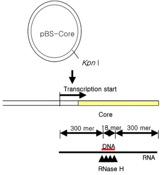
Figure 1. Method of RNA-DNA hybrid synthesis for RNase H enzyme assay. pBS-Core was pBluescript with HBV core gene.
Transcription was carried by T7 RNA polymerase.
pBS-Core
Transcription start
Core Kpn I
300 mer 18 mer 300 mer
DNA
RNA RNase H
pBS-Core
Transcription start
Core Kpn I
300 mer 18 mer 300 mer
DNA
RNA RNase H
(NEB, Beverly, USA)을 이용하여 정제하였다.
RNase H 효소 활성 확인. Kpn I으로 절단된 pBS-core 1μg을 32P-rCTP (50μCi; Amesham, England)와 함께 T7 RNA polymerase 10 unit로 37oC에서 전사하여 약 600 mer 의 RNA를 제작하고(Fig. 1), RNA free DNase I (Promega, NJ, USA) 20 unit를 37oC에서 10분간 처리하여 DNA를 제거한 후 에탄올 침전을 하였다. 침전된 RNA를 20μL 의 증류수로 회수한 후 core 유전자의 300 bp 부위에 결 합하는 18 mer oligonucleotide 20 pmol을 첨가하여 80oC 에서 5분간 변성시킨 후 온도를 50oC까지 천천히 내린 다음 4oC에 10분 이상 방치하여 RNA-DNA hybrid를 제 작하였다. 제작된 RNA-DNA hybrid 50 ng과 재조합 RNase H 단백질을 0.5 mM MnCl가 포함된 50 mM Tris-HCl (pH 7.0)에 첨가하여 37oC에서 1시간 동안 반응 시켰다. 반응액을 8% polyacrylamide gel에서 전기영동한 후 gel drier로 말리고 X-AR필름에 실온에서 12시간 동안 노출시켜 자동현상기로 현상하였다.
ScFv 형태의 human naive antibody phage display library 제작. Antibody phage display library의 제작은 Barbas 등(17)의 방법을 사용하였으며 구체적인 방법은 다음과 같다. 20대 정상 성인 50인의 말초혈액을 10 mL 씩 채취하고 각각을 histopaque-1077 (Sigma, St. Louis, USA) 10 mL에 중층하고 400×g로 실온에서 30분간 원 심분리한 후 림프구층을 분리하고 10 mL PBS로 2회 세 척하였다. 분리된 림프구 1×10⁶ 세포로부터 Ultraspec II (BIOTEX, Houston, USA)로 total RNA를 추출하였다.
Total RNA 20μg을 4μg의 oligo (dT)12-18와 혼합하고 DEPC 처리된 증류수를 첨가하여 최종 96μL가 되도록 한 다음 72oC에서 10분간 반응시키고 4oC서 1분 이상 방 치하였다. cDNA 반응용액 56μL (10× PCR buffer 16μ L, 25 mM MgCl2 16μL, 10 mM dNTP 8μL, 100 mM DTT 16μL)를 첨가하고 42oC에서 5분간 반응시키고 8μL (200 U/μL)의 SUPERSCRIPT II 역전사 효소(Stratagene, CA, USA)를 첨가하여 42oC에서 1시간 동안 cDNA를 합 성하였다. 인간 항체 유전자 PCR 증폭에 사용한 primer 들은 Barbas 등(17)이 사용한 것과 동일한 것이다. PCR 증폭된 유전자는 2% agarose gel 전기영동으로 확인하고 Qiaex II (Qiagen, Valencia, USA)를 이용하여 정제한 후 적절한 VH chain, VL chain set로 2차 PCR을 실시하였다.
최종 PCR 산물을 Sfi I으로 제한효소 절단하고 Sfi I 절단 된 pComb3X 에 클로닝하였다. 클로닝된 유전자를 Gene Pulser II (Bio-Rad, Hercules, USA)로 ER2537 electrocom- petent cell에 형질전환하였으며 SOC 배지(tryptone 20 g, yeast extract 5 g, NaCl 0.5 g, KCl 186 g, glucose 20 mM /1 L)와 Super Broth 배지(SB 배지; MOPS 10 g, Tryptone 30 g, yeast extract 20 g/1 L)로 37oC에서 진탕배양하였다.
Ampicillin을 사용하여 형질전환된 대장균만 선택배양하
였으며 phage의 제작에는 VCSM13 helper phage를 사용 하였다. 제작된 phage display library는 PEG 침전 (PEG8000 4% (w/v), NaCl 3% (w/v))하고 1% bovine se- rum albumin in tris buffered saline으로 회수하였다.
RNase H를 인지하는 항체 클론의 선택. 제작된 항체 library에서 RNase H 단백질을 인지하는 항체 클론의 선 택은 BIAcore 2000 (Biacore AB, Uppsala, Sweden)을 사용 하였다. RNase H 단백질의 고정에는 CM5 chip과 amine coupling 방법을 사용하였다. RNase H 단백질이 고정된 flow cell에 항체 library phabs를 2μL/min으로 흘려 결합 시킨 후 해리되는 시간별로 6시간 동안 회수하였다. 6시 간 후 regeneration buffer (25 mM NaOH, 0.5 M NaCl)를 사용하여 해리되지 않은 phage를 회수하였다.
회수된 phage들을 대수 증식기의 ER2537에 감염시킨 후 50μg/mL의 ampicillin이 첨가된 LB 고형 배지에서 배 양하여 집락을 형성하게 하였다. 96 well 세균배양 plate 에 형성된 각 집락을 SB 배지로 배양하고 VCSM13을 감 염시켜 phage를 제작하였다. 제작된 각각의 phage를 RNase H 단백질이 고정된 flow cell에 다시 흘려 각 클론 의 RNase H 단백질에 대한 결합 강도를 확인하여 강하 게 결합하는 클론들을 선택하였다. 선택된 phage 클론을 대장균 Top10F' (Invitrogen, CA, USA)에 감염시키고 37oC SB 배지에서 배양하고 600 nm에서 흡광도가 0.9∼
1.0일 때 IPTG를 넣고 8시간 더 배양함으로써 항체분절 의 수용성 발현을 유도하였다. 대장균을 수확하여 lysis buffer (10 mM Na2HPO4 pH 8.0, 500 mM NaCl, 10 mM imidazole)에 부유한 후 초음파로 세포를 분쇄하였다. 원 심분리한 상청액에서 항체분절은 Ni-NTA column (Qia- gen, Valencia, USA)과 imidazole buffer를 이용하여 정제 하고 microcon으로 농축하여 사용하였다.
항체의 친화도 측정. BIAcore 2000을 사용하여 정제된 항체분절의 RNase H에 대한 친화도를 측정하였으며 이 때 CM5 chip에 고정한 RU 값은 2000이었다. 정제된 항 체분절을 농도별로 5μL/min 속도로 흘려 결합과 해리를 측정하고 Biaevaluation software (Biacore AB, Uppsala, Sweden)로 Kon과 Koff를 측정하고 KD 값을 계산하였다.
염기서열 분석과 CDR 확인. ABI prism 3100을 사용하 여 각 항체 클론의 염기서열을 분석하였으며 VH 유전자 의 분석을 위하여 VH 유전자의 경우 HRML-F (5' ggt ggt tcc tct aga tct tcc 3')를, VL 유전자의 분석을 위하여 ompseq (5' aag aca gct atc gcg att gca g 3')를 primer로 각각 사용하였다. 염기서열 분석 결과는 Genetyx를 사용 하여 아미노산 서열을 유추하였으며 VH와 VL의 CDR을 확인하였다.
RNase H 효소 활성 중화 실험. 제작된 항체분절의 RNase H 효소 활성 중화의 확인에 사용한 RNA-DNA hybrid는 RNase H 단백질의 효소 활성 확인을 위하여 제
작한 RNA에 500 mer의 DNA를 결합시켜 사용하였다.
RNase H 0.5μg과 항체분절을 혼합한 후 RNA-DNA hybrid 100 ng이 포함된 반응액을 최종 50μL가 되게 첨 가하여 37oC에서 1시간 동안 반응시켰다. 반응액을 에탄 올 침전하여 분해된 RNA 조각을 제거하고 liquid scintillation counter (MicroBeta TriLUx; Perkin elmer, Wel- lesley, USA)로 방사선량을 측정하여 항체의 RNase H 효 소 활성 중화 정도를 확인하였다.
결 과
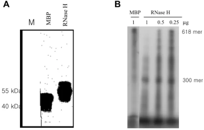
대장균에서 발현 정제된 RNase H. HBV의 RNase H를 중화하는 항체를 제작하기 위해서는 효소활성을 가진 RNase H의 정제가 필요하다. 본 연구에서는 MBP와 결 합된 RNase H를 대장균에서 발현하였으며 그 효소활성 도 확인하였다. 대장균에서 발현된 RNase H는 56 kDa로 anti-MBP 항체에 강하게 반응하였다(Fig. 2A). RNA- DNA hybrid를 사용한 효소활성 검사 결과 대조군으로 사용한 MBP를 반응한 lane에서는 효소 활성을 확인할 수 없었으며 제작된 RNase H를 반응한 lane에서는 300 mer의 분해 산물을 확인할 수 있었다(Fig. 2B). 대장균에 서 발현시킨 HBV RNase H는 적절한 효소활성을 지니고 있어 중화항체의 제작에 사용할 항원으로 적절하였다.
Human naive antibody phage display library. 인간 단세 포군 항체를 제작하는 경우 원하는 항원에 대한 항체를 가진 대상이 없는 경우 대부분의 연구자들은 실험동물 을 면역하여 제작된 항체를 인간화하는 과정을 주로 사 용하고 있다. 본 연구에서는 동물항체의 인간화의 어려 움과 실패가능성을 고려하여 면역되지 않은 사람으로부 터 naive library를 제작하여 항체를 제작하고자 하였다.
HBV 항체가 없는 20대 성인 50명의 말초 혈구를 분리하 여 scFv 형태의 library를 제작하였다. 모든 중쇄와 경쇄
들을 PCR 증폭하기 위하여 가능한 모든 primer 조합으로 PCR을 실시하였으며 조합에 따라 다른 정도의 증폭 정 도를 보였다(Fig. 3A, B, C). 각 조합의 경쇄, 중쇄를 모은 후 single chain으로 결합시키기 위하여 overlapping PCR 을 실시하였으며 원하는 900 bp 정도의 특이 band를 확 인하였다(Fig. 3D). Single chain의 형태로 결합된 항체 유 전자를 phage 발현 벡터인 pComb3X에 클로닝하여 여러 차례의 electroporation한 결과 2.41×10⁹ cfu 크기의 kappa chain과 heavy chain을 연결한 library와 2.05×10⁹ cfu 크 기의 lambda chain과 heavy chain을 연결한 library를 제작 하였다. 또한 Bst O1 제한 효소에 의한 fingerprinting을 통 하여 제작된 library의 다양성을 확인할 수 있었다(Fig. 4).
RNase H에 대한 인간 단세포군 항체. BIAcore 2000의 recover 기능을 사용하여 제작된 library로부터 RNase H 를 인지하는 RN41과 RN56의 2개 클론을 선택하였다.
각 클론의 항체분절 발현을 확인하기 위하여 대장균 Top10F'에서 IPTG로 발현을 유도한 결과 34 kDa의 anti- His 항체와 강하게 반응하는 특이 band를 SDS-PAGE와 western blot에서 확인하였다(Fig. 5). Ni-NTA column을 사
Figure 2. Purification and enzyme assay of RNase H in E. coli.
MBP and RNase H was purified with amylose resin column chromatography. (A) Immunoblot with rabbit anti-MBP polysera and HRP-conjugated gout anti-rabbit IgG. (B) RNase H enzyme assay.
1 1 0.5 0.25 µg
MBP RNase H
B A
RNase H
MBP
618 mer
300 mer
M
55 kDa 40 kDa
Figure 4. Bst O1 fingerprinting of antibody library. M is 100 bp ladder marker. Lane 1 to lane 16 were Bst O1 digested PCR products of 16 different clones selected randomly from library.
M
1 2 3 4 5 6 7 8 9 10 11 12 13 14 15 16
Figure 3. RT-PCR products and overlapping PCR products of light and heavy chain segments. (A) Light lambda chain segments, (B) Light kappa chain segments, (C) Heavy chain segments, (D) Overlapping PCR products (lane 1, kappa and heavy chains; lane 2, lambda and heavy chains). M is 100 bp ladder marker. All unlabelled lanes were PCR products by different primer sets.
M
M
M
M A
B
C
D 1 2
용하여 항체분절을 정제하고 BIAcore로 RNase H에 대한 친화도를 측정하였다. RN41은 Kon 값이 1.9×10⁴ 1/Ms, Koff 값이 8.6×10⁻³ 1/s로 KD는 4.5×10⁻⁷ M였으며 RN56 은 Kon 값이 2.4×10⁴ 1/Ms, Koff 값이 3.7×10⁻³ 1/s로 KD
는 1.9×10⁻⁷ M이었다(Fig. 6). 염기서열 분석을 통하여 각 클론의 아미노산 서열을 비교하였다. 경쇄의 CDR은 서로 유사하였으나 중쇄의 CDR은 상이하였다(Table I).
RNase H 효소 활성 중화 실험. 1μg에서부터 2배씩 희 석한 RN41과 RN56을 각각 RNase H 500 ng과 혼합한 후 100 ng의 RNA-DNA hybrid에 반응시켰다. RNase H에 의 해 분해된 RNA 조각을 에탄올 침전으로 제거한 후 방사 능을 측정하고 RNase H만 반응한 것을 100%로 하여 항 체 양에 따라 분석하였다(Fig. 7). 두 클론 모두 항체 양과 중화 정도는 비례하였으며 1μg에서 최대 47% (RN41)와 41% (RN56)의 중화 능력을 확인하였다. RN56에 비하여 RN41이 보다 높은 중화능력을 보였다.
고 찰
HBV의 복제에 있어 필수적인 과정은 pregenomic RNA의 역전사와 second-strand DNA의 합성이며(2,18), 이 과정을 수행하는 것이 바로 P 단백질이다. Pregenomic Figure 5. Expression of antibody fragments at selected clones.
(A) SDS-PAGE and (B) immunoblot with mouse anti-HA mono- clonal antibody (lane 1, 1E4 anti-preS1 clone; Lane 2, RN41;
Lane 3, RN56; Lane 4, non-transfected Top10F').
M 1 2 3 4
A B
1 2 3 4
45 31 66
21.5
Figure 6. Affinity of purified antibody fragments (A) RN41 and (B) RN56 with BIAcore 2000. Each curves were serial 2 fold dilution of antibody fragments from 10μg/ml.
A
B
-5 0 5 10 15 20 25
200 250 300 350 400 450 500 550 600
-50 0 50 100 150 200 250
200 250 300 350 400 450 500 550 600
Resonance unit (RU)Resonance unit (RU)
Time (s) Time (s) (5 µg/mL)
(1.25 µg/mL) (2.5 µg/mL)
(0.625 µg/mL)
(10 µg/mL) (5 µg/mL)
(1.25 µg/mL) (0.625 µg/mL) (2.5 µg/mL)
(10 µg/mL)
Table I. CDR of light and heavy chains variable regions of RN41 and RN56
ꠚꠚꠚꠚꠚꠚꠚꠚꠚꠚꠚꠚꠚꠚꠚꠚꠚꠚꠚꠚꠚꠚꠚꠚꠚꠚꠚꠚꠚꠚꠚꠚꠚꠚꠚꠚꠚꠚꠚꠚꠚꠚꠚꠚꠚꠚꠚꠚꠚꠚꠚꠚꠚꠚꠚꠚꠚꠚꠚꠚꠚꠚꠚꠚꠚꠚꠚꠚꠚꠚꠚꠚꠚꠚꠚꠚꠚꠚꠚꠚꠚꠚꠚꠚꠚꠚꠚꠚꠚꠚꠚꠚꠚꠚꠚꠚꠚꠚꠚꠚꠚꠚꠚꠚꠚꠚꠚꠚ
Clone CDR1 CDR2 CDR3
ꠏꠏꠏꠏꠏꠏꠏꠏꠏꠏꠏꠏꠏꠏꠏꠏꠏꠏꠏꠏꠏꠏꠏꠏꠏꠏꠏꠏꠏꠏꠏꠏꠏꠏꠏꠏꠏꠏꠏꠏꠏꠏꠏꠏꠏꠏꠏꠏꠏꠏꠏꠏꠏꠏꠏꠏꠏꠏꠏꠏꠏꠏꠏꠏꠏꠏꠏꠏꠏꠏꠏꠏꠏꠏꠏꠏꠏꠏꠏꠏꠏꠏꠏꠏꠏꠏꠏꠏꠏꠏꠏꠏꠏꠏꠏꠏꠏꠏꠏꠏꠏꠏꠏꠏꠏꠏꠏꠏ
VL RN41 RASQSISNWLA QASKLES QQYYSTPLT
RN56 RASQSISNYLN AASSLQS QQSYNTPFT
VH RN41 GGSISSSSYY SMFYGGSAYYNPSLKS GGDYYGSGTSAEYFQL
RN56 GFTFSNYAMS GIHNDALTTYYPESVKG LRGAPVTHYHFDY
ꠏꠏꠏꠏꠏꠏꠏꠏꠏꠏꠏꠏꠏꠏꠏꠏꠏꠏꠏꠏꠏꠏꠏꠏꠏꠏꠏꠏꠏꠏꠏꠏꠏꠏꠏꠏꠏꠏꠏꠏꠏꠏꠏꠏꠏꠏꠏꠏꠏꠏꠏꠏꠏꠏꠏꠏꠏꠏꠏꠏꠏꠏꠏꠏꠏꠏꠏꠏꠏꠏꠏꠏꠏꠏꠏꠏꠏꠏꠏꠏꠏꠏꠏꠏꠏꠏꠏꠏꠏꠏꠏꠏꠏꠏꠏꠏꠏꠏꠏꠏꠏꠏꠏꠏꠏꠏꠏꠏ
Figure 7. RNase H activity inhibiting assay. RNase H 500 ng was mixed with each purified antibody fragments and reacted with RNA-DNA hybrid 100 ng at 37oC for 1 hour. Reaction mixture precipitated with ethanol for removing RNA degradants and counted radio-isotope activity with liquid scintillation counter.
Antibody fragments (µg)
RNase H enzyme activity (%)
0 20 40 60 80 100
RN41 RN56
1 0.5 0.25
0.125 0.065
RNA로부터 만들어진 P 단백질은 cis-acting으로 작용하 며, TP, spacer, RT/POL 그리고 RNAse H의 4개 기능 부 위를 가지고 있다(2). P 단백질의 분석과 중요 기능부위 를 밝히기 위하여 여러 연구자들이 재조합 단백질로 발 현시켰다(19,20). P 단백질의 크기가 94 kDa 이어서 대장 균에서 전체 단백질을 재조합 단백질로 발현시키면 그 발현율이 낮다. 본 연구에서는 전체 P 단백질 중 RNase H 기능 부위만을 MBP 융합단백의 형태로 발현시켜 정 제하였다. 정제된 RNase H 기능부위 재조합 단백질 (pMRN)은 역전사 효소 활성을 가지고 있었다(Fig. 2). 본 연구를 통하여 제작된 pMRN 재조합 단백은 HBV P 단 백질의 RNase H 효소 활성을 억제하는 항체 제작에 있 어 panning과 screening에 적합하다고 판단된다.
일반적인 단세포군 항체는 실험동물을 특정항원으로 면역하여 제작하였다. 이 경우 진단이나 분석과 같이 생 체외적(in vitro)으로 사용하는 데 있어 아무런 문제가 없 지만, 치료나 진단의 목적으로 인체 내 투여하면 이종단 백으로 인지되어 면역반응을 유발한다. 그러므로 인체 내에서의 반감기가 감소되고 재차 사용 시 과민 반응을 일으킬 수 있다. 이러한 단점을 극복하기 위하여 여러 연구자들이 인간 단세포군 항체를 개발하고자 하였다.
인간 B 림프구를 in vitro immunization한 후 하이브리도 마의 제작, 항체의 불변영역이 인간 유전자로 치환된 transgenic mouse의 이용, 마우스 단세포군 항체의 humanization 그리고 phage display 기법을 이용한 human antibody library의 제작이 그 예들이다(21-24). In vitro immunization을 이용하는 방법은 하이브리도마의 제작 에 있어 Epstain-Barr virus를 이용한 불멸화의 성공률이 1∼3%로 낮다는 단점이 있으며, transgenic mouse를 이용 하는 경우 면역이나 하이브리도마를 쉽게 만들 수 있으 나 transgenic mouse를 가지고 있어야 한다는 단점을 가 지고 있고, 마우스 단세포군 항체의 humanization은 각각 의 항체에 따라 다른 방식이 요구된다는 단점을 가지고 있다. Phage display 기법은 항체에서 항원을 인지하는 분 절만을 filamentous phage의 표면에 발현시키는 기법으로 phage의 life cycle을 이용하여 in vitro panning이 가능하 다. In vitro panning이 가능함으로 하여 면역을 거치지 않고도 단세포군 항체를 제작할 수 있어 naive library나 synthetic library가 활용되고 있다. HBV가 감염된 환자에 서 P 단백질에 대한 항체가 만들어지지 않기 때문에 본 연구에서도 naive antibody library를 사용하였으며, 50인 의 정상 성인 말초혈액 림프구로부터 naive antibody phage display library를 4.46×10⁹ cfu의 크기로 제작하였 다. Synthetic library의 경우 1010 이상의 크기가 보고되고 있으나 완전한 naive library 보고는 아직 없다.
항체 library에서 특정 항원에 결합하는 클론을 선택하 기 위하여 다양한 방법이 사용된다. 가장 많이 사용되는
방법은 고전적인 panning이다(17). 이외에도 column이나 BIAcore sensorchip 같은 고형 보조물에 고정된 항원 (25-28), biotinylated 항원(29), 원핵세포에 고정된 항원 (30), 진핵세포에 고정된 항원(31), 조직절편이나 단편 (32) 등을 이용한 방법들이 사용된다. Surface plasmon resonance에 기반을 둔 BIAcore는 생물특이적인 상호작 용을 실시간으로 분석이 가능하여, 항원-항체반응의 역 동학적 측면을 이용한 항체의 선택을 가능하게 한다. 본 연구에서도 BIAcore를 항체의 선택에 사용하였으며 Koff
값이 높은 클론을 선택하기 위하여 6시간의 해리시간을 사용하였으나 10-7 M 정도의 친화도를 가진 RN41과 RN56만을 찾아낼 수 있었다. 일반적인 항체의 친화도가 10-8 M 이상인 점을 고려하면 101 정도 낮은 친화도이 다. 그러나 일반적인 library size에 비해 항체 친화도가 102 정도 낮다는 점을 고려할 때 본 연구로 제작된 RN41 과 RN56의 친화도는 적절한 것으로 분석된다. 이를 해 결하기 위해서는 library size를 1010 이상으로 증가시킬 필요가 있을 것이다(현재 진행 중). RN41과 RN56의 RNase H 효소 활성 중화 능력이 RNase H의 1.5배 농도 인 1μg에서 41∼47%이었다. 친화도가 더 높은 RN56이 RN41보다 낮은 중화 능력을 보인 것은 중쇄의 CDR이 다른 점으로 볼 때 RNase H의 인지 부위가 다르기 때문 으로 분석된다. 그리고 이 항체들의 항원에 대한 친화도 를 chain shuffling이나 CDR mutagenesis로 증가시킨다면 보다 높은 중화 능력을 보일 것이다.
본 연구를 통하여 naive library로 HBV의 RNase H를 중화하는 항체의 제작 가능성을 확인하였으며 이를 기 반으로 보다 높은 중화 능력을 가진 항체를 제작하고 세 포내 항체로 발현시킨다면 HBV 감염의 새로운 치료법 이 될 수 있을 것이다.
참 고 문 헌
1. Lau JY, Wright TL: Molecular virology and pathogenesis of hepatitis B. Lancet 342;1335-1340, 1993
2. Ganem D: Hepadnaviridae and their replication. In: Fields BN eds.: Fields virology 3rd ed. p1199-1233, Philadelphia Lippincott- Raven, 1996
3. Wong DK, Cheung AM, O'Rourke K, Naylor CD, Detsky AS, Heathcote J: Effect of alpha-interferon treatment in patients with hepatitis B e antigen-positive chronic hepatitis B. A meta-analy- sis. Ann Intern Med 119;312-323, 1993
4. Perrillo RP, Schiff ER, Davis GL, Bodenheimer HC, Jr., Lindsay K, Payne J: A randomized, controlled trial of interferon alfa-2b alone and after prednisone withdrawal for the treatment of chronic hepatitis B. The Hepatitis Interventional Therapy Group.
N Engl J Med 323;295-301, 1990
5. Krogsgaard K, Bindslev N, Christensen E, Craxi A, Schlichting P, Schalm S: The treatment effect of alpha interferon in chronic hepatitis B is independent of pre-treatment variables. Results based on individual patient data from 10 clinical controlled trials.
European Concerted Action on Viral Hepatitis (Eurohep). J Hepatol 21;646-655, 1994
6. Lai CL, Ching CK, Tung AK, Li E, Young J, Hill A: Lamivudine
is effective in suppressing hepatitis B virus DNA in Chinese hepatitis B surface antigen carriers: a placebo-controlled trial.
Hepatology 25;241-244, 1997
7. Bartholomew MM, Jansen RW, Jeffers LJ, Reddy KR, Johnson LC, Bunzendahl H, Condreay LD, Tzakis AG, Schiff ER, Brown NA: Hepatitis-B-virus resistance to lamivudine given for recur- rent infection after orthotopic liver transplantation. Lancet 4;349:
20-22, 1997
8. Tipples GA, Ma MM, Fischer KP, Bain VG, Kneteman NM, Tyrrell DL: Mutation in HBV RNA-dependent DNA polymerase confers resistance to lamivudine in vivo. Hepatology 24;714-717, 1996
9. Blum HE, Galun E, von Weizsacker F, Wands JR: Inhibition of hepatitis B virus by antisense oligodeoxynucleotides. Lancet 337;1230, 1991
10. Offensperger WB, Offensperger S, Walter E, Teubner K, Igloi G, Blum HE, Gerok W: In vivo inhibition of duck hepatitis B virus replication and gene expression by phosphorothioate modi- fied antisense oligodeoxynucleotides. EMBO J 12;1257-1262, 1993
11. Carman W, Thomas H, Domingo E: Viral genetic variation:
hepatitis B virus as a clinical example. Lancet 341;349-353, 1993 12. von Weizsacker F, Wieland S, Kock J, Offensperger WB, Offen- sperger S, Moradpour D: Gene therapy for chronic viral hepa- titis: ribozymes, antisense oligonucleotides, and dominant nega- tive mutants. Hepatology 26;251-255, 1997
13. Winter G, Milstein C: Man-made antibodies. Nature 349;293-299, 1991
14. McCafferty J, Griffiths AD, Winter G, Chiswell DJ: Phage antibodies: filamentous phage displaying antibody variable do- mains. Nature 348;552-554, 1990
15. Bird RE, Hardman KD, Jacobson JW, Johnson S, Kaufman BM, Lee SM: Single-chain antigen-binding proteins. Science 242;423- 426, 1998
16. Radziwill G, Tucker W, Schaller H. Mutational analysis of the hepatitis B virus P gene product: domain structure and RNase H activity. J Virol 64;613-620, 1990
17. Barbas CF III, Burton DR, Scott JK, Silverman GJ: Phage display, a laboratory manual. p9.23-11.15 CSHL press, 2001 18. Tsurimoto T, Fujiyama A, Matsubara K: Stable expression and
replication of hepatitis B virus genome in an integrated state in a human hepatoma cell line transfected with the cloned viral DNA. Proc Natl Acad Sci USA 84:444-448, 1987
19. Jeong JH, Kwak DS, Rho HM, Jung G: The catalytic properties of human hepatitis B virus polymerase. Biochem Biophys Res
Commun 223:264-271, 1996
20. Seifer M, Standring DN: Recombinant human hepatitis B virus reverse transcriptase is active in the absence of the nucleocapsid or the viral replication origin, DR1. J Virol 67;4513-4520, 1993 21. Winter G, Harris WJ: Humanized antibody. Immunol Today 14;
139-143, 1993
22. Andrzejewski C, Young PJ, Goldman J: Production of human IgM warm reacting red cell monoclonal autoantibodies by Epstein- Barr virus transformation. Transfusion 101;196-202, 1989 23. Lonberg N, Taylor LD, Harding FA: Antigen-specific human
antibodies from mice comprising four distinct modifications.
Nature 368;856-859, 1994
24. Winter G, Griffiths AD, Hawkins RE: Making antibodies by phage display technology. Ann Rev Immunol 12:433-455, 1994 25. Marks JD, Hoogenboom HR, Bonnert TP, McCafferty J,
Griffiths AD, Winter G: By-passing immunization. Human anti- bodies from V-gene libraries displayed on phage. J Mol Biol 222;
581-597, 1991
26. Clackson T, Hoogenboom HR, Griffiths AD, Winter G: Making antibody fragments using phage display libraries. Nature 352:624- 628, 1991
27. Griffiths AD, Williams SC, Hartley O, Tomlinson IM, Water- house P, Crosby WL: Isolation of high affinity human antibodies directly from large synthetic repertoires. EMBO J 13;3245-3260, 1994
28. Malmborg AC, Duenas M, Ohlin M, Soderlind E, Borrebaeck CA: Selection of binders from phage displayed antibody libraries using the BIAcore biosensor. J Immunol Methods 198;51-57, 1996
29. Hawkins RE, Russell SJ, Winter G: Selection of phage antibodies by binding affinity. Mimicking affinity maturation. J Mol Biol 226;889-896, 1992
30. Bradbury A, Persic L, Werge T, Cattaneo A: Use of living columns to select specific phage antibodies. Biotechnology (NY) 11;1565-1569, 1993
31. Cai X, Garen A: Anti-melanoma antibodies from melanoma patients immunized with genetically modified autologous tumor cells: selection of specific antibodies from single-chain Fv fusion phage libraries. Proc Natl Acad Sci USA 92;6537-6541, 1995 32. Van Ewijk W, de Kruif J, Germeraad WT, Berendes P, Ropke
C, Platenburg PP, Logtenberg T: Subtractive isolation of phage-displayed single-chain antibodies to thymic stromal cells by using intact thymic fragments. Proc Natl Acad Sci USA 94;
3903-3908, 1997