ISSN 0378-6471 (Print)⋅ISSN 2092-9374 (Online)
http://dx.doi.org/10.3341/jkos.2015.56.11.1810
Case Report
내경정맥 혈전증으로 인한 안압 상승 1예
Internal Jugular Vein Thrombosis Presenting with Elevated Intraocular Pressure
최연정⋅김윤택
Yeon Jung Choi, MD, Yun Taek Kim, MD, PhD
이화여자대학교 의학전문대학원 안과학교실
Department of Ophthalmology, Ewha Womans University School of Medicine, Seoul, Korea
Purpose: To report a case of elevated intraocular pressure (IOP) caused by internal jugular vein thrombosis.
Case summary: A 58-year-old male diagnosed with diabetic retinopathy visited our clinic for a regular checkup. On ophthalmic examination, IOP was 30 mm Hg in the right eye and 28 mm Hg in the left eye. Slit lamp examination showed chemosis, con- junctival injection and slight corneal edema in both eyes. Additionally, gonioscopic examination showed open angle. We ob- served face edema that started 1 month prior and he was diagnosed with internal jugular vein thrombosis on the right side, in- ternal jugular vein and innominate vein stenosis on the left side approximately 2 months ago. The patient underwent percuta- noeus transluminal angioplasty for dilating stenosed vessel. Four days after the procedure, his IOP was 15 mm Hg in the right eye and 12 mm Hg in the left eye based on Goldman applanation tonometer and was well maintained.
Conclusions: Internal jugular vein thrombosis on both sides can cause an increase in IOP.
J Korean Ophthalmol Soc 2015;56(11):1810-1816
Key Words: Episcleral vein pressure, Internal jugular vein thrombosis, Intraocular pressure
■Received: 2015. 5. 8. ■ Revised: 2015. 7. 8.
■Accepted: 2015. 8. 21.
■Address reprint requests to Yun Teak Kim, MD, PhD Department of Ophthalmology, Ewha Womans University Mokdong Hospital, #1071 Anyangcheon-ro, Yangcheon-gu, Seoul 07985, Korea
Tel: 82-2-2650-2818, Fax: 82-2-2650-4334 E-mail: jjongofhim@ewha.ac.kr
ⓒ2015 The Korean Ophthalmological Society
This is an Open Access article distributed under the terms of the Creative Commons Attribution Non-Commercial License (http://creativecommons.org/licenses/by-nc/3.0/) which permits unrestricted non-commercial use, distribution, and reproduction in any medium, provided the original work is properly cited.
안압은 홍채 뒤쪽의 섬모체라는 조직에서 생성되는 방수 의 양과 눈의 전방에서 빠져나가는 방수의 유출에 의한 저 항 간의 균형 및 상공막 정맥압(Episcleral vein pressure)에 의해 결정된다.1 따라서 방수의 생성 과다와 불충분한 배출, 맥락막(Choroidal plexus)의 혈액량 증가, 중심정맥압의 상 승은 안압 상승을 일으킬 수 있다. 그 외에도 외안근의 긴 장력 증가, 안구에 대한 외부 압력의 증가 및 높은 공막 경 축(Scleral rigidity) 등에 의해서도 안압은 높아질 수 있다.2 내경정맥 혈전증(Internal jugular vein thrombosis)은 매우
희귀한 질환으로 두개 내 내경정맥과 쇄골하정맥 사이 혈관 내에 혈전이 생긴 경우를 말하며, 중심정맥압(Central venous pressure)을 증가시킴으로써 뇌압상승, 경부나 안면의 부종과 통증 등이 나타날 수 있고, 안과적 합병증으로는 드물게 시력 소실과 유두 부종이 나타날 수 있다고 보고된 바 있다.2
지금까지 국내외에 내경정맥 혈전증으로 인한 안압 상승 에 대해서는 보고된 바가 없어, 이에 저자들은 다른 안과적 특이 소견이 없고 양측 내경정맥 혈전증으로 인한 혈관 폐 쇄 및 협착이 있는 환자에서 양안의 안압 상승을 보였다가 피부경유혈관성형술(Percutanoeus transluminal angioplasty) 로 혈관 확장 후 안압의 정상화를 경험하였기에 이를 보고 하고자 한다.
증례보고
기저 질환으로 당뇨, 고혈압이 있으며, 5년 전부터 말기
- 최연정⋅김윤택 : 내경정맥 혈전증으로 인한 안압 상승 -
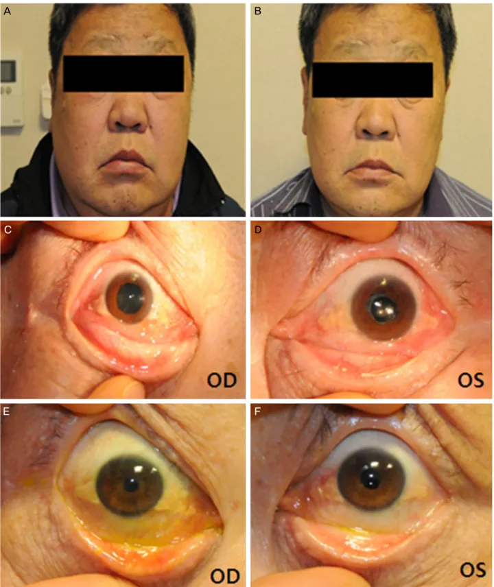
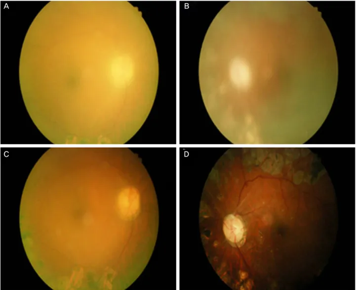
신질환으로 혈액 투석을 받고 있는 58세 남자 환자가 정기적 인 검진을 위해 본원 안과에 내원하였다. 환자는 6년 전 양안 증식성 당뇨 망막병증으로 양안 범망막광응고술(Panretinal photocoagulation), 좌안 유리체 출혈로 경평면부 유리체 절 제술(Trans pars plana vitrectomy) 및 초음파수정체 유화술 (Phacoemulsification)과 후방 인공수정체 삽입술(Posterior intraocular lens [IOL] implantation)을 받고 경과 관찰 중이 었다. 시력은 나안시력으로 우안 0.4 , 좌안 0.7 정도였으며 지난 6년 동안 측정된 평균 안압은 우안 12.78 ± 2.56 (range 11-20) mmHg, 좌안 14.61 ± 2.25 (range 9-17) mmHg로 양안 모두 정상 범위를 유지하고 있었다. 내원 당 일 환자의 나안 시력은 우안 0.3, 좌안 0.7이었고, 안압은 골드만압평안압계로 우안 30 mmHg, 좌안 28 mmHg로 측 정되었다. 세극등 현미경으로 시행한 전안부 검사상 양안 결막 부종, 충혈 및 경미한 각막 부종 소견(Fig. 1C, D)을 보였으며, 전방은 모두 깊었고 염증 소견은 관찰되지 않았 다. 전방각경 검사상 360도 개방되어 있었으며, 쉴렘관에 혈액은 관찰되지 않았고 우안 백내장 외에 다른 특이 소견 은 보이지 않았다. 양안 안저 검사상 우안에 비해 좌안의 시신경 유두가 다소 창백해 보였으며(Fig. 2A, B) 황반부는 안저 검사 및 빛간섭단층 촬영상 모두 이상 소견이 관찰되 지 않았다(Fig. 3). 환자는 1달 전부터 얼굴이 심하게 붓기 시작했다고 하였으며, 내원 당시에도 육안상으로 뚜렷한 안면 부종 및 결막 충혈, 부종을 관찰할 수 있었다(Fig. 1A, C, D). 환자의 1년 전 시행한 영상검사에서 Arteriovenous Shunt Arteriography 상 좌측 완두정맥(Brachiocephalic vein or innominate vein)의 부분적인 협착이 관찰되었고, 2달 전 시행한 경부 전산화 단층촬영에서는 우측 내경정맥 혈전증 및 좌측 내경정맥 협착이 의심되어 정맥조영(Venography) 을 권유 받은 상태였으며(Fig. 4), 최근 타 병원 혈관외과에 서 좌측 완두 정맥의 부분적 폐쇄를 진단 받고 피부경유혈 관성형술(Percutaneous transluminal angioplasty) 시행 예정 인 상태였다. 우측 내경정맥 혈전증으로 인한 내경정맥 폐 쇄와 좌측 내경정맥 협착 및 완두정맥 폐쇄로 인한 안압 상 승으로 진단하고 높아진 안압에 대해 하루 2회 안압하강제 (Cosopt®, Merck & Co. Inc., Whitehouse Station, NJ, USA) 점안을 시작하였으며, 시술 후 본원 안과 외래 경과 관찰하 기로 하였다. 환자는 2주 뒤 타 병원에서 좌측 완두정맥 폐 쇄에 대해 혈관 확장 시술을 받았으며(Fig. 5) 시술 후 4일 째 본원 안과에 내원하여 안압을 측정하였을 때 골드만 압 평안압계로 우안 15 mmHg, 좌안 12 mmHg로 측정되었다.
이에 사용하던 Cosopt® (Merck & Co. Inc.)를 중단하였으 며 시술 후 9일째 시력은 우안 나안시력 0.4, 좌안 나안시력 0.6으로 측정되었으며, 안압은 우안 15 mmHg, 좌안 12
mmHg였다. 시술 후 2주째 측정된 안압은 우안 14 mmHg, 좌 안 12 mmHg, 2달째 우안 15 mmHg, 좌안 11 mmHg로 안압 하강제 중단 후에도 정상 안압이 유지되고 있으며, 안면 부 종 및 각막, 결막 부종도 뚜렷하게 호전된 것을 관찰할 수 있었다(Fig. 1B, E, F, 2C, D).
고 찰
내경정맥 혈전증(Internal juglar vein thrombosis)은 임상 적으로 보기 드문 질환으로 과거에는 두경부 급성 감염의 합병증으로 주로 발생하였으나, 최근에는 중심정맥카테더 삽입, 주사제제 남용으로 인한 혈전증이 대부분을 차지한 다. 이러한 원인 외에도 암, 앞목추간반절제술(anterior cer- vical discectomy), 방사선치료, 혈액응고장애로 인해 생길 수 있으며, 자연적으로도 발생할 수 있다.3,4 발병 기전에 대 해서는 아직 명확히 밝혀지지 않았으나, 주로 비정상적인 혈액흐름, 혈액의 구성 성분 및 혈관 내피의 이상에 의해 유발될 수 있으며, 특히 중심정맥 도자법(central venous catheterization)과 같이 혈관 벽을 파괴시킬 만한 선행 요인 이 있으면 발생할 위험이 높아진다고 알려져 있다.3 특히 본 증례 환자의 경우처럼 혈액 투석을 위한 카테터 삽입 시 술을 받은 경우, 26%에서 내경정맥 혈전증이 발생할 수 있 다는 연구 결과가 보고된 바 있다.5
진단을 위해 과거에는 정맥촬영술(IV venography)이 이 용되었으나, 최근에는 비침습적인 진단 도구인 경부 전산 화단층촬영법, 경부 자기공명영상 및 초음파 검사 등으로 확진이 가능해졌다.6 증상 및 징후로는 발열, 백혈구 증가, 경부통 및 경부의 부종 등이 보고된 바 있으나 비특이적이 고, 대부분 특이한 증상이나 징후를 보이지 않는 경우가 많 아 진단이 지연되고 그 예후에 대해서도 잘 알려져 있지 않 다. 내경정맥 혈전증의 가장 심각한 합병증은 폐색전증이 며, 그 외에도 감염 혈전과 패혈증 등이 발생할 수 있다.7 안과적 합병증으로는 지금까지 Gutteridge et al8에 의해 정 맥울혈망막증으로 인한 시력 소실과 Masood and While9에 의해 양측 내경정맥 혈전증으로 인한 유두부종이 보고된 바 있으며, 두 증례 모두 양측 안구 및 대뇌로부터 경정맥 환류에 이상이 생겨 뇌내압상승과 안구 정맥 고혈압(ocular venous hypertension)이 유발되어 위와 같은 증상이 나타날 수 있다고 설명하고 있다.
안구의 주요 정맥배출계는 위눈정맥(Superior ophthalmic vein)과 아래눈정맥(Inferior ophthalmic vein)으로 구성되며 두 정맥 모두 최종적으로 해면정맥굴(carvernous sinus)로 배출된다.10 그중에서도 위눈정맥은 두개내(Intracranial)와 두개외(extracranial)의 정맥계와 연결되어 있기 때문에 두
Figure 1. Face and anterior segment photograph before and after percutaneous transluminal angioplasty. (A, C, D) Clinical photog-
raphy shows prominent face edema and chemosis before percutaneous transluminal angioplasty (PTA). (B, E, F) Recovery of face swelling and chemosis was observed after PTA at left innominate vein (or brachiocephalic vein). OD = oculus dexter; OS = oculus sinister.개내압과 두개외의 정맥배출로(venous outflow tract)의 저 항력에 영향을 받는다. 따라서 해면정맥굴 정맥계의 압력이
증가하거나 두개내외의 정맥배출로(venous outflow tract)에 협착이나 차단을 일으키는 질환이 발생하게 되면 경정맥
A B
C D
E F
- 최연정⋅김윤택 : 내경정맥 혈전증으로 인한 안압 상승 -
Figure 2. The changes of fundus photography according the changes in intraocular pressure. (A, B) Fundus photography shows that
optic disc was seen a little pale in the left eye but, the retina and optic disc in both eyes was dimly visible (IOP 30/28 mm Hg). (C, D) Two weeks after PTA, fundus photography shows that the retina and optic disc is seen more clearly (IOP 14/12 mm Hg). IOP= intraocular pressure; PTA = percutanoeus transluminal angioplasty.
역류(jugular venous reflux)가 일어나게 되고 순차적으로 위 눈정맥의 혈류가 역류하게 된다. Hsu et al11 역시 경정맥 역류가 있는 경우 눈과 뇌의 순환계 역류를 일으킬 수 있다 고 하였으며, 본 증례에서도 내경정맥 혈전증이 있는 환자 에서 혈관의 협착 혹은 폐쇄가 발생함으로써 경정맥 역류 가 야기되어 비정상적인 압력 경사가 만들어지고 결국 안 구와 대뇌정맥의 혈액 유출을 방해하게 되었다고 생각해 볼 수 있다.12 하지만 한쪽에만 병변이 있는 경우에는 동측 에 측부 혈관이 발달하여13 정맥 혈류 흐름에 대한 기능이 유지되거나 반대측 내경정맥이 정상적인 기능을 한다면, 특별한 증상이 나타나지 않을 수 있는데, 본 증례에서는 우 측 내경정맥의 완전 폐쇄와 좌측 내경정맥의 부분 폐쇄 및 완두 정맥(brachiocephalic vein or innominate vein)의 폐쇄
로 인하여 양측에서 정맥 흐름의 차단이 발생하였을 것이 라고 생각된다.
내경정맥 혈전증은 상공맥정맥압(episcleral vein pres- sure)을 높일 수 있는 여러 가지 원인 중의 한 가지가 될 수 있으며, 상공막정맥압의 상승은 일반적으로 안압의 상승과 관련이 있다고 알려져 있다. 안압은 방수가 생성되는 양과 유출에 의한 저항 간의 균형, 그리고 상공막 정맥압에 의해 결정된다. 대부분의 방수는 섬유주쉴렘관 유출로를 통해 배출되고 그 후 상공막 정맥을 거쳐 정맥계로 유입되며 이 정맥들의 압력을 나타내는 상공막정맥압의 상승은 동일한 만큼의 안압 상승을 보이게 된다.14 상공막정맥압과 안압과 의 관련성에 대하여 Teng et al15은 경정맥의 수축이 안압을 상승시킬 수 있다는 가설을 제시한 바 있다. 또한 이와 관
A B
C D
A B
C D
E F
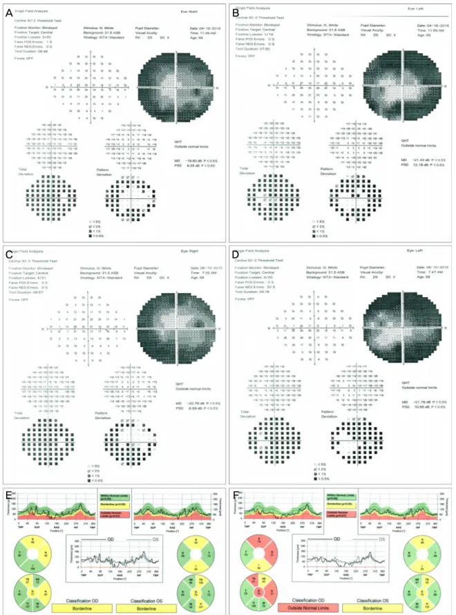
Figure 3. Visual field examination and optical coherence tomography (OCT) before and after percutaneou transluminal angioplasty
(PTA). (A-D) Humphrey visual field testing revealed a central island of vision in the right eye and showed clover leaf pattern in the left eye. (E) Before PTA, OCT dose not showed significant RNFL thinning in both eyes. (F) But, RNFL thinning in the nasal region in TSNIT graph after PTA. OD = oculus dexter; OS = oculus sinister; RNFL = retinal nerve fiber layer; TSNIT = temporal-supe- rior-nasal-inferior-temporal.- 최연정⋅김윤택 : 내경정맥 혈전증으로 인한 안압 상승 -
Figure 5. Before and after digital subtraction angiography (DSA) image of Percutaneous transluminal angioplasty. Images show (A)
segmental stenosis on the innominate vein (or brachiocephalic vein, white arrow) and internal jugular vein (black arrow) in left side.(B) Percutanoeus transluminal angioplasty with balloon dilation catheter was done through left forearm graft fisula. (C) DSA dem- onstrates improved flow of contrast agent through the left innominate vein.
Figure 4. CT images of internal jugular vein thrombosis. Post-contrast neck CT images of coronal section (A) and sagittal view (B)
showed a complete thrombosis of the right internal jugular vein without any flow (white arrows) and (C) partial stenosis of the left internal jugular vein (black arrow) and the innominate vein (white arrow). CT = computed tomography.련하여 Jeon et al16은 폐암에 의한 상대정맥증후군(superior vena cava syndrome) 환자에서 안허혈로 인해 발생한 신생 혈관 녹내장에 대해 보고한 바 있는데, 림프절 팽창으로 인 해 동측의 상대정맥의 압박, 경정맥의 확장, 상대적인 경동 맥직경의 감소로 인해 상공막정맥압이 상승되고 그 결과 안압 상승과 안혈류 공급의 장애가 발생하여 안허혈 증후 군이 발생하였다고 설명하였다. 혈류의 장애가 단측에서 일어난 Jeon et al16의 증례와 달리 본 증례에서는 양측 내경 정맥의 협착과 폐쇄로 안구와 대뇌로부터 빠져나가는 혈액 의 흐름이 완전히 차단됨으로써 상공맥 정맥압이 증가하여
이차적으로 안압 상승이 발생하였다고 생각하였으며, 내경 정맥의 협착에 대한 치료를 통해 안압의 정상화를 가져옴 으로써 경정맥 혈전증이 안압 상승을 일으킨 주요 원인이 었음을 알 수 있다.
본 증례에서는 경과 관찰 기간 동안 환자의 갑작스런 안 압 상승을 설명할 만한 안과적 특이 소견이 없음에도 불구 하고 안압이 상승되어 있었으며 시야 검사상 녹내장성 변 화를 보였다. 또한 환자는 혈액 투석을 위해 양측에 중심 정맥 도관을 삽입한 과거력이 있었으며 이전부터 경부 전 산화단층 촬영상 우측 내경정맥 혈전증으로 인한 혈관 폐
C
C
A B
A B
= 국문초록 =
내경정맥 혈전증으로 인한 안압 상승 1예
목적: 내경정맥 혈전증(Internal juglar vein thrombosis) 환자에서 나타난 안압 상승에 대하여 피부 경유혈관성형술 후 안압이 정상화 된 1예를 경험하였기에 이를 보고하고자 한다.
증례요약: 58세 남자 환자가 당뇨 망막병증으로 정기 검진을 위해 본원 안과에 내원하였다. 내원 당일 측정된 안압은 우안 30 mmHg, 좌안 28 mmHg였으며, 세극등 현미경으로 시행한 전안부 검사상 결막 부종, 충혈 및 경도의 각막 부종이 관찰되었고 전방각경 검사상 개방각 소견을 보였다. 1달 전부터 시작된 안면 부종을 관찰할 수 있었고, 환자는 2달 전 시행한 경부 전산화단층촬영에서 우측 내경 정맥 혈전증과 좌측 내경정맥 및 완두정맥 폐쇄를 진단 받았다. 이에 대해 피부경유혈관성형술(percutanoeus transluminal angioplasty)을 시행 받았으며, 시술 후 4일째 골드만압평안압으로 우안 15 mmHg, 좌안 12 mmHg로 안압은 정상화되었고 이후 안압 하강제 없이도 정상 안압이 유지되었다.
결론: 내경정맥 혈전증(Internal jugular vein thrombosis) 환자에서 안압 상승 가능성에 대해 염두에 두어야 할 것이다.
<대한안과학회지 2015;56(11):1810-1816>
쇄와 좌측 내경정맥 및 완두정맥의 협착이 관찰되었고 안 압이 상승한 후에 안과적으로 안압을 낮추기 위한 지속적 인 약물 치료 혹은 시술을 시행하지 않았음에도 불구하고 좌측 완두 정맥을 확장하는 혈관성형술을 받은 후 자연적 으로 안압이 정상화된 것을 관찰할 수 있었다.
결론적으로, 양측 내경정맥의 혈전증은 이차적으로 안압 상승을 일으킬 수 있음을 시사하며, 내경정맥 혈전증 외에 도 내경정맥의 협착이나 폐쇄를 일으킬 만한 질환이 있는 경우에는 이를 고려해야 한다.
참고문헌
1) Kwon YH, Fingert JH, Kuehn MH, Alward WL. Primary open-an- gle glaucoma. N Engl J Med 2009;360:1113-24.
2) Murphy DF. Anesthesia and intraocular pressure. Anesth Analg 1985;64:520-30.
3) Uduma FU, Yarouda M, Wali M. An unusual presentation of bi- lateral internal jugular venous thrombosis: a case report. Glob J Health Sci 2011;3:237-41.
4) Gallanos M, Hafner JW. Posttraumatic internal juglar vein throm- bosis presenting as a painfulneck mass in a child. Pediatr Emerg Care 2008;24:542-5.
5) Wilkin TD, Kraus MA, Lane KA, Trerotola SO. Internal jugular vein thrombosis associated with hemodialysis catheters. Radiolo- gy 2003;228:697-700.
6) Fishman EK, Pakter RL, Gayler BW, et al. Jugular venous
thrombosis: diagnosis by computed tomography. J Comput Assist Tomogr 1984;8:963-8.
7) Ahmed N. Thrombosis after central venous cannulation. Med J Aust 1976;1:217-20.
8) Gutteridge IF, Royle JP, Cockburn DM. Spontaneous internal ju- glar vein thrombosis and venous-stasis retinopathy. Stroke 1987;
18:808-11.
9) Masood I, While A. Bilateral jugular vein thrombosis: a rare cause of papilloedema. Eye (Lond) 2006;20:249-50.
10) Dutton JJ. Atlas of Clinical and Surgical Orbital Anatomy, 2nd ed.
Philadelphia: WB Saunders, 2011;83-98.
11) Hsu HY, Chao AC, Chen YY, et al. Reflux of jugular and retro- bulbar venous flow in transient monocular blindness. Ann Neurol 2008;63:247-53.
12) Chung CP, Hsu HY, Chao AC, et al. Jugular venous reflux affects ocular venous system in transient monocular blindness. Cerebr- ovasc Dis 2010;29:122-9.
13) Jayaraman MV, Boxerman JL, Davis LM, et al. Incidence of ex- trinsic compression of the internal jugular vein in unselected pa- tients undergoing CT angiography. AJNR Am J Neuroradiol 2012;
33:1247-50.
14) Kollarits CR, Gaasterland D, Di Chiro G, et al. Management of a patient with orbital varices, visual loss, and ipsilateral glaucoma.
Ophthalmic Surg 1977;8:54-62.
15) Teng C, Gurses-Ozden R, Liebmann JM, et al. Effect of a tight necktie on intraocular pressure. Br J Ophthalmol 2003;87:946-8.
16) Jeon JH, Cho KJ, Chang KC, Chang MH. Neovascular glaucoma with ocular ischemia in superior vena cava syndrome. J Korean Ophthalmol Soc 2012;53:1346-51.