298
DOI: 10.4046/trd.2010.68.5.298
ISSN: 1738-3536(Print)/2005-6184(Online) Tuberc Respir Dis 2010;68:298-300
CopyrightⒸ2010. The Korean Academy of Tuberculosis and Respiratory Diseases. All rights reserved.
성인에서 횡격막마비로 오인한 우엽간 횡격막탈장 1예
1
남양주한양병원 내과,
2원광대학교 의과대학 내과학교실
박정현1, 황기은2, 김소영2, 김학렬2, 양세훈2, 김휘정2, 정은택2
Diaphragmatic Hernia of the Right Hepatic Lobe Mistaken for Diaphragmatic Paralysis in Adult
Jung Hyun Park, M.D.
1, Ki Eun Hwang, M.D.
2, So Young Kim, M.D.
2, Hak Ryul Kim, M.D.
2, Sei Hoon Yang, M.D.
2, Hwi Jung Kim, M.D.
2, Eun Taik Jeong, M.D.
2Department of Internal Medicine,
1Namyangju Hanyang Hospital, Namyangju,
2Wonkwang University College of Medicine, Iksan, Korea
Diaphragmatic paralysis can be demonstrated through diaphragmatic elevation on chest X-ray after thoracic lung surgery or the placement of chest tubing. Additional causes of diaphragmatic paralysis are iatrogenic, mass, atelec- tasis, etc. For the diagnosis of diaphragmatic paralysis, it required some studies (fluoroscopy, computed tomography [CT], magnetic resonance imaging). Diaphragmatic hernia of the liver is a rare clinical entity, usually found after trauma in adults. Congenital diaphragmatic hernia in neonates requires surgery. Non-traumatic diaphragmatic hernia of the liver in an adult is a rare right-sided diaphragmatic hernia. On developing any symptoms, surgery must be performed. When diaphragmatic hernia is incidentally found in adults without trauma, it is placed under observation for a time period. We diagnosed the diaphragmatic herniation of a right hepatic lobe by 16-slice CT scan without surgery.
Key Words: Hernia; Diaphragm; Liver
Supported by a grant from Wonkwang University in 2010.
Address for correspondence: Jung Hyun Park, M.D.
Department of Internal Medicine, Namyangju Hanyang Medical Hospital, 570, Onam-ri, Onam-eub, Namyangju 472- 883, Korea
Phone: 82-31-510-0114, Fax: 82-31-510-0123 E-mail: parkaiver@hanmail.net
Received: Apr. 16, 2010 Accepted: Apr. 21, 2010
서 론
횡격막 마비는 임상에서 자주 접하는 소견이지만, 횡격 막 탈장은 흔히 접하는 임상소견은 아니다. 간의 선천성 횡격막탈장은 소아에서 주로 관찰되며, 소아에서는 여러 합병증으로 수술을 요하는 경우가 많다. 성인의 경우에는 주로 외상에 의해 일반적으로 관찰되며 비외상성으로 관찰 되는 경우는 드물다. 이럴 경우 우측 횡격막의 상승으로 인해 횡격막마비로 오인되는 경우가 있다. 만약 꼬임에 의
한 증상이 있을 경우 수술을 요하며, 무증상일 경우 관찰을 요한다. 저자들은 성인에서 횡격막마비로 오인한 우엽간 횡격막탈장 1예를 경험 하였기에 보고 하는 바이다.
증 례
환 자: 김○○, 76세, 여자 주 소: 흉부방사선 이상
현병력: 환자는 2개월 전부터 한방병원에 입원하여 목 부위 통증으로 보존적치료 해오던 환자로, 내원 10여 일 전부터 기침과 객담 발생하여 시행한 흉부방사선사진 소 견상 우측 횡격막의 상승 소견 보여 정밀검사위해 본원 내원하였다.
과거력: 2002년 개인병원에서 우측 무릎 치환술 및 양 측 어깨관절 수술을 시행하였다. 그 외에 사고나 외상의 경력은 없었다.
개인력: 비흡연자였으며 특이사항 없었다.
Image of the Month
Tuberculosis and Respiratory Diseases Vol. 68. No. 5, May 2010
299
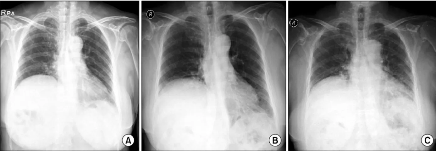
Figure 1. Right diaphragm elevation on Chest X-ray. (A) 5 years ago. (B) Full inspiration at admission. (C) Full expiration at admission.Figure 2. Right diaphrag- matic herniation of liver (Bi- fid liver) on abdomino-pel- vic CT. Rt: right; Lt: left.
가족력: 특이사항 없었다.
이학적 소견: 내원 당시 혈압은 120/80 mm Hg, 맥박수 84회/분, 체온 36.5oC, 호흡수 20회/분이었다. 흉부 청진 에서 우측 폐하부의 폐음이 좌측에 비해 감소되어 있었다.
다른 심장, 복부, 사지 등의 진찰 소견은 정상이었다.
검사 소견: 일반 혈액검사와 생화학검사 및 소변검사는 정상이었다. 폐기능 검사상 노력성 폐활량(FVC)은 1.27 L (정상 예측치의 63%), 1초간 노력성 폐활량(FEV1)은 0.92 L (정상 예측치의 62%), 1초간 노력성 호기량의 노력 성 폐활량에 대한 비(FEV1/FVC)는 73%였고, 폐확산능
JH Park et al: Congenital diaphragmatic herniation, diaphragmatic paralysis
300
(DLco)은 8.7 L/min/mm Hg (예측치의 56%)였다. 심전 도는 정상이었다.
방사선 소견: 과거 5년 전 수술 당시의 단순 흉부 방사 선 사진 및 내원 시 실시한 단순 흉부 방사선 검사에서 우측 횡격막선의 상승이 현저하였다(Figure 1). 흉부 투시 검사에서 우측 횡격막이 비정상으로 상승되었으나 호흡 에 따라 조금씩 움직이는 양상을 보였다. 흉부 전산화 단 층촬영에서 우측 횡격막의 상승과 간의 우측엽이 횡격막 상방으로 상승되어 있는 소견이 관찰되었다(Figure 2).
치료 및 경과: 과거 사진과 비교하여 선천적인 우측간 의 횡격막 상방으로의 탈장이 관찰되나 임상증상이 없어 서 관찰하기로 하였다.
고 찰
횡격막은 경추 척추신경 3, 4 그리고 5번의 지배를 받는 가로막신경(격막신경; phrenic nerve)에 의해 조절된다.
가로막신경이 손상되면 횡격막의 단측 마비가 주로 오며, 일부에서는 양측의 마비가 오기도 한다. 가로막신경 손상 은 주로 수술에 의해 발생하며, 1963년 성인의 심장수술 후 합병증으로 처음 알려졌다. 인공호흡기의 기간, 입원 기간, 항생제의 사용과 연관이 있으며, 이는 호흡기능에 영향을 미치게 된다1. 동물실험에서 인공호흡기에 의해 횡격막 기능장애가 발생할 수 있으며, 조절환기법(con- trolled mandatory ventilation) 모드와 일부 연관이 있다 고 보고하고 있다2. 횡격막마비는 탈장을 유발할 수 있으 며 선천성 횡격막탈장은 소아에서 주로 관찰되며, 소아에 서는 여러 합병증(특히 호흡곤란)으로 수술을 요하는 경 우가 많다3. 선천성 횡격막탈장은 흉복막관(pleuroperito- neal ducts)의 폐쇄장애로 발생하는데, 이런 소아는 폐 발 달장애가 많이 동반되며 폐동맥고혈압이 발생할 수 있다.
이로 인해 체외순환(extra-corporeal membrane oxygen- ation)이 필요한 경우가 있으며 사망률이 50%에 이른다4. 성인의 횡격막탈장은 주로 외상에 의해 일반적으로 관 찰되며 비외상성으로 관찰되는 경우는 드물다. Bochda- lek hernia는 70∼90%가 좌측에 발생하며, 남자에 많고, 위, 비장, 대장, 소장, 복막 등이 흉부로 탈장되는 경우가 많다. Morgagni hernia은 횡격막의 우측 앞쪽에 생기는 경우로 여자에 많으며, 간, 담낭, 췌장, 콩팥, 후복막 지방 이 탈장되기도 한다. 이럴 경우 횡격막의 상승으로 인해 횡격막마비로 오인되는 경우가 있다. 꼬임(strangulation) 이 약 30%에서 발생하는 데 이로 인한 증상이 있을 경우 수술을 요하며, 무증상일 경우 관찰을 요한다5,6. 간의 발
달장애는 간생성이 결핍되거나 과생성되는 경우로 나뉘 는데, 간의 좌엽의 발달이 안 되면 위염전(gastric volvu- lus)을 일으킬 수 있고, 우엽이 발달이 안 되면 간문맥고혈 압을 일으킬 수 있다. 드물게 간생성이 결핍되어 우측 횡 격막으로 장탈장이 일어나기도 한다. 또한 간의 과다생성 은 부간엽(accessory lobes)을 만들며 이는 비틀림(tor- sion)을 일으킬 수 있다7,8. 횡격막마비는 흉부방사선에서 횡격막의 상승으로 관찰되는 데, 횡격막탈장을 정확히 진 단하기 위해서는 컴퓨터단층촬영이나 자기공명영상을 실 시하여야 한다9. 성인에서 선천성 횡격막탈장이 무증상으 로 진단되는 경우는 드물며, 대부분 증상(복통, 가슴답답 함, 호흡곤란 등)을 호소하여 진단되어 치료하는 경우가 대부분이다. 본 증례는 우연히 흉부방사선상 횡격막마비 로 오인하여 발견한 선천적 우간엽 횡격막탈장 환자로, 특별한 치료 없이 지속적인 경과 관찰중이다.
참 고 문 헌
1. Ross Russell RI. C 3, 4 and 5, keep the diaphragm alive. Intensive Care Med 2006;32:1109-11.
2. Vassilakopoulos T, Petrof BJ. Ventilator-induced dia- phragmatic dysfunction. Am J Respir Crit Care Med 2004;169:336-41
3. McGuigan R, Azarow KS. Living on the edge: current concepts in the management of congenital diaphrag- matic hernia. Curr Surg 2005;62:390-5.
4. Robertson DJ, Harmon CM, Goldberg S. Right congeni- tal diaphragmatic hernia associated with fusion of the liver and the lung. J Pediatr Surg 2006;41:e9-10.
5. Losanoff JE, Sauter ER. Congenital posterolateral dia- phragmatic hernia in an adult. Hernia 2004;8:83-5.
6. Luo HF, Lei T, Wang HJ, Tan G, Wang ZY. Non-trau- matic diaphragmatic hernia of the liver in an adult: a case report. Hepatobiliary Pancreat Dis Int 2007;6:219- 21.
7. Daver GB, Bakhshi GD, Patil A, Ellur S, Jain M, Daver NG. Bifid liver in a patient with diaphragmatic hernia.
Indian J Gastroenterol 2005;24:27-8.
8. Zenda T, Kaizaki C, Mori Y, Miyamoto S, Horichi Y, Nakashima A. Adult right-sided Bochdalek hernia facili- tated by coexistent hepatic hypoplasia. Abdom Imaging 2000;25:394-6.
9. Sadeghi N, Nicaise N, DeBacker D, Struyven J, Van Gansbeke D. Right diaphragmatic rupture and hepatic hernia: an indirect sign on computed tomography. Eur Radiol 1999;9:972-4.