Journal of Korean Arthroscopy Society abbreviation (by Index Medicus): J Korean Arthrosc Soc Volume 16, Number 1, February, 2012
Introduction
Tibial plateau fractures comprise a low percentage of all fractures but the severe consequences can result from improper diagnosis or treatment. The magnitude of the force, the position of the extremity
at the time of injury,1)and the degree of osteopenia2,3) can all help determine the fracture type and extent of soft tissue damage. The treatments for tibial plateau fractures ranged from non-operative treatments to arthroscopy-assisted internal fixation, with pros and cons for each option.4-7)Due to the obvious proximity to the knee joint, tibial plateau fractures may accom- pany a variety of soft tissue injuries8,9) including injuries to the menisci, collateral and cruciate liga- ments, arteries, and nerves. The reported incidence of associated soft tissue injury in cases of tibial plateau fractures diagnosed by physical examination or operative findings was 7% to 50%.8,9)Cruciate liga- ment injuries may occur as avulsion, midsubstance, or attenuation injuries, and meniscal injuries may
경
경골 골 고 고평 평부 부 골 골절 절의 의 반 반월 월상 상 연 연골 골 파 파열 열 양 양상 상
차의과학대학교 분당차병원 정형외과1, 차의과학대학교 구미차병원 정형외과2 이동훈1∙김병국2∙김재화1∙정주환1∙이인성1∙이준구1∙이순철1
Characteristics of Meniscus Tear in Tibial Plateau Fractures
Dong-Hoon Lee, M.D.
1, Byung-Kuk Kim, M.D.
2, Ju-Hwan Chung, M.D.
1, In-Sung Lee, M.D.
1, Jun-Ku Lee, M.D.
1, Soon-Chul Lee, M.D.
1Department of Orthopaedic Surgery, CHA Bundang Medical Center, CHA University of Korea1, Department of Orthopaedic Surgery, CHA Gumi Medical Center, CHA University of Korea2
Purpose: Tibial plateau fractures cause a variety of problems in the knee joint. The purpose of this study was to investigate the characteristics of the meniscus injuries in tibial plateau fracture arthroscopically.
Materials and Methods: Thirty-three out of 39 consecutive patients diagnosed with tibial plateau fractures underwent arthroscopy between March 2007 and March 2010. According to Schatzker classification, there were 1 type I (3.3%), 19 type II (53.3%), 4 type III (13.3%), 2 type IV (6.6%), 2 type V (6.6%) and 5 type VI (20%) fracture patterns in 33 patients.
Results: Twenty-five cases (75.8%) had lateral meniscus tears. There were 18 meniscal tears in 19 cases of Schatzker type II frac- tures (94.7%), 3 meniscal tears out of 4 cases of Schatzker type III fractures (75%) and 4 meniscal tears out of 5 cases of Schatzker type VI fractures (80%). The most commonly affected site (22/25) was the anterior horn of the lateral meniscus. Of the 25 document- ed meniscal tears, all but one were vertical longitudinal tear at meniscocapsular junction so most cases are amenable to arthroscopic repair.
Conclusion: Arthroscopy for the meniscal injuries in tibial plateau fractures is a valuable diagnostic and treatment tool, we recom- mend arthroscopy in tibial plateau fracture.
KEY WORDS: Tibial plateau fractures, Soft tissue injury, Meniscus tear, Arthroscopy
�Address reprint request to Byung-Kuk Kim, M.D.
Department of Orthopaedic Surgery, CHA Gumi Medical Center, CHA University,
855 Hyungkok-dong, Gumi-si, Gyeongsangbuk-do 730-040, Korea Tel: 82-31-780-5289, Fax: 82-31-708-3578
E-mail: [email protected]
접수일: 2011년 10월 28일 게재심사일: 2011년 11월 14일 게재승인일: 2012년 2월 14일
appear as peripheral, radial, or flap tears.8,9) The meniscus can be especially prone to injuries due to its attachment to the tibial plateau via the joint capsule.
Injuries of the meniscus may be subtle compared to obvious injuries as ones to the cruciate ligaments, but the consequences from meniscal injury could be as serious as osteoarthritis of the knee joint due to the loss of meniscus load sharing role. The purpose of this study was to assess the incidence and charac- teristics of meniscus injury with the use of arthroscopy in tibial plateau fracture.
Materials and Methods
We found 39 consecutive patients diagnosed with tibial plateau fractures between March 2007 and March 2010. Out of them, 6 patients were excluded because 4 patients showed nondisplaced fracture without any other suspected pathology in the knee joint who had gone through conservative treatment and 2 patients refused surgical treatment. Therefore, our study included 33 consecutive patients who had undergone arthroscopic-assisted osteosynthesis.
There were 14 men and 19 women with 19 injuries to the right knees and 14 injuries to the left knees. The average age of the patients was 51.6 years (range, 29~81 years). A total of 20 patients (60.6%) were injured by traffic accidents (motorcycle and motor vehicle accidents) and 8 patients (24.2%) were injured by falling accidents. The remaining 5 patients were injured from slippage on the floor. The tibial fractures were analyzed and graded according to Schatzker’s classification (Table 1).
All patients underwent anteroposterior (AP) and lateral plain radiography and computed tomography
(CT) of their injured knees. Fractures with articular incongruity of 3 mm or greater were considered for operative treatment.10) Calcaneal traction was applied to severely swollen knees if needed.
Although surgery was delayed until swelling of the leg had subsided, all the operations could be per- formed within 2 weeks from the date of injury. Once the swelling was reduced, a formal arthroscopy- assisted surgery was performed with minimal strip- ping and dissection. All operations in this study were performed by the senior author. In all cases, inflow pressure is maintained with the use of gravity only instead of infusion pump. The tourniquet was applied to all patients and the mean tourniquet pressure we used was 300 mmHg. Arthroscopic findings of asso- ciated soft tissue injury in the 33 patients with tibial plateau fracture were recorded, and the relationship between fracture type and soft tissue injury was then analyzed.
1. Surgical Techniques
The patients were placed in a supine position on the operating table with the knee flexed to a 90�
angle. A pneumatic tourniquet was applied on the proximal thigh and a kidney bar was used instead of a leg holder because the “figure 4”position enabled us to freely maneuver the arthroscope. Anterolateral and anteromedial portals were created to accommo- date a viewing and working portal, respectively. A thorough irrigation was performed until a clear view was achieved. All debris such as blood clots, bone fragments and denuded articular cartilage was removed. Next, a systemic evaluation to examine the medial and lateral compartments of the knee for
Table 1. Schatzker Classification of Fracture Types and Associated Soft Tissue Injuries
Meniscus ACL PCL MCL LCL
Type I 1*
Type II 18 1* 1*
Type III 03
Type IV 1*
Type V 1* 1*
Type VI 04 1*
Total 25 4* 3* 0 0
*: Avulsion fracture
ACL: anterior cruciate ligament, PCL: posterior cruciate ligament, MCL: medial collateral ligament, LCL: lateral collateral ligament.
possible meniscal or ligamentous injuries was per- formed. All soft tissue lesions were recorded and analyzed according to fracture types.
In order to first reduce the depressed fragment, a bony window was created on the anteromedial or anterolateral aspect of the tibial condyle depending on the fracture location as determined by fluoro- scopic examination and an impactor was introduced (Fig. 1A). By hammering the impactor on the depressed fragment, reduction was achieved. Allo- bone chip and blocks (freezed dried human allograft) were implanted to fill up the defect of the tibial condyle. We then confirmed reduction by arthroscopy, and subsequent fixation by buttress plate (LCP Proximal Tibial Plate; Synthes, Inc., PA, USA) was performed (Fig. 1B).
Intra-articular pathologies discovered during the
diagnostic arthroscopy were treated in an appropri- ate sequence after fixation of the fracture (Table 2).
For the meniscal repair, 2 methods, as described by Ahn et al.,11,12)were used: the modified outside-in and inside-out suture technique for anterior and midbody meniscal lesions (Fig. 2). Anterior cruciate ligament (ACL) injuries were treated in the usual manner by arthroscopy-assisted fixation of the ACL avulsion fracture in a first-stage surgical procedure using suture wires and second-stage reconstruction after fracture healing for complete ACL tears (Fig. 3).
2. Clinical Assessment
Clinical assessment to validate the arthroscopy included extra time for completing measurements required for the procedure in addition to fixation of
Fig. 1. Intraoperative fluoroscopic images. (A) Impactor was introduced through anterolateral tibial condyle. (B) Temporary fixation with plate using two K-wires after fracture site reduction.
A B
Table 2. Treatment of Associated Soft Tissue Injuries in 33 Tibial Plateau Fractures
Soft tissue injuries No. of injuries Treatment
Meniscal tear 23 Arthroscopic suture repair
01 Arthroscopic partial menisectomy
01 Conservative treatment
ACL avulsion Fx 03 Arthroscopically assisted fixation by pullout suture ACL rupture 01 Second stage reconstruction following fracture healing PCL avulsion Fx 02 Arthroscopically assisted fixation by pullout suture PCL rupture 01 Second stage reconstruction following fracture healing
MCL rupture 01 Conservative treatment
ACL: anterior cruciate ligament, Fx: fracture, PCL: posterior cruciate ligament, MCL: medial collateral ligament.
the fracture. Additionally, the complications such as vascular or neurological injury, articular cartilage injury, hemarthrosis, deep vein thrombosis, infection, compartment syndrome caused by the arthroscopy alone were investigated.
3. Statistical method
Statistical analysis was conducted using a Chi- square test for discrete variables with SPSS version 9.0 (SPSS Inc., Chicago, IL, USA). The level of sig- nificance was set at P<0.05.
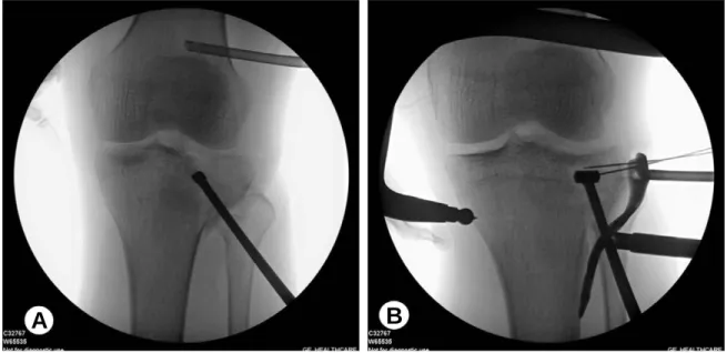
Fig. 2. Meniscus repair. (A) Outside-in technique. (B) Inside-out technique.
A B
Fig. 3. Tibial plateau fracture (Schatzker type V) with anterior cruciate ligament (ACL) avulsion fracture. Preoperative (A) antero- posterior (AP) and (B) lateral plain x-ray. (C) Arthroscopy-assisted fixation of the ACL avulsion fracture using suture wire.
Postoperative (D) AP and (E) lateral plain radiograph.
A B C
D E
Results 1. Fracture types
According to Schatzker classification, there were 1 type I (3.3%), 19 type II (53.3%), 4 type III (13.3%), 2 type IV (6.6%), 2 type V (6.6%) and 5 type VI (20%) fracture patterns in 33 patients. There was one open fracture (Gustilo-Anderson open type I) of Schatzker type VI. Two cases were accompanied by other con- comitant fractures. In one case of Schatzker type II, there was a concomitant medial femoral condyle fracture. The other case was complicated by com- minuted femoral shaft and ankle fractures in the same extremity in a Schatzker type V fracture. The mean articular depression of patients was 5.5 mm (range, 3.3~11.7 mm) on the coronal image of CT evaluation. The mean interval between the injury and surgery was different depending on the fracture type. In type I, II and III fracture, the interval was 3.5 days (1 to 7 days) compared to 11.7 days (3 to 14 days) in higher type IV, V, and VI fracture.
2. Meniscal tears
Among the patients in our study, there were 25 lateral meniscus tears (73%), and no medial menis- cus tear was observed. Of the 19 Schatzker type II fractures, 18 showed meniscus tears. Three meniscus tears were seen in Schatzker type III fractures and 4 in Schatzker type VI. Meniscus tears were found at a statistically higher rate in Schatzker type II frac- tures (P<0.01). Of the 25 documented tears, all but one were vertical longitudinal tear at the menisco- capsular junction. No other tear pattern was found.
In the majority of the meniscal tears, repairs were
performed instead of menisectomy (23 versus 1).
Regardless of Schatzker classification, anterior horn was most commonly involved (Table 3).
3. Other soft tissue injuries
There were total of 4 ACL related injuries and 3 posterior cruciate ligament (PCL) injuries (Table 1).
The cruciate ligament-related injuries were treated in an appropriate time sequence. We performed arthroscopy-assisted first-stage fixation for the ACL or PCL avulsion fracture, and second-stage reconstruction following fracture healing for com- plete ACL or PCL tears.
The collateral injury showed low incidence in our patients.
4. Time related to arthroscopic procedures & postopera- tive complications
Extra time spent on specific procedures was mea- sured as a part of an attempt to assess disadvantages of the arthroscopic procedure. The average time taken for diagnostic arthroscopy was 7.2 minutes (range, 5~10 minutes). The average time of menis- cus-related arthroscopic procedures was 34.3 min- utes (range, 17~52 minutes). These procedures included meniscal repairs as well as meniscectomies.
The average time for other soft tissue procedures such as ACL or PCL reconstruction was 38.1 minutes (range, 32~45 minutes). Total tourniquet time which include bony and soft tissue procedures was 53.4 minutes (range, 33~131 minutes). Complications specifically related to arthroscopic procedures such as compartment syndrome and neurovascular injury were not noted. No postoperative infections or cases
Table 3. Relationship between Fracture Type and Meniscal Injury and its Location
Fracture type No. of patients Location of the meniscal tear
Anterior Midbody Posterior horn
Type I
Type II 18 15* 9* 3*
Type III 03 03* 2*
Type IV Type V
Type VI 04 04* 3* 2*
*: Involving more than two tear locations
of deep vein thrombosis were seen.
Discussion
As with any other intra-articular fracture, the tib- ial plateau fracture is always challenging for orthopaedic surgeons because of its widely varying trauma, and associated soft-tissue injuries add fur- ther complexity. With regard to meniscal injury, Honkonen13)reported a 50% rate of meniscal lesions, mainly in the lateral menisci. Vangsness et al.14) arthroscopically evaluated 36 tibial plateau fractures and discovered 17 (47%) meniscal tears. Abdel- Hamid et al.15)reported a 71% frequency of associated soft-tissue injuries among tibial plateau fractures (70/98), and found that the menisci were injured in 57% of the subjects (56/98) patients.
In 2005, recent report of Gardner et al.16)showed a higher incidence of meniscus tears compared to pre- vious report, 91% of lateral meniscus and 44% of medial meniscus. Similar with this report we found a higher incidence (73%) of meniscus tears compared to previous studies. We believe that this high inci- dence of intra-articular pathology should be encour- aging evidence for arthroscopic intervention for every tibial plateau fracture amenable to surgical interventions.
Abdel-Hamid et al.15)reported that the peripheral tear was the most common type of meniscal injury.
In their series, it occurred at a higher rate (37/98, 37.8%) than radial (18/98, 18.4%) or flap tears (1/98, 1%, P=0.08). In fact, we observed only longitudinal tears in our series. No radial or flap tears were noted.
We believe that this is due to the nature of the injury.
As the peripheral portion of the tibial condyles is depressed or split, tension is applied to the tethered meniscus which easily results in the peripheral lon- gitudinal type of injury. While previous studies only reported the incidence of meniscal tears,15)we found that the most commonly injured portion within the meniscus was from the anterior horn to midbody of the lateral meniscus. In our series, meniscal tears were repairable in all but 2 patients because the tears were fresh with clear margin, longitudinal tear and located in peripheral area within red-red zones.17)All these characteristics of the tear increase the likeli- hood of recovery. We performed a partial menisecto-
my in one patient because the tear was located more than 5 mm from the periphery. In another patient, the tear size was small enough to conservatively manage.
To properly diagnose and treat such a variety of soft tissue injuries, many have used arthroscopy.
This provides a safe, quick, and accurate method of diagnosis and treatment. It is difficult to visualize the posterior horn of the contralateral meniscus through medial or lateral arthrotomy, and the cruci- ate ligaments can be adequately examined only through an anterior incision.14)However, these can be easily examined using an arthroscope. The entire articular surface may be visualized without the extensive dissection required for traditional open reduction and internal fixation (ORIF).18)The arthro- scope allows for evacuation of hemarthrosis and any fracture debris. In addition, arthroscopic treatment of meniscal and ligamentous injuries is often superior to repairing or reconstruction using larger, open incisions.18,19)
Although the theoretic danger of compartment syndrome complicating arthroscopy for tibial plateau fracture warrants some caution, only 1 case of com- partment syndrome in the leg after arthroscopic examination of a tibial plateau fracture has been reported.20) Arthroscopic surgery for tibial plateau fractures can be a technically demanding procedure which requires a much steeper learning curve than open techniques. However, considering the lower rate of complications associated with arthroscopic evaluation and subsequent treatments along with lit- tle time added to the whole surgical time, most tibial plateau fractures seem to be good candidates for arthroscopic intervention. Vangsness et al.14)recom- mended arthroscopy as a safe, quick and precise means of diagnosing associated soft tissue injuries with no specific complications. In all of our cases, we observed substantial swelling in the knee joint ini- tially, but it subsided significantly within 2 weeks, making arthroscopic intervention possible.
Therefore, we recommend arthroscopic examination for every tibial plateau fracture which needs opera- tive management.
The limitation of this study is its small number of patients. Furthermore, only the patients who require surgical intervention were included. This means the
scope of our study was limited to tibial plateau frac- tures that were considered for operative treatment.
Soft tissue injuries in non-displaced tibial plateau fractures are not addressed in this study.
Conclusion
The most common locations of injury were anterior to the midbody of the meniscus and meniscocapsular junction. Since most of the meniscal tears, vertical longitudinal tear, associated with tibial fractures have been amenable to arthroscopic repair, we believe that most cases which require surgical inter- ventions should be considered for arthroscopic eval- uation.
REFERENCES
01. Kennedy JC, Bailey WH. Experimental tibial-plateau frac- tures. Studies of the mechanism and a classification. J Bone Joint Surg Am. 1968;50:1522-34.
02. Foltin E. Bone loss and forms of tibial condylar fracture.
Arch Orthop Trauma Surg. 1987;106:341-8.
03. Foltin E. Osteoporosis and fracture patterns. A study of split-compression fractures of the lateral tibial condyle. Int Orthop. 1988;12:299-303.
04. Blokker CP, Rorabeck CH, Bourne RB. Tibial plateau fractures. An analysis of the results of treatment in 60 patients. Clin Orthop Relat Res. 1984;(182):193-9.
05. Jensen DB, Rude C, Duus B, Bjerg-Nielsen A. Tibial plateau fractures. A comparison of conservative and surgi- cal treatment. J Bone Joint Surg Br. 1990;72:49-52.
06. Koval KJ, Sanders R, Borrelli J, Helfet D, DiPasquale T, Mast JW. Indirect reduction and percutaneous screw fixa- tion of displaced tibial plateau fractures. J Orthop Trauma.
1992;6:340-6.
07. Mallik AR, Covall DJ, Whitelaw GP. Internal versus external fixation of bicondylar tibial plateau fractures.
Orthop Rev. 1992;21:1433-6.
08. Tscherne H, Lobenhoffer P. Tibial plateau fractures.
Management and expected results. Clin Orthop Relat Res.
1993;(292):87-100.
09. Hung SS, Chao EK, Chan YS, et al. Arthroscopically assisted osteosynthesis for tibial plateau fractures. J Trauma. 2003;54:356-63.
10. Honkonen SE. Indications for surgical treatment of tibial condyle fractures. Clin Orthop Relat Res. 1994;(302):199- 205.
11. Ahn JH, Wang JH, Oh I. Modified inside-out technique for meniscal repair. Arthroscopy. 2004;20 Suppl 2:178-82.
12. Ahn JH, Wang JH, Yoo JC, Kim SK, Park JH, Park JW.
The modified outside-in suture: vertical repair of the ante- rior horn of the meniscus after decompression of a large meniscal cyst. Knee Surg Sports Traumatol Arthrosc.
2006;14:1288-91.
13. Honkonen SE. Degenerative arthritis after tibial plateau fractures. J Orthop Trauma. 1995;9:273-7.
14. Vangsness CT Jr., Ghaderi B, Hohl M, Moore TM.
Arthroscopy of meniscal injuries with tibial plateau frac- tures. J Bone Joint Surg Br. 1994;76:488-90.
15. Abdel-Hamid MZ, Chang CH, Chan YS, et al.
Arthroscopic evaluation of soft tissue injuries in tibial plateau fractures: retrospective analysis of 98 cases.
Arthroscopy. 2006;22:669-75.
16. Gardner MJ, Yacoubian S, Geller D, et al. The incidence of soft tissue injury in operative tibial plateau fractures: a magnetic resonance imaging analysis of 103 patients. J Orthop Trauma. 2005;19:79-84.
17. DeHaven KE. Meniscus repair. Am J Sports Med.
1999;27:242-50.
18. Buchko GM, Johnson DH. Arthroscopy assisted operative management of tibial plateau fractures. Clin Orthop Relat Res. 1996;(332):29-36.
19. Gill TJ, Moezzi DM, Oates KM, Sterett WI. Arthroscopic reduction and internal fixation of tibial plateau fractures in skiing. Clin Orthop Relat Res. 2001;(383):243-9.
20. Belanger M, Fadale P. Compartment syndrome of the leg after arthroscopic examination of a tibial plateau fracture.
Case report and review of the literature. Arthroscopy.
1997;13:646-51.
목적: 경골 고평부 골절은 슬관절에 다양한 문제를 발생시킨다. 본 연구는 관절경하 경골 고평부 골절에서 반월상 연골 손상 양상을 확인하고자 한다.
대상 및 방법: 2007년 3월부터 2010년 3월까지 경골 고평부 골절을 진단 받은 39명의 환자 중 33명 환자를 대상으로 골 절 고정 시내 관절경을 시행하였다. Schatzker 분류에 따르면 1명의 I형(3.3%), 19명의 II형(53.3%), 4명의 III형(13.3%), 2명의 IV형(6.6%), 2명의 V형(6.6%), 5명의 VI형(20%) 소견을 보였다.
결과: 25명의 환자(75.8%)에서 외측 반월상 연골 파열 소견을 보였다. Schatzker II형 환자 19명 중 18명의 환자 (94.7%)가, Schatzker III형 환자 4명 중 3명의 환자(75%)가, Schatzker VI형 환자 5명 중 4명의 환자(80%)가 반월상 연골 파열 소견을 보였다. 반월상 연골 중 가장 많이 손상된 부분은 외측 연골의 전각부분이다(22/25). 25명의 반월상 연골 파 열 환자 중, 한 명을 제외한 모든 환자에서 반월상 연골 변연부 수직 종파열 양상을 보였으며, 이는 대부분의 경우에 있어 관절경하 봉합술을 시도할 수 있었다.
결론: 경골 고평부 골절에 있어 관절경하 반월상 연골 손상에 대한 파악 및 치료에 유용하며, 경골 고평부 골절환자에서 관절경을 권한다.
색인 단어: 경골 고평부 골절, 연부조직손상, 반월상 연골 파열, 관절경 초 록