© 2018 The Korean Ophthalmological Society
This is an Open Access article distributed under the terms of the Creative Commons Attribution Non-Commercial License (http://creativecommons.org/licenses /by-nc/3.0/) which permits unrestricted non-commercial use, distribution, and reproduction in any medium, provided the original work is properly cited.
Original Article
An increasing number of patients want to alter the perior- bital changes associated with aging. Clinical features of lower eyelid aging include protrusion of the lower eyelid fat pads,
elongation of the tear trough, and redundancies at the inferior margin of the orbicularis muscle. The orbit appears to enlarge vertically as midface volumes descend and exert downward traction on the septum and arcus marginalis, exposing the tear trough and eventually the inferior orbital rim. Lower eyelid fat becomes prominent due to the loss of malar fat [1].
The lower eyelid serves important roles in tear distribu- tion and drainage. Blinking pushes tears along the eyelid margin, toward the punctum and lacrimal excretory sys-
Received: November 21, 2017 Accepted: January 26, 2018
Corresponding Author: Helen Lew, MD, PhD. Department of Ophthalmol- ogy, CHA Bundang Medical Center, CHA University, #59 Yatap-ro, Bun- dang-gu, Seongnam 13496, Korea. Tel: 82-31-780-5330, Fax: 82-31-780- 5333, E-mail: [email protected]
Changes in Tear Meniscus Height Following Lower Blepharoplasty as Measured by Optical Coherence Tomography
Sang Min Lee, Sok Joong Chung, Jong Seo Park, Helen Lew
Department of Ophthalmology, CHA Bundang Medical Center, CHA University, Seongnam, Korea
Purpose: The lower eyelid serves important roles in tear distribution and drainage. The purpose of this study was to measure the tear meniscus height (TMH) with anterior segment optical coherence tomography after lower blepharoplasty.
Methods: A total of 52 eyes from 26 patients treated between July 2012 and June 2015 were included in the study. A transcutaneous or transconjunctival approach was performed, depending on whether (1) the sup- portive lower lid orbicularis oculi muscle was tightened, (2) the middle lamella was elongated, (3) minimal fat was removed or transpositioned, and (4) lateral canthal support was established. Marginal reflex distance 2 and marginal nose distance were analyzed with Image J software. TMH was measured with anterior segment optical coherence tomography. A paired t-test and Wilcoxon signed-rank test were used for statistical compar- isons.
Results: Marginal reflex distance 2 decreased and marginal nose distance increased with both surgical tech- niques. TMH decreased from 337.3 ± 117.9 to 289.3 ± 69.1 µm (p = 0.024) in patients who had transcutaneous lower blepharoplasty, but increased from 186.5 ± 35.5 to 274.8 ± 58.3 µm (p = 0.000) in patients who had transconjunctival lower blepharoplasty. Medial and lateral TMHs decreased significantly from 228.8 ± 80.7 to 152.7 ± 42.1 µm (p = 0.008) in patients with transcutaneous lower blepharoplasty. TMH was significantly re- stored after lower blepharoplasty with either approach.
Conclusions: Enhancing the lower eyelid position combined with orbicularis muscle tightening and lateral can- thal support can normalize the TMH following lower eyelid blepharoplasty.
Key Words: Blepharoplasty, Eyelids, Optical coherence tomography, Tears
345 tem, which drains tears via a pump mechanism driven by
the dynamics of the orbicularis muscle [2].
Previous studies have reported tear problems after lower blepharoplasty. The incidence of dry eye after this proce- dure has been reported to range from 0% to 21.4% [3-5].
Surgically modifying the lower eyelid can alter the lower eyelid position and eyelid blink force, and excising the or- bicularis muscle may decrease blink rate, which promotes tear evaporation. These factors may produce dryness of the ocular surface [5,6].
Optical coherence tomography (OCT) is a safe, cross-sec- tional imaging method that uses infrared radiation to cap- ture high-resolution images of tissue microstructures [7]. It is a noninvasive and objective tool to measure tear volume [8-10]. With this technique, the mean normal tear meniscus height (TMH) was previously reported to be 290.86 ± 62.20 µm at our institution [11].
In the current study, anterior segment-OCT was used to measure changes in lower TMH after lower blepharoplasty in order to evaluate the effects of this procedure on tear fluid level.
Materials and Methods
This retrospective, observational case series comprised patients who underwent lower blepharoplasty at the Ocu- loplasty Service of Bungdang CHA Hospital, Seongnam, South Korea from July 2012 to June 2015. The case series consisted of 52 eyes (26 patients) without epiphora or eyelid malposition. This study was approved by the institutional review board of Bungdang CHA Hospital (CHAMC IRB 2017-08-053) and was performed in accordance with the te- nets of the Declaration of Helsinki. Informed consent was obtained from all study participants.
Preoperative evaluations were performed on all subjects.
Slit-lamp examinations were conducted to identify the punc- tual status and exclude any corneal disorders. Patient histo- ries were reviewed to exclude dry eye syndrome and anteri- or segment disorders. Lower eyelid laxity was measured with the snap back test and categorized from 0 (absent) to 3 (very severe) [12]. Fluorescein dye disappearance and lacri- mal irrigation tests were performed to evaluate the lacrimal system. Exclusion criteria included eyelid malposition, com- bined surgery with upper blepharoplasty, and nasolacrimal duct obstruction with epiphora. The choice of treatment was
made to ensure that (1) the supportive lower lid orbicularis oculi muscle was tightened, (2) the middle lamella was elon- gated, (3) minimal fat was removed or transpositioned, and (4) lateral canthal support was established.
Measurement of the lower TMH
All patients were tested between 10 a.m. and 5 p.m. The temperature and humidity of the test room were main- tained at 23 ± 2°C and 40 ± 5%, respectively. All measure- ments were performed by two experienced technicians.
None of the subjects used eye drops 1 hour before testing to avoid eye drop effects. The patients blinked two or three times before images were captured to maintain consistency.
Patients were asked to look straight ahead at a fixation light with no background illumination and were allowed to blink spontaneously during the examination. Each eye was ex- amined; however, the contralateral eye remained open during image capture. A stable ambient room light was maintained during the examination. All patients were scheduled for examinations preoperatively, 1 week postop- eratively, and at the last follow up.
For repeatability and reproducibility of the TMH mea- surements, 10 patients who did not undergo blepharoplasty and were not diagnosed with ophthalmologic disease un- derwent TMH measurement by two experienced techni- cians. Each patient had TMH measured by two examiners independently on the same day.
Three point measurements of TMH
An anterior segment module, vertical raster, single-line, high-resolution scan was performed with a Spectralis OCT system (Heidelberg Engineering, Heidelberg, Germany).
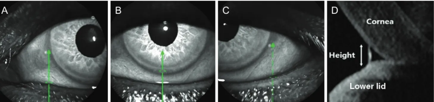
The scans were performed twice for each patient. All im- ages were read by a single ophthalmology specialist. The OCT pattern used to scan the tear meniscus was a 6-mm vertical line centered at three points (medial, central, and lateral). The vertical axis of the corneal center was used as a central point that crossed the lower eyelid. The medial limbus was used for the medial point, and the lateral lim- bus was used for the lateral point. An optical coherence to- mography, vertical line scan cross-sectional image of the tear meniscus was taken with spectral-domain OCT. TMH was measured from the cornea–meniscus junction to the lower eyelid-meniscus junction (Fig. 1A-1D) [11].
Measurement of lower eyelid position
The marginal reflex distance 2 (MRD2) and marginal nose distance (MND) were analyzed with Image J soft- ware (National Institutes of Health, Bethesda, MD, USA) (Fig. 2). Informed consent was obtained from all study participants before taking photos. The MRD2 is the dis- tance between the center of the pupil and lower eyelid margin, and MND is the distance between the lower eyelid margin and horizontal extension line of the nose tip. All patients were scheduled for photographs preoperatively, 1 week postoperatively, and at the last follow-up.
Assessment of subjective symptoms related to tearing Patient symptoms were retrospectively assessed on a scale of -2 to 2 based on the last follow-up. A grade of -2 indicated severe dryness with severe gritty symptoms that
required artificial tear drops, a grade of -1 indicated mild dryness with only gritty symptoms, a grade of 0 indicated no symptoms, a grade of 1 indicated mild tearing only dur- ing windy conditions, and a grade of 2 indicated constant tearing even indoors.
Surgical techniques
All patients underwent lower blepharoplasty by a single surgeon. When planning the lower blepharoplasty, the TMH was measured before surgery, and the surgeon se- lected a transcutaneous or tranconjunctival approach based on TMH and tearing results.
1) Transcutaneous approach
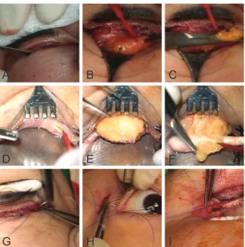
After local anesthesia with 2% lidocaine and 1 : 100,000 epinephrine, a skin incision was created 1 mm below the lower eyelid margin. The skin and orbicularis muscle flap were dissected inferior to the orbital margin (Fig. 3A). The orbital septum was then incised, and protruding orbital fat was removed (Fig. 3B-3F). The surgical procedure was used to correct horizontal lid laxity for 14 eyelids. Orbicu- laris muscle tightening and lateral canthopexy were per- formed in patients with grade 1 laxity (n = 2) (Fig. 3G), lateral canthal suspension was performed in patients with grade 2 laxity (n = 10) (Fig. 3H), and lateral tarsal strip surgery was performed in patients with grade 3 laxity (n = 2) (Fig. 3I). A medial spindle excision procedure was per- formed in patients with severe medial lower lid laxity.
2) Transconjunctival approach
Lidocaine (2%) with 1:100,000 epinephrine was injected into the palpebral conjunctiva. After everting the lower eyelid, a conjunctival incision was made 3 to 4 mm below Fig. 1. Lower eyelid tear meniscus height was measured by anterior segment optical coherence tomography with a Spectralis optical co- herence tomography. (A) Medial point, (B) central point, (C) lateral point, and (D) cross-sectional view. Green arrows indicate the vertical line centered at three points (medial, central, and lateral).
A B C D
Fig. 2. Measurement of clinical parameters of lower eyelid posi- tion. Marginal reflex distance 2 (MRD2) and marginal nose dis- tance (MND) were analyzed with ImageJ software.
347 the tarsal plate (Fig. 3D). An incision to the orbital septum
was made, and orbital fat was removed through the open- ing (Fig. 3E, 3F). This surgical procedure was used to cor- rect the horizontal lid laxity for six eyelids. Orbicularis muscle tightening and lateral canthopexy were performed in patients with grade 1 laxity (n = 2), lateral canthal sus- pension for patients with grade 2 laxity (n = 2), and a later- al tarsal strip for patients with grade 3 laxity (n = 2). A me- dial spindle excision procedure was performed in patients with severe medial lower eyelid laxity.
Statistical analyses
All data are expressed as mean ± standard deviation. Pa- rameters were compared with paired t-tests and Wilcoxon signed-rank tests. IBM SPSS Statistics ver. 21.0 (IBM Corp., Armonk, NY, USA) was used for all statistical analyses. A level of p < 0.05 was considered statistically significant. In- traclass correlation analyses were used to verify the reliabil- ities of the examinations.
Results
Demographic characteristics of the patients
This study included 52 eyes of 26 consecutive patients who underwent lower blepharoplasty. Seventeen patients (65.3%) were female. The mean age at the time of surgery was 62.2 ± 13.1 years. The mean follow-up period after lower blepharoplasty was 10.2 ± 4.5 weeks. Lower blepha- roplasty was performed through a transcutaneous approach for 34 eyes from 17 patients and through a transconjuncti- val approach for 18 eyes from nine patients.
Repeatability and reproducibility of the TMH measure- ments
Intraclass correlation analyses were performed for 10 eyes to verify the examination repeatability and reliability between the two technicians. The correlation coefficients ranged from 0.889 to 0.951.
Measurement of eyelid position
Eyelid position changed significantly after surgery.
MRD2 (5.13 ± 1.21 mm for a transconjunctival approach and 5.29 ± 1.12 mm for a transcutaneous approach) de- creased significantly at 1 week after surgery (4.89 ± 0.81 mm for a transconjunctival approach and 4.97 ± 0.69 mm for a transcutaneous approach) (p = 0.001 and p = 0.001, respectively) and was maintained at the last follow-up (4.90
± 0.79 mm for a transconjunctival approach and 4.95 ± 0.71 mm for a transcutaneous approach) (p = 0.003 and p = 0.005, respectively). The MND (61.71 ± 11.35 mm for a transconjunctival approach and 60.37 ± 13.27 mm for a transcutaneous approach) increased significantly at 1 week after surgery (63.73 ± 15.71 mm for a transconjunctival ap- proach and 63.49 ± 14.63 mm for a transcutaneous ap- proach) (p = 0.001 and p = 0.003, respectively) and at the last follow-up (63.41 ± 15.09 mm for a transconjunctival ap- proach and 62.50 ± 14.65 mm for a transcutaneous ap- proach) (p = 0.005 and p = 0.005, respectively). In summa- ry, MRD2 and MND changed significantly with both approaches (Table 1).
A B C
D E F
G H I
Fig. 3. Surgical procedures used in lower blepharoplasty. (A-C) The transcutaneous approach. (A) An incision was made 1 mm below the lower eyelid margin, (B) followed by incision of the or- bital septum, and (C) removal of orbital fat. (D-F) The transcon- junctival approach. (D) An incision was made at the conjunctiva 3 to 4 mm below the tarsal plate, (E) followed by an incision at the orbital septum, and (F) removal of orbital fat. (G-I) Lateral canthal tightening procedures, (G) orbicularis muscle tightening and lateral canthopexy, (H) lateral canthal suspension, and (I) lateral tarsal strip.
348
Preoperative
Preoperative Preoperative Preoperative
1 wk Last Preoperative
1 wk Last Preoperative
1 wk Last 500
400 300 200 100 0 (µm)
Medial Central Lateral
1 wk Last 1 wk Last 1 wk Last
500 400 300 200 100 0 (µm)
Medial Central Lateral
500 400 300 200 100 0 (µm)
Medial Central Lateral
A B
Preoperative
Preoperative
Preoperative Preoperative Preoperative
Preoperative Preoperative 1 wk Last
Preoperative 1 wk Last
Preoperative
1 wk Last 500
400 300 200 100 0 (µm)
Medial Central Lateral
1 wk Last 1 wk Last 1 wk Last
500 400 300 200 100 0 (µm)
Medial Central Lateral
1 wk Last 1 wk Last 1 wk Last
500 400 300 200 100 0 (µm)
Medial Central Lateral Preoperative
Preoperative
Preoperative Preoperative Preoperative
Preoperative Preoperative 1 wk Last
Preoperative 1 wk Last
Preoperative 1 wk Last 500
400 300 200 100 0 (µm)
Medial Central Lateral
1 wk Last 1 wk Last 1 wk Last
500 400 300 200 100 0 (µm)
Medial Central Lateral
1 wk Last 1 wk Last 1 wk Last
500 400 300 200 100 0 (µm)
Medial Central Lateral
C
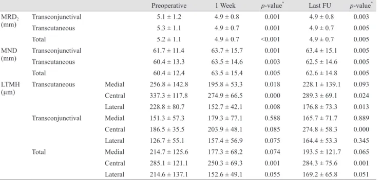
Table 1. Eyelid position and lower tear meniscus heights after lower blepharoplasty
Preoperative 1 Week p-value* Last FU p-value*
MRD2
(mm) Transconjunctival 5.1 ± 1.2 4.9 ± 0.8 0.001 4.9 ± 0.8 0.003
Transcutaneous 5.3 ± 1.1 4.9 ± 0.7 0.001 4.9 ± 0.7 0.005
Total 5.2 ± 1.1 4.9 ± 0.7 <0.001 4.9 ± 0.7 0.005
MND(mm) Transconjunctival 61.7 ± 11.4 63.7 ± 15.7 0.001 63.4 ± 15.1 0.005
Transcutaneous 60.4 ± 13.3 63.5 ± 14.6 0.003 62.5 ± 14.6 0.005
Total 60.4 ± 12.4 63.5 ± 15.4 0.005 62.6 ± 14.8 0.005
LTMH(µm) Transcutaneous Medial 256.8 ± 142.8 195.8 ± 53.3 0.018 228.1 ± 139.1 0.093 Central 337.3 ± 117.8 274.9 ± 66.5 0.000 289.3 ± 69.1 0.024
Lateral 228.8 ± 80.7 152.7 ± 42.1 0.008 176.8 ± 73.3 0.013
Transconjunctival Medial 151.3 ± 57.3 179.3 ± 77.1 0.588 165.7 ± 71.7 0.889
Central 186.5 ± 35.5 203.9 ± 48.1 0.085 274.8 ± 58.3 0.000
Lateral 126.7 ± 55.1 157.4 ± 56.9 0.075 164.4 ± 53.3 0.345
Total Medial 214.7 ± 125.6 177.3 ± 68.2 0.074 193.5 ± 121.7 0.065
Central 285.1 ± 121.1 250.3 ± 69.3 0.001 284.3 ± 75.6 0.001 Lateral 214.6 ± 137.1 152.6 ± 49.1 0.055 169.2 ± 65.8 0.051 Values are presented as the mean ± standard deviation.
MRD2 = marginal reflex distance 2; MND = marginal nose distance; LTMH = lower eyelid tear meniscus height; FU = follow-up.
*Wilcoxon signed-rank test.
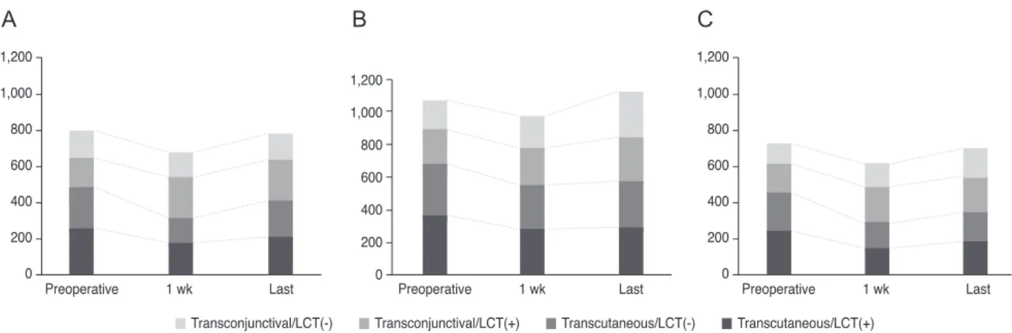
Fig. 4. The changes of lower eyelid tear meniscus height after lower blepharoplasty. (A) Transcutaneous approach (n = 34, Wil- coxon signed-rank test; *p < 0.05), (B) transconjunctival approach (n = 18, Wilcoxon signed-rank test; *p < 0.05), (C) total patients (Wilcoxon signed-rank test, *p < 0.05).
349 Subjective symptoms
The subjective symptom score was -0.3 ± 0.8 for patients with a transcutaneous approach, 0.4 ± 0.7 for patients with a transconjunctival approach, and 0.5 ± 1.2 for all patients.
Tearing scores were associated with changes in TMH fol- lowing lower blepharoplasty. Overall, most patients had a score within +1 or -1, indicating that patient eyes were nei- ther dry nor tearing after lower blepharoplasty.
Changes in lower TMH
After transcutaneous lower blepharoplasty, the mean preoperative TMH at the central point (337.3 ± 117.8 µm) was decreased at 1 week after surgery (274.9 ± 66.5 µm, p
= 0.000) and at the last follow-up (289.3 ± 13.9 µm, p = 0.024) (Fig. 4A and Table 1). The mean preoperative TMH at the medial point (256.8 ± 142.8 µm) was significantly de- creased at 1 week after surgery (195.8 ± 53.3 µm, p = 0.018).
The mean preoperative TMH at the lateral point (228.8 ± 80.7 µm) was significantly decreased at 1 week after sur- gery (152.7 ± 42.2 µm, p = 0.008) and at last follow-up (176.8
± 73.4 µm, p = 0.013).
In patients treated with a transconjunctival approach, the mean preoperative TMH (186.5 ± 35.5 µm) was increased, though not significantly, at 1 week after surgery (203.9 ± 48.1 µm, p = 0.085; Wilcoxon signed-rank test) and was sig- nificantly increased at the last follow-up (274.8 ± 15.6 µm, p
= 0.000; Wilcoxon signed-rank test) (Fig. 4B and Table 1).
The mean preoperative TMH at the central point (285.11
± 121.05 µm) was significantly decreased at 1 week after surgery (250.3 ± 69.2 µm, p = 0.001; Wilcoxon signed-rank test) and recovered at the last follow-up (284.3 ± 16.7 p = 0.001; Wilcoxon signed rank test) (Fig. 4C and Table 1). The mean preoperative TMH at the medial point (214.7 ± 125.6 µm) was significantly reduced at 1 week after blepharo- plasty (177.3 ± 68.3 µm, p = 0.044; Wilcoxon signed-rank test). The mean preoperative TMH at the lateral point (214.6
± 137.0 µm) was significantly reduced at the last follow-up (169.3 ± 65.8 µm, p = 0.044; Wilcoxon signed rank test).
Lower eyelid TMH did not change significantly after a lateral canthal tightening procedure. Preoperative TMH at the central point was significantly higher in patients treated with a lateral canthal tightening procedure through a transconjunctival approach (217.3 ± 30.1 vs. 171.1 ± 27.6 µm, p = 0.011) (Table 2 and Fig. 5A-5C).
Discussion
Eyelid position after lower eyelid surgery is a source of debate. Taban et al. [13] reported that the change in lower eyelid position was not statistically significant after a transconjunctival lower blepharoplasty with or without a skin pinch. Sultan et al. [14] reported that transcutaneous skin-muscle flap lower eyelid surgery increased the dis- tance between the pupil and lower eyelid margin. Segal et al. [15] reported that transconjunctival blepharoplasty with or without skin resurfacing did not induce lid retraction but elevated the lower lid in the majority of patients. Elevating the lower lid can reduce or eliminate inferior scleral show, providing further cosmetic advantage. In a study of Kore- ans, Sung et al. [16] reported that MRD2 decreased and MND increased after lower eyelid blepharoplasty. In the present study, lower eyelid position likely changed MRD2
and MND because horizontal eyelid laxity was corrected in 20 of 52 eyes with concurrent lower blepharoplasty.
The usefulness and reproducibility of TMH measure- ments using OCT have been confirmed in previous studies [11,17]. Previous studies reported normal TMH values that ranged from 194 to 345 µm [11,18-23]. The mean normal TMH at our institution was 290.86 ± 62.20 µm [11].
The present study reports quantitative changes in tear volumes after lower blepharoplasty. The TMH decreased immediately after surgery, then recovered by the last fol- low-up. Dry eye is a common complication after lower blepharoplasty, with an incidence from 0% to 24% [3-5].
Surgically modifying the lower eyelid can alter the lower eyelid position and eyelid blink force. Lower blepharoplas- ty may also injure innervations of the orbicularis muscle, which could affect the blink rate and promote evaporative tear loss [5]. These changes can result in dry eye after low- er blepharoplasty.
The TMH results differed for the two surgical approach- es. The TMH increased with a transconjunctival approach and decreased with a transcutaneous approach. In the early postoperative period, we presumed that a transcutaneous approach excised the orbicularis muscle and weakened the lacrimal pump function, while a transconjunctival ap- proach induced conjunctival irritation and conjunctival chemosis, resulting in tear hypersecretion. However, the mean TMH at 3 months after surgery was similar to nor- mal TMH values for both approaches. The surgical proce- dures in this study were performed through different ap-
Table 2. Lower eyelid tear meniscus height after lower blepharoplasty with and without lateral canthal tightening MedialCentralLateral LCT(+)LCT(-)Totalp-value* LCT(+)LCT(-)Totalp-value* LCT(+)LCT(-)Totalp-value*
Transcutan- eous (µm)
Preoperative259.5 ± 163.8
228.2 ± 111.6
256.8 ± 142.8
0.543363.0 ± 113.8
314.4 ± 119.8
337.3 ± 117.8
0.235241.7 ± 119.9
214.9 ± 88.3
228.8 ± 80.7
0.498 1 Week176.1 ± 65.9
(0.009)†
137.0 ± 3.1
(0.225)†
195.8 ± 53.3
0.138284.0 ± 80.9
(0.001)†
266.8 ± 51.6
(0.017)†
274.9 ± 66.5
0.461152.1 ± 45.4
(0.009)†
137.0 ± 65.1
(0.180)†
152.7 ± 42.1
0.800 Last FU209.2 ±
164.9 (0.155)
†
203.6 ±
104.4 (0,260)
†
228.1 ± 139.1
0.926291.3 ± 10.8
(0.617)†
287.3 ± 16.3
(0.879)†
289.3 ± 69.1
0.474188.2 ± 90.5
(0.173)†
156.2 ± 49.5
(0.022)†
176.8 ± 73.3
0.345
Transconju- nctival (µm)
Preoperative158.0 ± 84.1
148.6 ± 48.6
151.3 ± 57.3
0.844217.3 ± 30.1
171.1 ± 27.6
186.5 ± 35.5
0.011159.7 ± 67.7
109.5 ± 42.7
126.7 ± 55.2
0.271 1 Week226.0 ± 68.1
(0.068)†
137.0 ± 33.0
(0.593)†
179.3 ± 77.1
0.073226.8 ± 58.4
(0.463)†
192.5 ± 39.9
(0.084)†
203.9 ± 48.1
0.234197.0 ± 55.1
(0.068)†
129.5 ± 54.1
(0.144)†
157.4 ± 56.9
0.086 Last FU221.7 ± 28.9
(0.144)†
146.6 ± 66.3
(0.917)†
165.7 ± 71.7
0.060266.6 ± 13.3
(0.028)†
278.9 ± 18.5
(0.002)†
274.8 ± 58.3
0.131200.5 ± 5.6
(0.465)†
152.0 ± 56.7
(0.109)†
164.4 ± 53.3
0.091
Total (µm)
Preoperative238.2 ± 154.5
197.6 ± 9.4
214.7 ± 125.6
0.325323.3 ± 117.8
257.1 ± 117.5
285.1 ± 121.1
0.051222.83 ± 114.5
182.9 ± 90.9
214.6 ± 137.1
0.220 1 Week189.4 ± 68.1
(0.272)†
137.0 ± 31.1
(0.208)†
177.3 ± 68.2
0.170268.4 ± 78.5
(0.005)†
237.1 ± 59.5
(0.271)†
250.3 ± 69.3
0.125164.1 ± 50.5
(0.056)†
131.4 ± 32.1
(0.753)†
152.6 ± 49.1
0.113 Last FU212.4 ±
141.9 (0.532)
†
176.8 ± 90.8
(0.281)†
193.5 ± 121.7
0.403284.6 ± 15.9
(0.249)†
284.0 ± 17.5
(0.030)†
284.3 ± 75.6
0.892192.0 ± 74.2
(0.552)†
154.6 ± 50.5
(0.184)†
169.2 ± 65.8
0.119 LCT = lateral canthal tightening; FU = follow-up. * Mann-Whitney U-test with and without lateral canthal tightening procedures; † Wilcoxon signed-rank test for the difference from preoperative lower eyelid tear meniscus heights.
351 proaches. Correction of lower eyelid laxity depended on its
severity. Improving lower eyelid laxity and recovery of dy- namic blinking are thought to cause efficient tear distribu- tion and drainage. Changes in the TMH were observed at three points in patients treated with a transcutaneous ap- proach, but only at the central point for patients treated with a transconjunctival approach. The results suggest that a more extensive change in periorbital anatomy with a transcutaneous approach changed tear level at all points in the lower eyelid.
We also evaluated the TMH at the medial and lateral points of the lower eyelid. The medial and lateral TMH changed significantly only in patients treated with transcu- taneous lower blepharoplasty. We investigated TMH resto- ration according to the presence of a lateral canthal tight- ening procedure and showed that TMH was restored regardless of this procedure. The results suggest that the most suitable technique was sufficient to improve the TMH. Previous studies by Shao et al. [6] reported that transcutaneous lower blepharoplasty affected the ocular surface and tear fluid, leading to dryness, teary eyes, and chemosis, and the symptoms resolved within 3 months.
Unlike our study, TMH did not differ statistically before and three months after surgery. However, that study had some differences from the present study. The authors only studied the transcutaneous approach and did not consider eyelid laxity when planning the lower blepharoplasty pro- cedure. In addition, the TMH was only measured at the central point. The patients were younger (50.53 ± 4.80 years) than in our study (62.2 ± 13.1 years), and the postop- erative follow-up was 3 months in that study compared to
10.2 ± 4.5 weeks in the present study.
The retrospective review of lower blepharoplasty was a limitation of the present study. We, therefore, could not di- rectly determine the effects of each surgical technique on tear volume changes. Because of the retrospective design, the preoperative TMH differed between groups. Future prospective studies with larger sample sizes and longer fol- low-up periods may help to determine the relationship be- tween blepharoplasty approach and TMH. Also, postopera- tive TMH was only evaluated in two dimensions in our present study. Future prospective studies with 3-dimen- sional dynamic analysis may help to identify the relation- ship between blepharoplasty and tear film stability. Despite this limitation, we think that a customized lower blepharo- plasty approach is effective in improving the ocular sur- face. Although we tried to exclude patients with dry eye disease, we did not examine all patients for this disorder. In addition, although we measured tear volume changes after lower blepharoplasty, tear film stability may have been as- sociated with other factors. For example, evaluating blink response is also important in characterizing orbicularis muscle function after lower blepharoplasty.
In conclusion, transcutaneous or transconjunctival lower blepharoplasty, depending on patient characteristics, is ef- fective in normalizing TMH and improving cosmetic out- comes. Therefore, eyelid laxity and the tear film should be evaluated before lower blepharoplasty, and tear film prop- erties should be considered when planning surgery.
Preoperative 1 wk Last
1,200 1,000 800 600 400 200
0 Preoperative 1 wk Last
1,200 1,000 800 600 400 200
Preoperative 1 wk Last 0
1,200 1,000 800 600 400 200 0
▒ Transconjunctival/LCT(-) ▒ Transconjunctival/LCT(+) ▒ Transcutaneous/LCT(-) ▒ Transcutaneous/LCT(+)
Fig. 5. Lower eyelid tear meniscus height at three points (A, medial; B, central; C, lateral) after lower blepharoplasty with all lateral can- thal tightening (LCT) procedures.
A B C
Conflict of Interest
No potential conflict of interest relevant to this article was reported.
References
1. Buchanan DR, Wulc AE. Contemporary thoughts on lower eyelid/midface aging. Clin Plast Surg 2015;42:1-15.
2. Tse DT, Erickson BP, Tse BC. The BLICK mnemonic for clinical-anatomical assessment of patients with epiphora.
Ophthalmic Plast Reconstr Surg 2014;30:450-8.
3. Prischmann J, Sufyan A, Ting JY, et al. Dry eye symptoms and chemosis following blepharoplasty: a 10-year retrospec- tive review of 892 cases in a single-surgeon series. JAMA Facial Plast Surg 2013;15:39-46.
4. Honrado CP, Pastorek NJ. Long-term results of lower-lid suspension blepharoplasty: a 30-year experience. Arch Fa- cial Plast Surg 2004;6:150-4.
5. Hamawy AH, Farkas JP, Fagien S, Rohrich RJ. Preventing and managing dry eyes after periorbital surgery: a retro- spective review. Plast Reconstr Surg 2009;123:353-9.
6. Shao C, Fu Y, Lu L, et al. Dynamic changes of tear fluid after cosmetic transcutaneous lower blepharoplasty mea- sured by optical coherence tomography. Am J Ophthalmol 2014;158:55-63.
7. Huang D, Swanson EA, Lin CP, et al. Optical coherence to- mography. Science 1991;254:1178-81.
8. Wang J, Aquavella J, Palakuru J, et al. Relationships between central tear film thickness and tear menisci of the upper and lower eyelids. Invest Ophthalmol Vis Sci 2006;47:4349-55.
9. Wang Y, Zhuang H, Xu J, et al. Dynamic changes in the lower tear meniscus after instillation of artificial tears. Cor- nea 2010;29:404-8.
10. Mainstone JC, Bruce AS, Golding TR. Tear meniscus measurement in the diagnosis of dry eye. Curr Eye Res 1996;15:653-61.
11. Park DI, Lew H, Lee SY. Tear meniscus measurement in nasolacrimal duct obstruction patients with Fourier-domain
optical coherence tomography: novel three-point capture method. Acta Ophthalmol 2012;90:783-7.
12. Robinson FO, Collin RO. Ectropion. In: Yanoff M, Duker JS, editors. Ophthalmology. 2nd ed. Edinburgh: Mosby;
2006. p. 676-83.
13. Taban M, Taban M, Perry JD. Lower eyelid position after transconjunctival lower blepharoplasty with versus without a skin pinch. Ophthalmic Plast Reconstr Surg 2008;24:7-9.
14. Sultan B, Genther DJ, Perkins SW. Measurement of change in lower eyelid position in patients undergoing transcuta- neous skin-muscle flap lower eyelid blepharoplasty. JAMA Facial Plast Surg 2016;18:429-35.
15. Segal KL, Patel P, Levine B, et al. The effect of transcon- junctival blepharoplasty on margin reflex distance 2. Aes- thetic Plast Surg 2016;40:13-8.
16. Sung YJ, Park JS, Lew H. Changes in lower eyelid positions after individualized lower blepharoplasty. J Korean Oph- thalmol Soc 2015;56:1831-9.
17. Zhou S, Li Y, Lu AT, et al. Reproducibility of tear meniscus measurement by Fourier-domain optical coherence to- mography: a pilot study. Ophthalmic Surg Lasers Imaging 2009;40:442-7.
18. Palakuru JR, Wang J, Aquavella JV. Effect of blinking on tear dynamics. Invest Ophthalmol Vis Sci 2007;48:3032-7.
19. Wang J, Aquavella J, Palakuru J, Chung S. Repeated mea- surements of dynamic tear distribution on the ocular sur- face after instillation of artificial tears. Invest Ophthalmol Vis Sci 2006;47:3325-9.
20. Johnson ME, Murphy PJ. The agreement and repeatability of tear meniscus height measurement methods. Optom Vis Sci 2005;82:1030-7.
21. Shen M, Li J, Wang J, et al. Upper and lower tear menis- ci in the diagnosis of dry eye. Invest Ophthalmol Vis Sci 2009;50:2722-6.
22. Bitton E, Keech A, Simpson T, Jones L. Variability of the analysis of the tear meniscus height by optical coherence tomography. Optom Vis Sci 2007;84:903-8.
23. Cui L, Shen M, Wang J, et al. Age-related changes in tear menisci imaged by optical coherence tomography. Optom Vis Sci 2011;88:1214-9.