Analysis of Tear Meniscus using Optical Coherence Tomography after Cataract Surgery
Chang Won Park
1and Hyojin Kim
2,3,
*1
Dept. of Optometry, Baekseok Culture University, Cheonan 31065, Korea
2
Dept. of Visual Optics, Division of Health Science, Baekseok University, Cheonan 31065, Korea
3
Dept. of Visual Optics, Graduate School of Health and Welfare, Baekseok University, Seoul 06695, Korea (Received June 17, 2018: Revised August 30, 2018: Accepted October 8, 2018)
···
Purpose: To investigate the effect of dry eye after cataract surgery, we analyzed the tear meniscus using anterior segment optical coherence tomography (AS-OCT) and compared the results to conventional dry eye tests (TBUT and Schirmer). Methods: We included 125 eyes that were to undergo cataract surgery and 104 eyes from normal subjects.
The height, depth, and area of the tear meniscus were measured using AS-OCT. These tear meniscus variables, as well as the conventional dry eye tests, were compared between the preoperative cataract and normal groups. The same tests were compared in patients with cataract before and 2 months after surgery. Subjective dry eye symptoms were evaluated using the McMonnies questionnaire before and after cataract surgery. Results: The tear meniscus variables and conventional dry eye tests did not differ significantly between the preoperative cataract and normal groups, but the tear meniscus area (TMA) reduced from 0.037±0.015 mm² in the preoperative cataract group to 0.015±0.010 mm² in the postoperative cataract group (p=0.001). The McMonnies questionnaire score rose from 10.28±3.93 to 13.32±4.82 points after cataract surgery (p=0.005). Conclusions: The TMA, as measured using AS-OCT, differed significantly between the pre- and postoperative cataract groups, suggesting that the parameter could be used to evaluate dry eye in patients with cataract.
Key words: Dry eye, Optical coherence tomography, Tear meniscus, Schirmer's test, Tear film break-up time
···
서 론
안구표면의 최외층인 눈물층은 점액층, 수성층, 지방층 으로 구성되어 있다.
[1]이러한 눈물층의 구성성분에 불균 형 또는 이상이 나타날 경우 눈물이 과도하게 증발하여 안구표면이 손상된다. 이로 인해 이물감, 충혈, 통증, 시력 저하 등의 증상을 느끼게 되며 이러한 증상이 심한 경우 를 안구건조증이라고 한다.
[2]안구건조증이 있으면 정상안 에 비해 눈물막의 불안정성이 심하며 눈물막의 파괴시간 이 빠르기 때문에 눈에 건조감 뿐만 아니라 불편함과 시 력의 질에도 영향을 미친다.
[3,4]안구건조증의 원인으로는 눈물막과 눈물샘의 문제, 안검의 형태 이상, 콘택트렌즈의 장시간 착용과 잘못된 안약 사용 등이 있다.
[5-7]이외에도 안구표면에 영향을 주는 익상편 제거수술, 초음파 유화 흡 인술, 레이저굴절교정수술 등의 안과적 수술도 안구건조 증의 발생과 관련이 있다.
[8-11]일반적으로 안구건조증의 진단에는 눈물층의 분비와
증발을 측정하는 쉬르머검사(Schirmer's test)와 눈물막 파 괴시간(tear film break up time, TBUT), 그리고 자각적 증상을 평가하는 설문지가 사용된다. 이외에도 눈물띠 (tear meniscus)의 높이, 각막표면염색, 결막압흔세포검사 등이 사용된다.
[12]그러나 이들 방법은 검사용지의 침습 적 방법으로 인한 재현성과 오차 발생, 검사자의 주관성 개입으로 객관적으로 건성안을 평가하고자 하는 시도
[13-18]가 지속적으로 있었다. 최근 빛간섭단층촬영장치 (optical coherence tomography, OCT)를 이용하여 전체 눈물부피의 양을 객관적이며 비침습적으로 반복하여 측 정하는 검사법이 개발되어 눈물띠의 높이, 넓이, 부피를 분석하여 안구건조증의 진단에 사용될 수 있음이 보고
되었다.
[12,19]눈물띠는 눈 깜박임 이후 생성된 눈물막과 위, 아래 눈 꺼풀 가장자리가 만나는 곳에 있는 삼각형 모양의 오목한 저장소를 말한다. 이것은 안구표면에 노출된 전체 눈물양 의 약 75%에서 90%를 포함하기 때문에
[20,21]노출된 안구
<초청논문>
*Corresponding author: Hyojin Kim, TEL: +82-41-550-2841, E-mail: [email protected]
표면의 눈물을 반영한다고 볼 수 있다. 따라서 눈물띠 부 피의 수치는 전체 눈물 부피의 차이로 간접적으로 생각될 수 있다.
[22]최근 OCT를 이용하여 건성안 환자에서 정상 인보다 눈물띠가 감소되었다는 결과가 알려졌다.
[19]또한 전안부 OCT는 비침습적이며 빠르고 해상도가 높은 안구 표면 영상을 제공한다. 따라서 전안부 OCT는 피검사자의 안구표면에 대한 정보를 자세하고 그대로 얻을 수 있다는 장점이 있다.
[23-25]그러나 지금까지 비침습적이며 객관적인 검사법을 이용 하여 안구건조증을 검사한 이전 연구들은 주로 20에서 30 대의 젊은 연령층을 대상으로 진행되어
[18,26]백내장과 같 은 안과적수술로 인한 안구건조증의 연구는 부족한 실정 이다. 따라서, 본 연구에서는 영상 획득이 빠르며 해상도 가 높은 AS-OCT를 이용하여 50~89세의 연령을 대상으로 백내장수술 후에 발생하는 건성안을 평가하였고, 더불어 기존에 많이 사용되던 눈물막 파괴시간, 쉬르머검사, 그리 고 자각적 증상과의 비교를 시행하였다.
대상 및 방법
1. 대상
본 연구는 50~89세까지의 백내장수술을 받은 97명 125 안의 환자군과 89명 104안의 백내장이 없는 대조군을 대 상으로 하였다(Table 1). 환자군은 병원에서 동일한 처방 에 의해 초음파유화술과 인공수정체 삽입술을 받은 백내 장환자만 포함하였다. 각막질환이나 각막수술을 포함하여 기타 안과적 수술을 받은 자, 콘택트렌즈를 사용하는 자, 눈물관 이상자 등 눈물막에 영향을 끼칠 수 있는 자는 대 상자에서 제외하였다.
2. 방법
백내장 환자군의 수술 전과 후, 그리고 대조군에 대하여 눈물막 파괴시간, 쉬르머검사, AS-OCT(Visante™ Carl Zeiss Meditec, Dublin, CA)를 이용하여 건성안을 측정하 였다. 백내장 수술 후에 모든 건성안 검사는 술 후 안정기 인 2달째 진행하였다. 또한 백내장수술 전과 후에 설문지 를 이용하여 환자의 자각증상을 조사하였고, 구체적인 검 사도구는 다음과 같다.
1) 건성안 검사
눈물막 파괴시간은 눈물막의 뮤신의 부족으로 초래되는 눈물의 증발 정도와 관련이 있는 검사로써 세극등현미경 (VS/SL-7F, TOPCON, Japan) 의 코발트블루 조명에서 눈물 막층이 균열에 의해 마른점(dry spot)의 형태가 최초로 발 생하는 시간을 측정하였다.
[27]쉬르머검사는 검사지(Color BarTM, Eagle Vision, USA)를 하결막낭에 삽입하여 눈물 에 의해 적셔지는 부위를 측정하여 10 mm/5 min 이상을 정상으로 간주하는 방법을 사용하였고, 국소점안 마취제 를 사용하지 않은 상태에서 시행하였다.
[2,28]정확한 측정 값을 얻기 위해 검사 전 안검에 자극을 줄 수 있는 행동 을 자제시켰고 특히, 안신경이 많이 분포해 있는 중심부 각막에 검사지가 닿지 않도록 주의하였으며 심한 각막자 극으로 인해 반사눈물 분비가 많은 경우는 실험에서 제외 하였다.
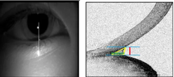
AS-OCT를 이용하여 눈물띠를 측정하였고, 눈물띠는 높 이(tear meniscus height, TMH), 깊이 (tear meniscus depth, TMD), 그리고 넓이(tear meniscus area, TMA)로 구분하였 다. Fig. 1은 눈물띠를 측정한 영상을 보여준다. 검사는 암 실에서 눈을 자연스럽게 깜박인 다음 3 sec 후에 촬영하
Fig. 1. Images of the lower tear meniscus and the position of the eye. Left: The white line shows the site of image capture and the
scanning direction. Right: The red line represents the tear meniscus height and the area within the yellow triangle
represents the tear meniscus area. The green line represents the tear meniscus depth.
였고, 검사실의 온습도가 각각 20~25
oC와 30~40%가 되도 록 유지하였다. 대상자들은 촬영동안 정면을 주시하도록 하였다. 각막의 6시 방향을 수직으로 지나는 6 mm 선으 로 하안검의 눈물띠를 스캔하였고, 스캔된 영상은 소프트 웨어에 의해 자동적으로 계산되었다.
마지막으로 맥모니설문지(Mcmonnies questionnaire)를 이용하여 안구건조증의 증상을 점수화하였다.
[29,30]이 설 문지는 안구건조증 증상의 위험인자에 관한 12가지 질문 으로 평가를 하는데 점수가 높을수록 건성안 증상이 심각 한 것을 의미하게 된다. Mcmonnies questionnaire의 질문 에 대한 반응은 다양한 타입과 점수로 표현된다. 예를 들 면 질문1은 대답의 방식은 3가지 예(2점), 아니오(0점), 불 확실한(1점)으로 구성된다. 반면에 질문9의 대답은 4가지 로 이루어져있으며 절대 아닌(0점), 때때로(1점), 자주(2점), 지속적으로(3점)으로 계산된다.
2) 자료분석
통계학적 분석은 SPSS 18.0 version 18.0 (SPSS Inc., Chicago, IL) 을 사용하였고, 안구건조증 평가의 각 파라미 터들간의 결과를 독립표본 t-test와 pearson 상관분석하여 p<0.05 인 경우를 통계적으로 유의한 것으로 간주하였다.
결과 및 고찰
1. 대상자의 일반적인 특성
환자군의 평균 연령은 62.61±7.15세로 남성이 43명, 여 성이 54명이었다. 대조군은 평균 65.47±6.52세로 남성과 여성 각각 44명과 45명이었고, 두 군 간에 통계적으로 연 령과 성별은 유의한 차이가 없었다(Table 1).
2. 환자군과 대조군의 눈물막 비교
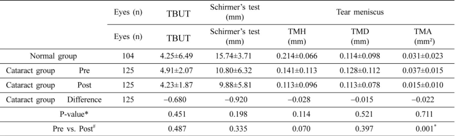
환자군에서 백내장수술 전에 눈물막 파괴시간과 쉬르머 검사 결과는 각각 4.91±2.07 sec와 10.80±6.32 mm로 대조 군의 4.25±6.49 sec, 15.74±3.71 mm와 통계적으로 유의한 차 이가 없었다. 눈물띠의 높이, 깊이, 넓이 또한 백내장 수술 전 에 각각 0.141±0.113 mm, 0.128±0.112 mm, 0.037±0.015 mm² 로 대조군의 0.214±0.066 mm, 0.114±0.098 mm, 0.031±0.023 mm²와 유의한 차이가 없었다(Table 2).
3. 백내장수술 전후의 눈물막 비교
환자군에서 백내장수술 후의 눈물막 파괴시간과 쉬르머 검사는 각각 4.23±1.87초와 9.88±5.81 mm로 수술 전과 차 이가 없었다. 빛간섭단층촬영 영상에 의한 평가에서 눈물
Table 1. Subjects’ demographics
Characteristics Normal group (n=104) Cataract group Pre (n=125)
Characteristics Normal group (n=104) Cataract group Post (n=125)
Eyes (n) 50
–59 y
ears (45) 50
–59 y
ears (53)
60
–69 y
ears (41) 60
–69 y
ears (45)
70
–79 y
ears (18) 70
–79 y
ears (27)
Age (years) 65.47±6.52 62.61±7.15
Sex (M/F) 44/45 43/54
Table 2. Comparison of TBUT, Schirmer’s test, and tear meniscus variables between the preoperative cataract and normal groups Eyes (n) TBUT Schirmer’s test
(mm) Tear meniscus
Eyes (n) TBUT Schirmer’s test
(mm) TMH
(mm) TMD
(mm) TMA
(mm²) Normal group 104 4.25±6.49 15.74±3.71 0.214±0.066 0.114±0.098 0.031±0.023 Cataract group Pre 125 4.91±2.07 10.80±6.32 0.141±0.113 0.128±0.112 0.037±0.015 Cataract group Post 125 4.23±1.87 9.88±5.81 0.113±0.096 0.113±0.078 0.015±0.010
Cataract group Difference 125 −0.680 −0.920 −0.028 −0.015 −0.022
P-value* 0.451 0.198 0.114 0.521 0.711
Pre vs. Post
#0.487 0.335 0.070 0.397 0.001
*Values are presented as mean ± SD
TBUT: tear film break-up time, TMH: tear meniscus height, TMD: tear meniscus depth, TMA: tear meniscus area
*Unpaired t-test between preoperative cataract and normal groups
#
Unpaired t-test between pre- and postoperative cataract groups
띠의 높이는 0.113±0.096 mm로 평균 0.028 mm 감소하였 고, 깊이는 0.113±0.078 mm로 평균 0.015 mm 감소하였으 나 통계적인 차이는 없었다. 그러나 눈물띠의 넓이는 수술 후에 0.015±0.010 mm²로 수술 전에 비해 평균 0.022 mm² 의 감소를 보였다(p=0.001)(Table 2).
4. 백내장수술 전후의 자각적 증상 비교
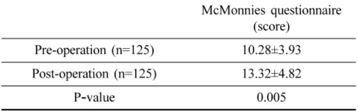
Mcmonnies questionnaire 를 이용하여 조사한 자각적 증 상에 대한 점수는 백내장수술 전에 10.28±3.93점에서 수 술 후에 13.32±4.82점으로 안구건조증에 대한 증상의 심 한 정도가 높아졌다(p=0.005)(Table 3).
5. 백내장수술 후 눈물막과 자각적 증상과의 상관관계 백내장수술 후에 환자의 눈물띠와 자각적 증상은 눈물 띠의 높이(r
2= −0.334, p=0.030)와 넓이(r
2= −0.523, p=0.010) 에서 자각증상의 정도와 통계적으로 유의한 음의 상관관 계가 있었다. 또한 눈물막 파괴시간이 짧고(r
2= −0.260, p=0.167), 쉬르머검사(r
2= −0.661, p=0.001) 결과가 짧을수 록 자각증상이 심했다(Table 4).
안구건조증은 눈물량의 부족 또는 과도한 증발로 인해 안구표면과 눈물층의 불안정성을 야기하는 복합적인 질환 으로 현대인에게 흔하게 볼 수 있는 안과질환 중 하나이
다. 눈물의 충분한 분비와 균일한 눈물층의 유지는 건강한 시생활을 유지하는데 필수적이다. 일반적으로 안과에서 안 구건조증을 진단하는 방법으로 쉬르머검사, 눈물막 파괴시 간, McMonnies questionnaire 등이 사용하고 있다.
[31,32]그러 나 이 세 가지 방법은 오랫동안 사용되어왔지만 검사자의 주관이 개입되고 침습적인 검사법에 따른 한계점이 있다.
쉬르머검사는 피검자의 하결막낭에 쉬르머 검사지를 삽입 하여 시행되는데 검사지 스트립 말단의 안신경의 자극으 로 인해 반사눈물량이 과도하게 생성되는 단점이 존재한 다. 또한 이들 안구건조증을 평가하는 방법은 주관적인 판 단을 통제하기 어렵다는 보고가 있었다.
[3,33,34]따라서 안구건조증의 진단을 객관화하는 과학적인 평가 법에 대한 연구가 시행되어 왔다. Monts-Mic 등은
[35]수차 계를 이용하여 눈물층의 역동적인 상태를 광학적으로 평 가하였고, Park 등
[26]은 눈물층의 변화, 불안정성에 대해 double-pass retinal imaging technique을 이용하여 건성안 의 정도를 정량화된 값으로 나타내었다. 본 연구에서는 최 근에 개발된 AS-OCT를 이용하여 눈물띠를 비침습적으로 측정하여 수치화 하였다. 눈물띠의 높이 및 넓이는 측정기 기에 내장된 소프트웨어에 의해 자동으로 계산된다. 이러 한 측정방법은 이전 연구를 통해 안구건조증을 진단하는 데 높은 진단력을 가졌다고 보고되었다.
[36,37]또 다른 이전 연구에서는 안과적수술 후에 인공눈물의 사용이 눈물층의 안정에 영향을 미쳤는지 보기 위해서 OCT를 활용하여 눈 물띠를 객관적으로 측정하는데 사용하였다.
[12]이렇듯 OCT를 이용한 눈물띠의 측정은 충분한 재현성과 정확도 를 가져 임상에서 안구건조증의 진단에 도움을 줄 수 있 다는 주장이 제기되었다.
[34,38,39]본 연구에서 백내장수술 전의 환자군과 대조군의 눈물 띠는 유의한 차이가 없었고, 백내장수술 후에는 수술 전에 비해 전반적으로 눈물띠의 높이, 깊이, 넓이 모두 감소하 는 경향을 보였다. Kim 등
[12]의 연구에서는 결막의 절개가 반드시 필요한 사시수술 환자를 대상으로 OCT를 이용하 여 눈물층의 불안전성을 관찰하였다. Cho 등
[39]의 연구에 서는 눈물막 파괴검사, 쉬르머검사, 눈물띠를 측정하여 백 내장수술 후에 안구건조증의 증상이 더욱 심해졌다고 보 고하였다. 그러나 이들 연구에서는 눈물띠를 슬릿램프로 만 측정하여 기존의 한계점을 보완하지 못 하였다. 또한 본 연구에서는 환자의 자각적 증상을 평가하는 Mcmonnies questionnaire와 눈물띠 결과에 대해 상관성을 추가적으로 분석하였는데 눈물띠의 높이, 넓이의 결과와 Mcmonnies questionnaire의 결과가 유의하게 음의 상관성을 띠었다 (r
2= −0.334, r
2= −0.523). 이는 실제로 환자가 호소하는 증 상의 정도가 각결막사이의 눈물띠 감소와 관련이 있음을 알 수 있었고 백내장 수술 후 환자들은 건조감을 포함하 Table 3. Comparison of subjective symptoms in the pre- and
postoperative cataract groups using the McMonnies questionnaire
McMonnies questionnaire
(score) Pre-operation (n=125) 10.28±3.93 Post-operation (n=125) 13.32±4.82
P
-value 0.005
Values are presented as mean±SD
Table 4. Correlations between TBUT, Schirmer‘s test, tear meniscus variables, and subjective symptoms using the McMonnies questionnaire in cataract surgery patients
McMonnies questionnaire
score(n=125)
r
2P
-value
TBUT −0.260 0.167
Schirmer’s test −0.661 0.001
TMH −0.334 0.030
TMD −0.208 0.096
TMA −0.523 0.010
TBUT: tear film break-up time, TMH: tear meniscus height,
TMD: tear meniscus depth, TMA: tear meniscus area
여 눈물량의 부족에 대한 뻑뻑함 증상, 충혈되는 증상을 대개 호소하였다. 다만 눈물띠의 깊이는 Mcmonnies questionnaire 결과와 관련성이 적고, 쉬르머검사가 눈물띠 의 넓이보다 자각증상과 더 높은 상관성을 보였다. 이것은 눈물띠의 깊이는 눈물막 높이, 면적에 비해서 개인의 해부 학적인 안검의 구조 및 형태에 영향을 받기 때문으로 사 료된다. 또한 눈물띠 넓이의 측정간극은 쉬르머검사 결과 치의 간극에 비해 크지 않기 때문으로 생각된다. 이렇듯 백내장수술 후에 안구건조증 증상을 호소하는 것은 안과 적수술로 인하여 각막의 지각이 감소한 것과 관련이 있다 고 생각되는데, 각막의 지각이 저하될 경우에 눈물 분비량 이 감소하고 순목반사가 감소하여 각막손상이 증가하고 상처 치유에도 영향을 미친다고 알려져 있다.
[40]Donnenfeld 등
[38]은 라식수술 환자를 대상으로 각막지각 의 변화를 관찰하였는데 라식 수술은 각막지각 저하를 발 생시키고 안구의 건조한 증상을 악화시킨다고 보고 하였 다. 또한 Roberts 등
[41]은 백내장수술 후에 유발되는 안구 건조증에 수술 시 각막의 절개방향이 영향을 미친다고 보 고 하였다. 이것은 안신경 세포의 손상과 밀접한 관련이 있다고 생각되는데 향후 연구에서는 단기적인 눈물막의 변화를 포함하여 각결막을 절개하는 여러 가지 안과적수 술에 따른 각막의 변화를 포함하여 객관적인 눈물막의 평 가가 필요할 것으로 보인다.
결 론
백내장수술 후에 눈물막 파괴검사, 쉬르머검사, 눈물띠 의 높이와 깊이는 통계적으로 유의한 차이가 없었다. 그러 나 빛간섭단층촬영을 이용한 눈물띠의 넓이는 백내장수술 전에 0.037±0.015 mm²에서 수술 후에 0.015±0.010 mm²로 유의하게 감소되어 향후 백내장수술에 따른 건성안 평가 에 유용하게 쓰일 수 있을 것이라 생각된다. 또한 McMonnies questionnaire를 이용하여 조사한 환자의 자각 증상도 수술 전에 10.28±3.93점에서 수술 후에 13.32±4.82 점으로 통계적으로 유의한 차이를 보였다. 본 연구는 국내 에서 AS-OCT를 이용하여 백내장수술에 따른 환자의 눈 물띠 분석을 최초로 시도한 논문으로 기존의 건성안 검사 법의 제한점(검사자의 주관성 개입, 침습적인 방법으로 인 한 반사눈물 영향 등)에서 자유롭고 검사자가 바뀌더라도 AS-OCT 검사장비에서 일정한 눈물띠 결과를 얻을 수 있 기에 상대적으로 객관적인 결과를 얻을 수 있다는 장점이 있다. 또한 AS-OCT를 이용한 눈물띠 검사법은 짧은 시간 내에 정량화 분석이 가능하였고, 침습적 검사방법에 대해 거부감이 심한 환자들에게도 대체 검사수단으로 적용할 수 있어 유용하게 활용될 것으로 기대된다.
감사의 글
이 논문은 2018년도 백석대학교 대학연구비에 의해 수 행되었습니다.
REFERENCES
[1] Kim JS, Shin JA, Lee OJ. Textbook of ocular anatomy and physiology, 1st Ed. Seoul: Chungkumunhwasa, 2005;
240-243.
[2] Lemp MA, Baudouin C, Baum J, Dogru M, Foulks GN, Kinoshita S et al. The definition and classification of dry eye disease: report of the definition and classification sub- committee of the International Dry Eye Workshop (2007).
Ocul Surf. 2007;5(2):75-92.
[3] Pflugfelder SC, Tseng SC, Sanabria O, Kell H, Garcia CG, Felix C et al. Evaluation of subjective assessments and objective diagnostic tests for diagnosing tear-film dis- orders known to cause ocular irritation. Cornea. 1998;
17(1):38-56.
[4] Byun YS, Jeon EJ, Chung SK. Clinical effect of cyclo- sporine 0.05% eye drops in dry eye syndrome patients. J Korean Ophthalmol Soc. 2008;49(10):1583-1588.
[5] Nathan E. Contact lens complications, 1st Ed. Seoul:
Elsevier Korea, 2008;54-69.
[6] Chung SH, Na KS, Kwon HG, Lee HS, Kim SY, Kim EC et al. Levels of severity in dry eye syndrome according to Delphi panel classification. J Korean Ophthalmol Soc.
2010;51(9):1179-1183.
[7] Mantelli F, Massaro-Giordano M, Macci I. Lambiase A, Bonini S. The cellular mechanisms of dry eye: from pathogenesis to treatment. J Cell Physiol. 2013;228(12):
2253-2256.
[8] Sullivan BD, Whitmer D, Nichols KK, Tomlinson A, Foulks GN, Geerling G et al. An objective approach to dry eye disease severity. Invest Ophthalmol Vis Sci. 2010;
51(12):6125-6130.
[9] Cher I. Fluids of the ocular surface: concepts, functions and physics. Clin Exp Ophthalmol. 2012;40(6):634-643.
[10] Nichols KK, Foulks GN, Bron AJ, Glasgow BJ, Dogru M, Tsubota K et al. The international workshop on mei- bomian gland dysfunction: executive summary. Invest Ophthalmol Vis Sci. 2011;52(4):1922-1929.
[11] Konomi K, Chen LL, Tarko RS, Scally A, Schaumberg DA, Azar D et al. Preoperative characteristics and a potential mechanism of chronic dry eye after LASIK. Invest Oph- thalmol Vis Sci. 2008;49(1):168-174.
[12] Kim JH, Kim CR, Kim SJ, Chung IY, Seo SW, Yoo JM.
Analysis of tear meniscus change after strabismus surgery using optical coherence tomography. J Korean Ophthal- mol Soc. 2016;57(12):1932-1938.
[13] Benito A, Pérez GM, Mirabet S, Vilaseca M, Pujol J,
Marín JM et al. Objective optical assessment of tear-film
quality dynamics in normal and mildly symptomatic dry eyes. J Cataract Refract Surg. 2011;37(8):1481-1487.
[14] Thibos LN, Hong X. Clinical applications of the Shack- Hartmann aberrometer. Optom Vis Sci. 1999;76(12):817- 825.
[15] Koh S, Maeda N, Kuroda T, Hori Y, Watanabe H, Fujik- ado T et al. Effect of tear film break-up on higher-order aberra- tions measured with wavefront sensor. Am J Ophthalmol.
2002;134(1):115-117.
[16] Maeda N. Clinical applications of wavefront aberrometry - a review. Clin Exp Ophthalmol. 2009;37(1):118-129.
[17] Choi SH, Shin YI. Changes in higher order aberration according to tear-film instability analyzed by continuous measurement using wavefront. J Korean Ophthalmol Soc.
2012;53(8):1076-1080.
[18] Lee YJ, Kim JM, Lee KJ. Analysis of accuracy of tear breakup time (TBUT) and non-invasive TBUT. Korean J Vis Sci. 2017;19(3):257-266.
[19] Jung NY, Baek JW, Shin SJ, Chung SK. Tear meniscus evaluation using optical coherence tomography in dry eye patients. J Korean Ophthalmol Soc. 2015;56(3):323-330.
[20] Tiffany JM. Surface tension in tears. Arch Soc Esp Oftal- mol. 2006;81(7):363-366.
[21] Yokoi N, Bron AJ, Tiffany JM, Maruyama K, Komuro A, Kinoshita S. Relationship between tear volume and tear meniscus curvature. Arch Ophthalmol. 2004;122(9):1265- 1269.
[22] Mainstone JC, Bruce AS, Golding TR. Tear meniscus measurement in the diagnosis of dry eye. Curr Eye Res.
1996;15(6):653-661.
[23] Wang Y, Zhuang H, Xu J, Wang X, Jiang C, Sun X. Dynamic change in the lower tear meniscus after instillation of arti- ficial tears. Cornea. 2010;29(4):404-408.
[24] Roh JH, Chi MJ. Efficacy of dye disappearance test and tear meniscus height in diagnosis and postoperative assessment of nasolacrimal duct obstruction. Acta Ophthalmol. 2010;
88(3):e73-e77.
[25] Chylack LT Jr, Wolfe JK, Singer DM, Leske MC, Bulli- more MA, Bailey IL et al. The lens opacities classifica- tion system III: the longitudinal study of cataract study group. Arch Ophthalmol. 1993;111(6):831-836.
[26] Park CW, Kim H. Comparison among the four examina- tion methods for dry eye (OQAS test, TBUT, Schirmer test, McMonnies test). J Korean Ophthalmic Opt Soc. 2015;
20(4):519-526.
[27 Calonge M, Diebold Y, Sáez V, Enríquez de Salamanca A, García-Vázquez C, Corrales RM et al. Impression cytology of the ocular surface: a review. Exp Eye Res.
2004;78(3):457-472.
[28] Albarrán C, Pons AM, Lorente A, Montés R, Artigas JM.
Influence of the tear film on optical quality of the eye.
Cont Lens Anterior Eye. 1997;20(4):129-135.
[29] Simpson TL, Situ P, Jones LW, Fonn D. Dry eye symp- toms assessed by four questionnaires. Optom Vis Sci.
2008;85(8):692-699.
[30] Begley CG, Caffery B, Chalmers RL, Mitchell GL. Use of the dry eye questionnaire to measure symptoms of ocular irritation in patients with aqueous tear deficient dry eye.
Cornea. 2002;21(7):664-670.
[31] Halberg GP, Berens C. Standardized Schirmer tear test kit. Am J Ophthalmol. 1961;51(5):840-842.
[32] Maurice D. The Charles Prentice award lecture 1989: the physiology of tears. Optom Vis Sci. 1990;67(6):391-399.
[33] Kojima T, Ishida R, Dogru M, Goto E, Takano Y, Matsu- moto Y et al. A new noninvasive tear stability analysis system for the assessment of dry eyes. Invest Ophthalmol Vis Sci. 2004;45(5):1369-1374.
[34] Zhou S, Li Y, Lu AT, Liu P, Tang M, Yiu SC et al. Repro- ducibility of tear meniscus measurement by Fourier- domain optical coherence tomography: a pilot study. Oph- thalmic Surg Lasers Imaging. 2009;40(5):442-447.
[35] Montés-Micó R, Alió JL, Muñoz G, Charman WN. Tem- poral changes in optical quality of air-tear film interface at anterior cornea after blink. Invest Ophthalmol Vis Sci.
2004;45(6):1752-1757.
[36] Ibrahim OM, Dogru M, Takano Y, Satake Y, Wakamatsu TH, Fukagawa K et al. Application of visante optical coherence tomography tear meniscus height measurement in the diagnosis of dry eye disease. Ophthalmology.
2010;117(10):1923-1929.
[37] Lee JA, Cho YK. The influence of preoperative meibo- mian gland disease on dryness after cataract surgery. J Korean Ophthalmol Soc. 2016;57(2):228-235.
[38] Donnenfeld ED, Solomon K, Perry HD, Doshi SJ, Ehren- haus M, Solomon R et al. The effect of hinge position on corneal sensation and dry eye after LASIK. Ophthalmol- ogy. 2003;110(5):1023-1029.
[39] Cho YK, Kim MS. Dry eye after cataract surgery and associated intraoperative risk factors. J Korean Ophthal- mol Soc. 2009;23(2):65-73.
[40] Holland EJ, Mannis MJ, Lee WB. Ocular surface disease:
cornea, conjunctiva and tear film: expert consult, 1st Ed.
Philadelphia: Elsevier saunders, 2013;29-33.
[41] Roberts CW, Elie ER. Dry eye symptoms following cata-
ract surgery. Insight. 2007;32(1):14-21.
빛간섭단층촬영 영상을 이용한 백내장수술 전후의 눈물띠 분석
박창원
1
, 김효진2,3,
*1
백석문화대학교 안경광학과, 천안, 31065
2
백석대학교 보건학부 안경광학과, 천안, 31065
3