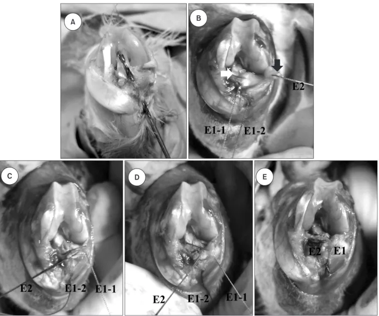
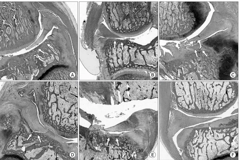
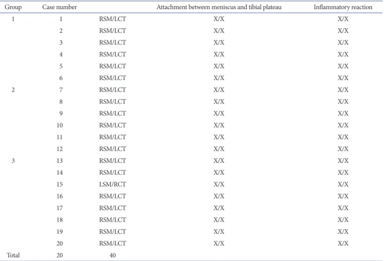
Repair of the Complete Radial Tear of the Anterior Horn of the Medial Meniscus in Rabbits: A Comparison between Simple Pullout Repair and Pullout Repair with Human Bone Marrow Stem Cell Implantation
7
0
0
전체 글
(2)
(3)
(4)
(5)
(6)
(7)
수치

관련 문서
After choosing the type of bike, the next step is the right bike size. the right size for you from
44 글의 첫 번째 문장인 The most important thing in the Boat Race is harmony and teamwork.을 통해 Boat Race에서 가장 중요한 것은 조 화와 팀워크임을
Now that you have the right bike for you, it is important to learn the right riding position.. A poor riding position can lead to injuries
44 글의 첫 번째 문장인 The most important thing in the Boat Race is harmony and teamwork.을 통해 Boat Race에서 가장 중요한 것은 조 화와 팀워크임을
Nonlinear Optics Lab...
12 that절 내의 주어를 문장의 주어로 쓰고 that절 내의 동사를 to부정사로 써 서 수동태 문장을 만들 수 있다... 13 반복되는 the reason을 관계대명사 which로 바꿔주고
The proposal of the cell theory as the birth of contemporary cell biology Microscopic studies of plant tissues by Schleiden and of animal tissues by Microscopic studies of
웹 표준을 지원하는 플랫폼에서 큰 수정없이 실행 가능함 패키징을 통해 다양한 기기를 위한 앱을 작성할 수 있음 네이티브 앱과
