introduction
Tibial plateau fractures account for 1% of all fractures and occur relatively frequently in the knee concomitantly with injuries to the meniscus and ligaments1).
In particular, the frequency of lateral tibial plateau fractures that cause metaphyseal split or depression of the articular surface is higher (55%–70%) than medial and bilateral tibial plateau frac
tures (10%–23% and 11%–31%, respectively). It has been report
ed that lateral tibial plateau fractures are significantly associated with lateral meniscal pathology (Schatzker type II, 45%; type III,
18%)2).
Therefore, identification and treatment of the concomitant le
sions in the lateral meniscus and ligaments are expected to affect the clinical outcome of the treatment of lateral tibial plateau frac
tures3).
However, there are insufficient treatment guidelines for lateral meniscal tears associated with tibial plateau fractures due to the lack of followup data on meniscal repair in such cases.
The purpose of this retrospective study was to assess the fre
quency of lateral meniscal tears in patients diagnosed with lat
eral tibial plateau fractures and evaluate the clinical outcome of meniscal repair.
Materials and Methods
1. patient group
We retrospectively reviewed 25 skeletally mature patients who were treated operatively under the diagnosis of lateral tibial plateau fractures at our institution between January 1, 2010 and February 28, 2016. The inclusion criteria were Schatzker type II or III and Arbeitsgemeinschaft für Osteosynthesefragen/Ameri
The Efficacy of Meniscal Treatment Associated with Lateral Tibial Plateau Fractures
HyunJoo Park, MD, HuiDu Lee, MD, and Jin Ho Cho, MD
Department of Orthopaedic Surgery, Inje University Ilsan Paik Hospital, Goyang, Korea
purpose: The purpose of this study was to examine the incidence of lateral meniscal tears associated with lateral tibial plateau fractures and report the clinical outcomes of meniscal treatment with internal fixation of fractures.
Materials and Methods: All lateral tibial plateau fractures (Schatzker types II and III) in skeletally mature patients treated operatively at our institution between January 2010 and February 2016 were included. All patients underwent open reduction and internal fixation using a buttress plate or cancellous screws. All meniscal tears were initially considered for repair using an allinside technique.
results: The incidence of lateral meniscal tears with lateral tibial plateau fractures was 64%. Ten patients underwent meniscal repair. In secondlook arthroscopy, normal healing was observed in all of the repaired lateral menisci. At the last followup, none of the 10 patients had clinical symptoms related to meniscal injuries. One of the 4 patients who had not undergone meniscal treatment although a lateral tear was suspected based on magnetic resonance imaging achieved stable bony union; however, due to the complaint of persisting knee pain, lateral meniscectomy was performed.
conclusions: Treatment of meniscal lesions associated with lateral tibial plateau fractures showed good clinical and secondlook arthroscopic results.
Therefore, we believe that recognition and treatment of a meniscal injury at the time of surgical fixation can improve clinical outcome.
Keywords: Knee, lateral tibial plateau, fracture, lateral meniscus, repair pISSN 2234-0726 · eISSN 2234-2451
Knee Surgery & Related Research
Received May 17, 2016; Revised (1st) August 29, 2016;
(2nd) September 20, 2016; Accepted October 11, 2016 Correspondence to: Jin Ho Cho, MD
Department of Orthopaedic Surgery, Inje University Ilsan Paik Hospital, 170 Juhwaro, Ilsanseogu, Goyang 10380, Korea
Tel: +82319109733, Fax: +82319107967 Email: [email protected]
137
This is an Open Access article distributed under the terms of the Creative Commons Attribution NonCommercial License (http://creativecommons.org/licenses/bync/4.0/) which permits unrestricted noncommercial use, distribution, and reproduction in any medium, provided the original work is properly cited.
Copyright © 2017 KOREAN KNEE SOCIETY www.jksrr.org
can Orthopaedic Trauma Association type 41B fractures (Fig.
1). Surgical treatment was not considered necessary for Schatzker type I fractures, and thus were excluded from the study.
Of the 25 patients, 17 were male and 8 were female. Their mean age was 49 years (range, 22 to 68 years).
Fracture patterns were classified into Schatzker type II in 15 patients and Schatzker type III in 10 patients. The cause of injury was a traffic accident in 19, a slip down in 3, a sports injury (ski
ing/football) in 2, and a fall from a height in 1.
All patients underwent preoperative magnetic resonance imag
ing (MRI) for identification of an intraarticular injury accompa
nying the fracture. Lateral meniscal tears were observed in 16 of the 25 patients, medial collateral ligament injuries in 6, anterior cruciate ligament (ACL) avulsion fractures in 4, posterior cruci
ate ligament (PCL) injuries in 4, and a medial meniscus tear in 1 patient (Table 1).
2. Surgical technique
The mean interval from injury to surgery was 3.5 days (range, 0 to 7 days). Under general or spinal anesthesia, the patient was placed in the supine position, and draping was done with a tourniquet applied on the thigh. Prior to fracture reduction, ar
throscopic examination was performed to inspect for lesions in the joint. Then, a 10 cm longitudinal skin incision was made on the anterolateral aspect of the knee to expose the joint and reduce the fracture. In all knees, internal fixation was performed with a buttress plate (TomoFix Lateral High Tibial Plate; DePuy Synthes, West Chester, PA, USA) or cancellous screws.
Reduction of the articular surface was confirmed in two differ
ent stages. During reduction of the fracture site, a Carm rotated by 15° was used to check the reduction of the tibial plateau in anteroposterior and lateral views. Then, during the repair of the lateral meniscus, the articular surface at the fracture site was di
rectly visualized with arthroscopy.
For patients with large bone defects or osteoporosis that could disrupt maintenance of reduction, bone grafts were used: an al
logeneic bone graft in 11 patients and an autologous bone graft obtained from the iliac crest in 2 patients.
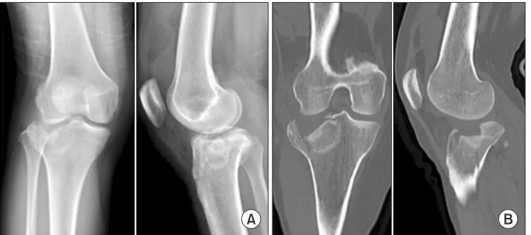
Following the fracture treatment, arthroscopic repair of the lateral meniscus was performed. Transvers and longitudinal tears in the anterior and central portions of the meniscus were Fig. 1. (A) Anteroposterior and lateral radiographs demonstrating Schatzker II tiabal plateau fracture of the right knee. (B) Coronal and Sagittal computed tomogra
phy of the same patient.
A B
Table 1. Demographic Data
Characteristic Data
No. of patients 25
Sex
Male 17 (68)
Female 8 (32)
Age (yr), mean (range) 49.28 (22–68)
Schatzker fracture type
II 15
III 10
Cause of injury
Traffic accident 19 (76)
Slip down 3 (12)
Sports injury 2 (8)
Fall down 1 (4)
Associated injury
Lateral meniscus tear 16 (64)
Medial collateral injury 6 (24)
ACL avulsion fracture 4 (16)
PCL injury 4 (16)
Medial meniscus tear 1 (4)
Values are presented as number (%).
ACL: anterior cruciate ligament, PCL: posterior cruciate ligament.
sutured with a modified allinside repair technique using an 18gauge spinal needle, whereas the allinside repair was per
formed through the posterolateral portal for posterior tears4). The procedure was performed using No.2 PDS (Ethicon, Somerville, NJ, USA) and knots were tied using the Samsung Medical Center sliding method. For lateral meniscus tears where suture repair was unfeasible or horizontal tears, partial meniscectomy was done (Fig. 2). Conservative treatment was prescribed for patients with medial collateral ligament injuries and those without joint instability in spite of ACL avulsion fractures or PCL injuries. For the medial meniscal tear, partial meniscectomy was performed based on the arthroscopic evidence of a horizontal tear.
3. postoperative rehabilitation
Patients who underwent surgical treatment were immobilized in a long leg cast for 1 week. Isometric quadriceps exercises were initiated from the 1st postoperative day. Range of motion (ROM) exercises were gradually carried out in the brace from the 7th postoperative day to obtain 90° of flexion/extension by 2–4 weeks
after surgery. Partial weight bearing was permitted when 90° flex
ion was achieved. Full weight bearing was allowed from the 8th postoperative week.
4. Second-look arthroscopy and Metal removal
Bony union was observed at the last followup in 18 patients who had been surgically treated prior to June 2015. For these patients, metal removal and diagnostic secondlook arthroscopy were performed. Metal removal was done at a mean of 14.8 months (range, 12 to 27 months) after surgery. In 11 patients with concomitant lateral meniscal injuries, diagnostic secondlook ar
throscopy was performed to assess any changes in the meniscus.
results
Intraoperative Carm images and postoperative plain radio
graphs of the anteroposterior and lateral views of the tibial plateau revealed anatomical reduction of the fracture (<1 mm articular stepoff). The fracture was found united in all knees at
Fig. 2. (A) Postoperative anteroposterior and lateral radiographs. (B) Arthroscopic image of a lateral meniscal tear after repair.
A
B
the last followup. In 2 patients, brisement under anesthesia was required due to restrictions on the ROM. There was no case of nonunion or delayed union necessitating a revision surgery. In
fection or loss of reduction did not occur in all knees.
The concomitant lateral meniscal tears were present in 16 of the 25 patients with tibial plateau fractures. Longitudinal tears were the most common (n=8), followed by radial tears (n=3), horizontal tears (n=3), and flap tears (n=2) (Table 2).
Of the 16 patients with lateral meniscal tears, suture repair was conducted in 10 and partial meniscectomy was performed in 2 because suture repair was not feasible in them. In the remaining 4 patients, additional procedure was not considered necessary because the extent of damage was small or minor.
During metal removal, a secondlook arthroscopy was per
formed in 11 patients with lateral meniscal tears, which showed complete healing of the lateral meniscus in all knees (Fig. 3).
Four patients were not treated surgically although lateral meniscal tears were suspected based on MRI and arthroscopic examinations. One of these patients complained of persistent pain in spite of achievement of bony union at the last follow
up. The presence of a lateral meniscal tear was suspected based on followup MRI. Thus, arthroscopic examination was done to
confirm the diagnosis. Partial meniscectomy was performed for treatment, which resulted in pain relief in the patient.
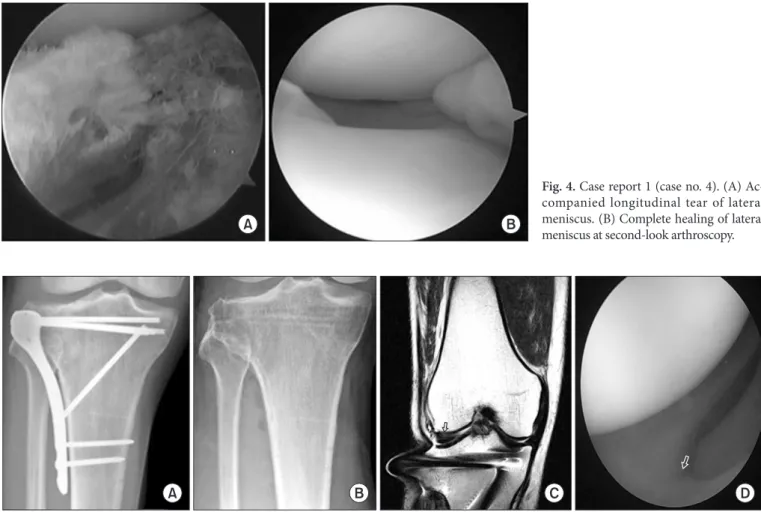
1. case report 1
A 48yearold female patient sustained a Schatzker type III lateral tibial plateau fracture in a pedestrian traffic accident. Con
comitant injuries to the lateral meniscus (Fig. 4A) and medial collateral ligament were observed. The fracture was treated with open reduction and internal fixation. For the longitudinal menis
cal tear, allinside repair4) using a spinal needle was performed.
The patient was ordered to wear a brace for the medial collateral ligament injury. At 1 year after surgery, secondlook arthroscopy and metal removal were performed, which showed complete healing of the meniscus (Fig. 4B).
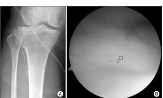
2. case report 2
A 60yearold male patient sustained a Schatzker type II lateral tibial plateau fracture in a vehicle accident. Although a lateral meniscal injury was present, the patient underwent only open re
duction and internal fixation (Fig. 5A) for the fracture. There was no procedure performed for the meniscal tear.
At 1 year after surgery, stable bony union was obtained at the fracture site (Fig. 5B). However, he complained of persistent knee pain. Followup MRI confirmed the presence of a lateral meniscal tear (Fig. 5C). At the time of metal removal, a second
look arthroscopy was performed. After partial meniscectomy, the symptom was relieved (Fig. 5D).
Fig. 3. (A) Post implant removal state. (B) Healed lateral meniscus (black open arrow:
previous tear site).
A B
Table 2. Lateral Meniscus Tear Type
Tear type No. (%)
Horizontal 3 (18.7)
Longitudinal 8 (50)
Radial 3 (18.7)
Flap 2 (12.5)
discussion
Tibial plateau factures are known to occur with meniscal tears in 36% to 61% of the cases59). In the current study, concurrent lateral meniscal tears were observed in 64% of patients with Schatzker type II or III fractures. Interestingly, it is common for surgeons not to report concomitant meniscal lesions in tibial pla
teau fractures treated without arthroscopy, indicating that menis
cal tears may remain untreated.
With improved understanding and treatment of meniscal tears in the field of orthopedic surgery, it has been well established that the appropriate use of meniscal repair or meniscectomy for preservation of the meniscus would contribute to prevention of cartilage loss.
The incidence of posttraumatic osteoarthritis of the knee ranges from 17% to 28.9%1013), and predisposing factors include articu
lar congruity, joint stability, meniscal injury, lower limb align
ment, and patient’s age14).
The most important factor of those listed above is articular
congruity, loss of which is termed as a stepoff15). The presence of lesions in the meniscus has also been associated with posttrau
matic osteoarthritis in the long term16).
In the current study, the secondlook arthroscopy revealed more satisfactory results in the knees where meniscal injuries, if present, were treated simultaneously at the time of reduction of lateral tibial plateau fractures. In particular, complete healing at the repair site was noted in knees where the lateral meniscus had been repaired.
In our opinion, the high success rate of meniscal repair in patients with combined lateral tibial plateau fractures can be at
tributed to the following three factors. First, meniscal repair was performed within the first week of injury. The meniscus was repaired during the surgery for the combined fracture, which was performed within 1 week of injury. Therefore, repair of an acute tear is expected to have a positive impact on healing. Second, the concurrent fracture reduction procedure facilitated bone marrow stimulation as in ACL reconstruction. Some studies have shown that ACL reconstruction combined with meniscal repair resulted
A B C D
Fig. 5. Case report 2. (A) The immediate postoperative radiograph showing fixation by lateral plating. (B) Stable bony union. (C) Postoperative mag
netic resonance imaging showing the healed tear site (black open arrow). (D) Secondlook arthroscopy of the healed tear site (white open arrow).
Fig. 4. Case report 1 (case no. 4). (A) Ac
companied longitudinal tear of lateral meniscus. (B) Complete healing of lateral meniscus at secondlook arthroscopy.
A B
in improved healing of the meniscus when compared to the re
sult of meniscal repair alone17,18). Therefore, simultaneous menis
cal repair and fracture reduction can be considered to contribute to enhanced meniscal healing. Third, restrictions on postopera
tive ROM were conducive to the desired environment for heal
ing of the repaired meniscus. Unlike the standard rehabilitation protocol prescribed after meniscal repair only, flexion was limited to 90° and weight bearing was progressively increased over the period of 8 postoperative weeks to stabilize reduction of the frac
ture, which also contributed to enhanced stability at the repair site.
Currently, computed tomography is a standard imaging modal
ity for the treatment of lateral tibial plateau fractures, whereas there are no guidelines on the use of MRI. However, it is our un
derstanding that early MRI scanning would be helpful consider
ing that the incidence of injuries to the meniscus and ligaments is high in patients with lateral tibial plateau fractures. In addition, it can be difficult to assess lesions on postoperative MRI scans due to the presence of internal fixation apparatus for fracture treat
ment.
The 60yearold male patient case demonstrated that lateral meniscal tears can be satisfactorily treated and these lesions can be responsible for persistent joint pain, swelling, and effusion un
less proper treatment is carried out.
Therefore, we suggest that 1) preoperative MRI should be an essential component of the management of lateral tibial plateau fractures and 2) treatment of a concomitant intraarticular lesion and fracture reduction should be performed simultaneously to improve clinical outcomes.
1. limitation
One of the limitations of this study include the small sample size precluding statistical analysis. Statistical significance of our findings should be evaluated in further studies involving a larger study population.
Another limitation was the short followup period. With a lon
ger followup, we could have demonstrated that the neglected meniscal lesions have an influence on the occurrence of posttrau
matic osteoarthritis, resulting in relatively poor clinical outcomes.
Lastly, compared to randomized controlled trials, there was an increased probability of selection bias of this retrospective study.
Since there was no control group for comparison, it was difficult to provide definitive results other than the clinical outcomes of repair of meniscal tears combined with lateral tibial plateau frac
tures.
conclusions
Schatzker type II and III fractures of the lateral tibial plateau fre
quently occur with lateral meniscal damage. Internal fixation and meniscal repair resulted in excellent clinical outcomes. Therefore, we believe preoperative MRI should be performed to identify in
traarticular lesions combined with lateral tibial plateau fractures.
Simultaneous repair of meniscal injuries would reduce the risk of posttraumatic osteoarthritis, eventually improving longterm clinical outcomes.
conflict of interest
No potential conflict of interest relevant to this article was re
ported.
references
1. Bennett WF, Browner B. Tibial plateau fractures: a study of associated soft tissue injuries. J Orthop Trauma. 1994;8:183
8.
2. Stahl D, SerranoRiera R, Collin K, Griffing R, Defenbaugh B, Sagi HC. Operatively treated meniscal tears associated with tibial plateau fractures: a report on 661 patients. J Or
thop Trauma. 2015;29:3224.
3. Tscherne H, Lobenhoffer P. Tibial plateau fractures: man
agement and expected results. Clin Orthop Relat Res.
1993;(292):87100.
4. Cho JH. Arthroscopic allinside repair of anterior horn tears of the lateral meniscus using a spinal needle. Knee Surg Sports Traumatol Arthrosc. 2008;16:6836.
5. AbdelHamid MZ, Chang CH, Chan YS, Lo YP, Huang JW, Hsu KY, Wang CJ. Arthroscopic evaluation of soft tissue in
juries in tibial plateau fractures: retrospective analysis of 98 cases. Arthroscopy. 2006;22:66975.
6. Chan YS, Chiu CH, Lo YP, Chen AC, Hsu KY, Wang CJ, Chen WJ. Arthroscopyassisted surgery for tibial plateau fractures: 2 to 10year followup results. Arthroscopy.
2008;24:7608.
7. Gill TJ, Moezzi DM, Oates KM, Sterett WI. Arthroscopic reduction and internal fixation of tibial plateau fractures in skiing. Clin Orthop Relat Res. 2001;(383):2439.
8. Kode L, Lieberman JM, Motta AO, Wilber JH, Vasen A, Yagan R. Evaluation of tibial plateau fractures: efficacy of MR imaging compared with CT. AJR Am J Roentgenol.
1994;163:1417.
9. Mustonen AO, Koivikko MP, Lindahl J, Koskinen SK. MRI of acute meniscal injury associated with tibial plateau frac
tures: prevalence, type, and location. AJR Am J Roentgenol.
2008;191:10029.
10. Honkonen SE. Degenerative arthritis after tibial plateau frac
tures. J Orthop Trauma. 1995;9:2737.
11. Lachiewicz PF, Funcik T. Factors influencing the results of open reduction and internal fixation of tibial plateau frac
tures. Clin Orthop Relat Res. 1990;(259):2105.
12. Scheerlinck T, Ng CS, Handelberg F, Casteleyn PP. Medium
term results of percutaneous, arthroscopicallyassisted osteosynthesis of fractures of the tibial plateau. J Bone Joint Surg Br. 1998;80:95964.
13. Mehin R, O’Brien P, Broekhuyse H, Blachut P, Guy P. End
stage arthritis following tibia plateau fractures: average 10
year followup. Can J Surg. 2012;55:8794.
14. Marsh JL, Buckwalter J, Gelberman R, Dirschl D, Olson S, Brown T, Llinias A. Articular fractures: does an anatomic
reduction really change the result? J Bone Joint Surg Am.
2002;84:125971.
15. Giannoudis PV, Tzioupis C, Papathanassopoulos A, Obak
ponovwe O, Roberts C. Articular stepoff and risk of post
traumatic osteoarthritis: evidence today. Injury. 2010;41:986
95.
16. Hunter DJ, Zhang YQ, Niu JB, Tu X, Amin S, Clancy M, Guermazi A, Grigorian M, Gale D, Felson DT. The as
sociation of meniscal pathologic changes with cartilage loss in symptomatic knee osteoarthritis. Arthritis Rheum.
2006;54:795801.
17. Horibe S, Shino K, Nakata K, Maeda A, Nakamura N, Mat
sumoto N. Secondlook arthroscopy after meniscal repair:
review of 132 menisci repaired by an arthroscopic insideout technique. J Bone Joint Surg Br. 1995;77:2459.
18. Tenuta JJ, Arciero RA. Arthroscopic evaluation of menis
cal repairs: factors that effect healing. Am J Sports Med.
1994;22:797802.