A retrospective clinical study of single short implants (less than 8 mm) in posterior
edentulous areas
Sang-Yun Kim1, Jeong-Kui Ku1, Hyun-Suk Kim1, Pil-Young Yun1, Young-Kyun Kim1,2*
1Department of Oral and Maxillofacial Surgery, Section of Dentistry, Seoul National University Bundang Hospital, Seongnam, Republic of Korea
2Department of Dentistry & Dental Research Institute, School of Dentistry, Seoul National University, Seoul, Republic of Korea
PURPOSE. The goal of this study was to evaluate the clinical outcome of single short implants, less than 8 mm in length, placed in the posterior area. MATERIALS AND METHODS. A total of 128 patients (75 male and 53 female, mean age: 52.6±11.2 years) with 154 implants participated. Implant marginal bone loss, and survival and success rates were measured. RESULTS. The mean follow-up period was 51.35±24.97 months. A total of 128 implants, 8 mm in length, were placed in patients who had mean marginal bone loss of 0.75 mm. These implants had a survival rate of 95.3%. Twenty-six implants, 7 mm in length, were placed in areas with a mean marginal bone loss of 0.78 mm and had a survival rate of 96.2%. Both marginal bone loss and survival rate were not statistically different among the groups. In the maxilla, 34 implants showed a mean marginal bone loss of 0.77 mm and a survival rate of 97.1%. In the mandible, 120 implants showed a mean marginal bone loss of 0.75 mm and a survival rate of 95.0%. The average marginal bone loss around all implants was 0.76±0.27 mm at the last follow-up review after functional loading. The survival rate was 95.6% and success rate was 93.5%.
CONCLUSION. In our study, single short implants less than 8 mm in length in the posterior areas had favorable clinical outcomes. [J Adv Prosthodont 2018;10:191-6]
KEYWORDS: Short implants; Single implant
INTRODUCTION
With the recent development of implant surface treatment and bone graft techniques, implants can be placed in the anterior and posterior areas with high predictability. However, the length of mandibular implants may be limited due to insufficient vertical height from the ridge to the inferior
alveolar nerve. This height deficiency results from alveolar bone resorption, commonly seen after long-term tooth loss.
As a result, several methods for placing short implants have been proposed. Short implants are advantageous for several reasons: 1) invasive surgical procedures such as ridge aug- mentation can be avoided in the posterior area; 2) overheat- ing during drilling is minimized; 3) maxillary sinus and infe- rior alveolar canal invasion is minimized; 4) root damage can be prevented in cases where adjacent teeth have large root curvatures; 5) labial bone perforation can be avoided in the areas where buccal undercut or concavity exists; and 6) surgeons are able to use simple instruments, shorten proce- dure times, and minimize bone grafting.1
Several studies have reported high failure rates with short implants less than 10 mm in length after load-bearing in the posterior area in partially edentulous patients. However, minimally invasive procedures using short implants may be the only treatment option for patients with unfavorable ana- tomical conditions such as insufficient alveolar ridge height or a reduced vertical distance to the inferior alveolar nerve.
Furthermore, the prognosis of these implants has been sig-
Corresponding author:
Young-Kyun Kim
Department of Oral and Maxillofacial Surgery, Section of Dentistry, Seoul National University Bundang Hospital
300 Gumi-dong, Bundang-gu, Seongnam 13620, Republic of Korea Tel. +82317872780: e-mail, [email protected]
Received May 27, 2017 / Last Revision December 23, 2017 / Accepted February 27, 2018
© 2018 The Korean Academy of Prosthodontics
This is an Open Access article distributed under the terms of the Creative Commons Attribution Non-Commercial License (http://creativecommons.
org/licenses/by-nc/3.0) which permits unrestricted non-commercial use, distribution, and reproduction in any medium, provided the original work is properly cited.
This study was supported by the Medical-Dental Convergence Research Program of Seoul National University (860-20140121).
nificantly improved due to recent improvements in implant designs and surface treatment techniques. Failure rates are higher in the maxilla than in the mandible because the max- illa has a lower bone density and is relatively softer.2-6
According to Misch, when two or more implants are placed, they must be connected.7 As such, it is generally accepted as a principle when two or more short implants are placed in the posterior area, they are splinted to distribute the stress among them. Many authors reported that the suc- cess rate of splinted short implants has been reported to be higher than that of non-splinted short implants.8-10 Antoun evaluated the outcomes of wide-diameter implants immedi- ately provisionalized with cement-retained single crowns in posterior molar sites. They suggested that wide-diameter (6 mm) implants can safely and successfully replace single posterior molars.11 Calandriello and Tomatis reported that the use of immediately loaded single lower molars support- ed by Branemark System Wide Platform TiUnite implants had favorable results. A total of 40 Brånemark System TiUnite Wide Platform MK III implants were placed. All implants were provided with provisional crowns in full centric occlu- sion at the time of surgery. Two implants failed so that the cumulative success rate at 5 years was 95.0%. The mean marginal bone remodeling (n = 38) expressed as an average of mesial and distal values was -1.17 mm (SD ± 0.90) at the 5-year time point.12
However, in 2004, Fugazzotto et al.13 reported a mean success rate of 95.1% during a period of 29.3 months for single implants less than 9 mm in length. Malmstrom et al.14 reported similar success rates among short implants 6-mm or 8-mm in length and long implants 11 mm in length and found no clinically significant difference between splinted implants and single crowns. Despite these studies, research on the clinical prognosis of single short implants in the pos- terior area is rare.
The goal of this study was to evaluate the clinical prog- nosis of single short implants less than 8 mm in length placed in the maxillary and mandibular posterior areas.
MATERIALS AND METHODS
Among patients who underwent implant placement in Seoul National University Bundang Hospital (SNUBH) between January 2006 and December 2014, 128 patients (75 male
and 53 female, mean age: 52.6 ± 11.2 years), who received short implants, were selected. The inclusion and exclusion criteria are as follows.
Inclusion criteria
1. Implants less than 8 mm in length 2. Cases treated by one surgeon
3. Cases placed in maxillary and mandibular posterior teeth
4. Products from 1 company Exclusion criteria
1. Inappropriate cases of medical records and radio- graphs
2. Cases with no follow-up observation
A total of 154 Implantium Superline implants (Dentium, Suwon, Korea) were placed by one surgeon. This study was performed with the permission of SNUBH Institutional Review Board (IRB) (No. B-1501/284-104). Implant length, width, placement area, primary and secondary stability, sur- vival and success rates, and surgical method, additional bone grafting, marginal bone loss and complications were investi- gated through a careful review of the patients’ medical records and radiographs. Implant stability was measured with using an Osstell Mentor device (Osstell, Gothenburg, Sweden) to determine ISQ values. Primary stability was measured at the time of implant placement and the secondary stability was measured at the time of prosthetic impression taking.
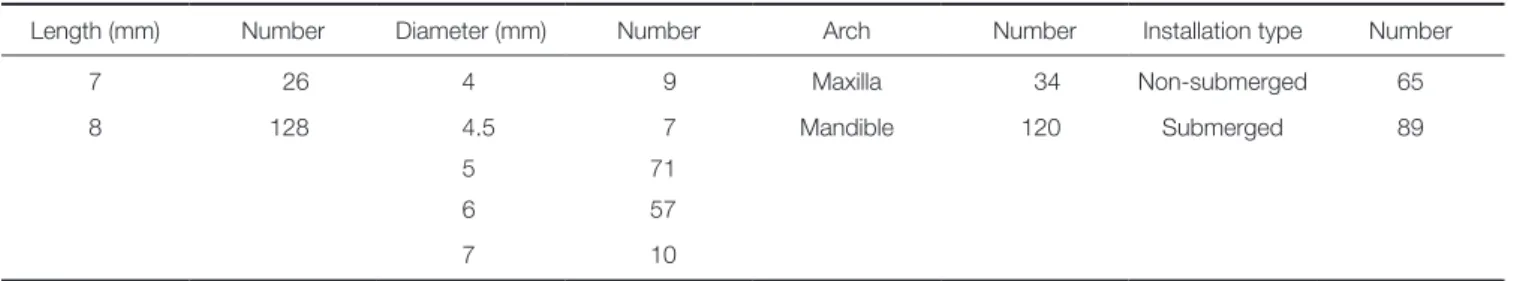
The implants were divided into a 7 mm group and an 8 mm group. There were 26 implants 7 mm-long and 128 implants 8 mm-long. The implants were also classified according to their width (4 mm, 4.5 mm, 5.0 mm, 6 mm, and 7 mm). There were 9 implants 4 mm-wide, 7 implants 4.5 mm-wide, 71 implants 5.0 mm-wide, 57 implants 6 mm- wide, and 10 implants 7 mm-wide. Also, the implants were classified according to the dental arch in which they were placed, and 34 implants were in the maxilla and 120 were in the mandible. When classified according to the placement method, it was found that non-submerged placement was used for 65 implants, and submerged placement was used for 89 implants (Table 1).
Successful implant placement was defined according to Albrektsson’s definition proposed in 198615:1) Individual unattached implant that is immobile when tested clinically;
2) radiography without evidence of peri-implant radiolucen- cy; 3) Bone loss less than 0.2 mm annually after the first
Table 1. Distribution of installed short implants
Length (mm) Number Diameter (mm) Number Arch Number Installation type Number
7 26 4 9 Maxilla 34 Non-submerged 65
8 128 4.5 7 Mandible 120 Submerged 89
5 71
6 57
7 10
post-operative year; 4) No persistent pain, discomfort, or infection; 5) By these criteria, success is defined as success rate of at least 85% at the end of a 5-year observation peri- od and 80% at the end of a 10 year period. The survival rate is the ratio of the number of retained implants from placement to the final examination regardless of loosening, inflammation, and bone loss, to the total number of implants placed.16
To measure the degree of alveolar bone resorption, peri- apical radiographs obtained immediately after the prosthesis placement were set as the baseline. Marginal bone loss was assessed by comparing periapical radiographs obtained using the paralleling technique 12 months after the functional load- ing and during the last visit to the hospital. Magnification was calculated by calculating the ratio of the length of the implant fixture to the length of the fixture on the periapical radiographs. The mean amounts of alveolar bone resorption in the mesial and distal implant surfaces were calculated.
Changes in primary and secondary stability were ana- lyzed with a paired t-test. The significance of the differenc- es in the amount of marginal bone loss as determined by implant width and length, dental arch, and placement meth- od were tested using an independent sample t-test at the sig- nificant probability of 95% (SPSS Inc., Chicago, IL, USA).
RESULTS
All 154 implants were placed in the posterior area as sin- gle restorations. The mean follow-up period was 51.35 ± 24.97 months (minimum 2.53 months; maximum 125.43 months). Of the 154 implants, 7 failed and were removed
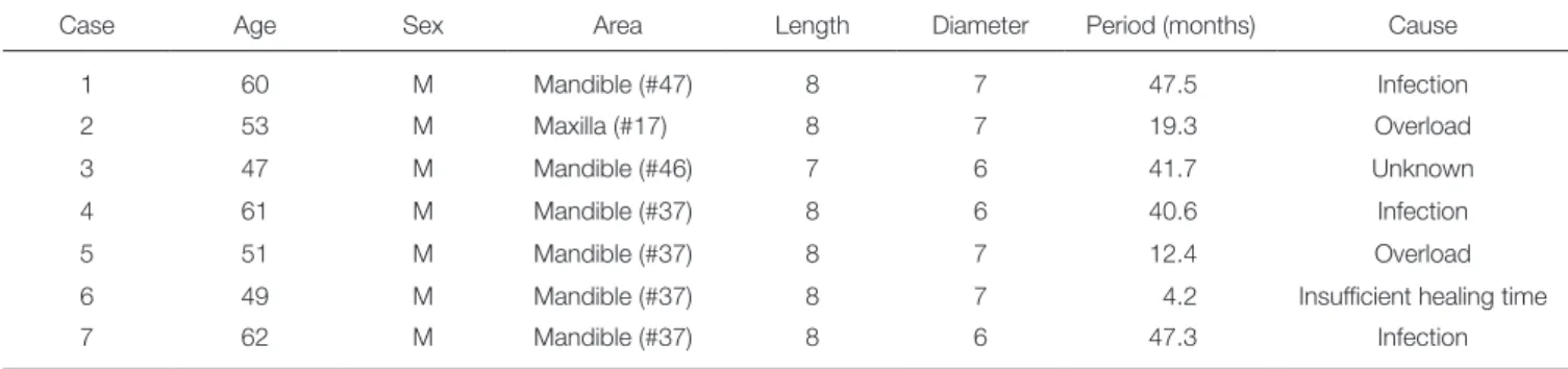
within 23.28 months after the functional loading, making their survival rate 95.6%. The cause of failure is unclear and various factors such as infection, overload, and insufficient healing time seem to have been involved (Table 2). Implant placement was unsuccessful in 10 implant cases due to exces- sive marginal bone loss, resulting in a success rate of 93.5%.
The mean marginal bone loss around the implants was 0.27 mm at 12 months after the functional loading and 0.76 mm on the last day of follow-up. Primary stability measured with Osstell device was 71.26 ± 16.07 and secondary stabili- ty was 77.83 ± 10.01. The increase in primary and second- ary stability was statistically significant. Of the 154 implants, 15 resulted in complications including screw loosening, fix- ture fracture, fixture surface exposure, peri-implantitis, and peri-implant gingivitis.
Of the 154 implants, 128 implants were 8 mm in length and 26 implants were 7 mm in length. In the 8 mm group, the mean period of follow-up was 52.1 months, and the mean marginal bone loss was 0.28 mm one year after the functional loading as compared to 0.75 mm at the last hos- pital visit. Of the 128 implants, 8 implants failed (6 were removed, and 2 had large bone loss), resulting in a success rate of 93.8% and a survival rate of 95.3%. In the 7 mm group, the mean period of follow-up was 47.4 months. The mean marginal bone loss was 0.25 mm one year after the functional loading and 0.78 mm at the last hospital visit. Of the 26 implants, 2 failed (1 was removed and 1 showed severe bone loss), resulting in a success rate of 92.3% and a survival rate of 96.2%. No significant differences in the amount of marginal bone loss, success rate, and survival rate were found between the two groups (P > .05) (Table 3).
Table 2. Removed implants
Case Age Sex Area Length Diameter Period (months) Cause
1 60 M Mandible (#47) 8 7 47.5 Infection
2 53 M Maxilla (#17) 8 7 19.3 Overload
3 47 M Mandible (#46) 7 6 41.7 Unknown
4 61 M Mandible (#37) 8 6 40.6 Infection
5 51 M Mandible (#37) 8 7 12.4 Overload
6 49 M Mandible (#37) 8 7 4.2 Insufficient healing time
7 62 M Mandible (#37) 8 6 47.3 Infection
Period: From implant placement to removal (months)
#: tooth number
Table 3. Comparison by implant length
Implant length (N) (mm) Survival rate (%) Success rate (%) Marginal bone loss (1 year) (mm) Marginal bone loss (Final F/U) (mm)
8 (128) 95.3 93.8 0.28 0.75
7 (26) 96.2 92.3 0.25 0.78
No significant differences in the amount of marginal bone loss, success rate, and survival rate were found between the two groups (P > .05).
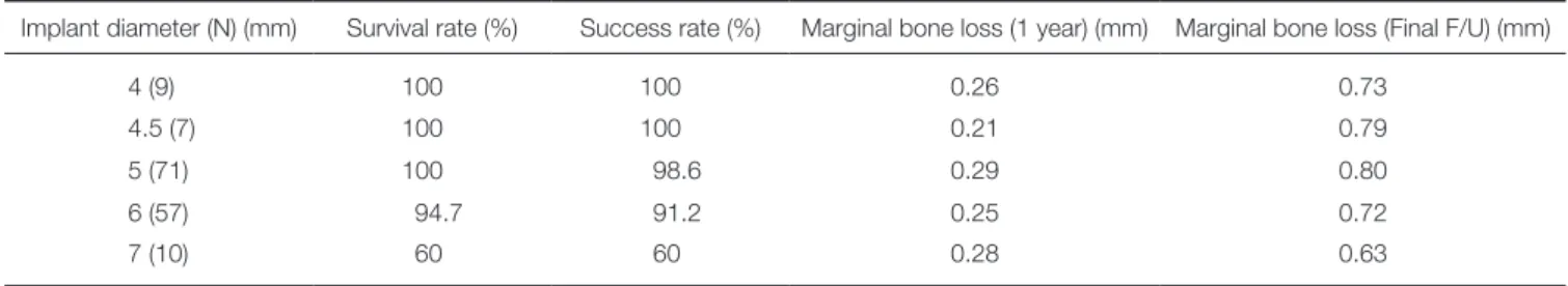
9 implants of 4 mm width, 7 implants of 4.5 mm width, 71 implants of 5.0 mm width, 57 implants of 6 mm width, and 10 implants of 7 mm width were placed. Regarding suc- cess and survival rates for each type of implants, the 4 mm- and 4.5 mm-wide implants had success rates and survival rates of 100%. None of the 5 mm-wide implants were removed, but 1 showed severe bone loss, resulting in a sur- vival rate of 100% and a success rate of 98.6%. Of the 6 mm-wide implants, 3 were removed, and 2 showed severe bone loss, resulting in a success rate of 91.2% and a survival rate of 94.7%. Four of the 7 mm-wide implants were removed, and the implants showed success and survival rates of 60%
(Table 4).
34 implants were placed in the maxilla and 120 in the mandible. In the maxilla group, the mean period of follow- up was 45 months. The mean marginal bone loss was 0.27 mm one year after functional loading and 0.7 mm at the last hospital visit. One implant was removed, resulting in suc- cess and survival rates of 97.1%. In the mandible group, the mean period of follow-up was 53.2 months. The mean mar- ginal bone loss was 0.27 mm one year after functional load- ing and 0.75 mm at the last hospital visit. 6 implants were removed, and 3 implants showed severe bone loss, resulting in a success rate of 92.5% and a survival rate of 95%. No significant differences were noted in the success rate, sur- vival rate, and the amount of marginal bone loss between the two groups (P > .05) (Table 5).
69 implants were non-submerged and 89 were submerged.
Primary stability was measured at the time of implant place- ment and the secondary stability was measured at the time
of prosthetic impression taking. A statistically significant difference in the primary and secondary stability measure- ments was found between the two groups (P < .05).
Primary stability ISQ values were 77.67 ± 7.46 and 67.95 ± 15.99 for the non-submerged and submerged group, respec- tively. Secondary stability ISQ values were 81.37 ± 8.93 and 75.57 ± 10.11, respectively. The mean amount of marginal bone loss one year later was 0.31 ± 0.16 mm in the non- submerged group and 0.25 ± 0.14 mm in the submerged group. The final amount of marginal bone loss was 0.77 ± 0.28 mm in the non-submerged group and 0.75 ± 0.26 mm in the submerged group. The non-submerged implants had a survival rate of 93.8% and a success rate of 92.3%. The submerged implants had a survival rate of 96.6% and a suc- cess rate of 94.4%. No significant differences in success rates and survival rates were found between the two groups (Table 6).
DISCUSSION
Herrmann et al.17 reported a success rate of 78.2% for short implants 7 mm in length and attributed the low success rate to the short implant length. In a study by Weng et al.,18 60%
of all implants that failed were short implants less than 10 mm in length, and the cumulative success rate of short implants was significantly lower than that of all implants.
According to Telleman et al.,19 the shortest implants were more likely to fail than short implants that were a bit longer in partially edentulous patients, and implants in maxilla had a greater failure rate than implants in mandible. However, in
Table 4. Comparison by implant diameter
Implant diameter (N) (mm) Survival rate (%) Success rate (%) Marginal bone loss (1 year) (mm) Marginal bone loss (Final F/U) (mm)
4 (9) 100 100 0.26 0.73
4.5 (7) 100 100 0.21 0.79
5 (71) 100 98.6 0.29 0.80
6 (57) 94.7 91.2 0.25 0.72
7 (10) 60 60 0.28 0.63
Table 5. Comparison by dental arch Arch (N) Survival
rate (%)
Success rate (%)
Marginal bone loss (1 year) (mm)
Marginal bone loss (Final F/U) (mm) Maxilla
(34) 97.1 97.1 0.27 0.77
Mandible
(120) 95 92.5 0.27 0.75
Table 6. Comparison by placement method Installation
method (N)
Survival rate (%)
Success rate (%)
Marginal bone loss (1 year) (mm)
Marginal bone loss (Final F/U)(mm) Non-submerged
(65) 93.8 92.3 0.31 0.77
Submerged
(89) 96.6 94.4 0.25 0.75
a 14-year cumulative study by Romeo et al.,20 in which 8 mm- and 10 mm-long implants were compared, no signifi- cant difference in the amount of marginal bone loss was found, and the success rate of short implant was found to be 97.9% vs 97.1 for the standard implants.
With improvements in implant surface treatment tech- niques and implant designs, positive prognosis of short implants has been reported. Maló et al.21 reported a high suc- cess rate of 96.2% for Brånemark implants 7 mm in length and 97.1% for Brånemark implants 8.5 mm in length in their study using 408 Brånemark implants. In 2012, Kim et al.22 published on the prognosis, prosthetic complications, and factors affecting these results following placement of single short implants in the maxillary and mandibular poste- rior areas. In a total of 87 patients, 96 single implants were placed. 6 implants failed in osseointegration until the last day of follow up, resulting in a survival rate of 91.1%. All of the failed implants had been placed in the mandibular second molar region. Screw loosening was the most com- mon prosthetic complication and was significantly associat- ed with mesiodistal cantilever. The mean amount of mar- ginal bone loss until the last day of follow-up was 0.2 mm.
In conclusion, the success of single implant placement in the maxillary and mandibular posterior areas depends on cantilever minimization and is affected by precise placement of the implant and consistent postoperative management.
Kim et al.23 published another retrospective cohort study in 2014 in which they reported the outcomes of single implant placement accompanied by sinus lifting in the maxilla. This study showed that maxillary sinusitis can affect the progno- sis of implant placement accompanied by sinus lifting in the maxilla. 8 mm long short implants also showed relatively good results. In this study, a total of 154 implants that mea- sured less than 8 mm in length were analyzed. Of all the implants, 7 led to fail in osseointegration, resulting in a mean survival rate of 95.6%. Three implants were consid- ered unsuccessful because they did not meet the standard regarding the amount of marginal bone loss, resulting in a mean success rate of 93.5%. The mean amount of bone absorption was 0.27 mm one year after the functional load- ing and 0.76 mm on the last day of follow-up. No signifi- cant difference in outcome was found between the 7 mm- and 8 mm-long single implants in this study. Reasons for implant removal were infection, overload, and insufficient healing period. Implant failure appeared to be insignificantly associated with implant length. During implantation, pre- ventive measures are important for preventing implant fail- ures, which include disinfecting the wound area following surgery, appropriate use of antibiotics, determination of an appropriate amount of healing time, and timing of func- tional loading.
Since this study only included single implants, it was not possible to compare them to splinted crowns. Of the total of 154 implants included in this study, 65 were placed using the one-stage approach and 89 using the two-stage approach.
Primary and secondary stability were found to be signifi- cantly higher in the implants using the one-stage approach.
However, there were no statistically significant differences between the groups with regard to the amount of marginal bone loss, survival rate, and success rate. The values for pri- mary stability and secondary stability were all within a stable range, and a notable increase in secondary stability was observed after a certain amount of time elapsed after the healing period. The primary stability of the submerged type implants was greater than that of the non-submerged type in this study; this was possibly because in the two-stage approach, submerged type implants are often placed when primary stability is low. Implant stability was observed to increase for all the implants over time, and no significant differences in survival rates and the amount of marginal bone loss were observed between the two placement meth- ods. Therefore, placement methods and healing time may not affect implant prognosis if the outcomes are deter- mined according to the bone quality and primary stability at the time of surgery.
The maxilla has a relatively low bone density and is soft.
As a result, the failure rate of short implants is higher in the maxilla than in the mandible.2,3,4 Therefore, it is advisable to perform the second surgery after a healing period of approximately 6 months, after which the prosthetic treat- ment in the maxilla can be completed. However, the man- dibular alveolar bone is dense and the initial fixation is firm, so implants can be placed by using the one-stage, non-sub- merged method. The prosthetic treatment can begin after a 2 - 3 month healing period. Various results have been report- ed with regard to changes in the success rate of short implants depending on whether one-stage or two-stage placement was performed. Gentile et al.5 reported higher success rates for short implants placed through the two-stage placement method. However, Sun et al.6 reported no difference in suc- cess rates between one-stage and two-stage placement methods.
Several studies have reported that the diameter of an implant plays an important role in primary stability and implant prognosis.24,25 In this study, while no significant dif- ference in the amount of marginal bone loss was observed according to implant diameter, the survival and success rates of the 6 mm- and 7 mm-wide implants were lower than those of the 5 mm-wide implants. In this study, the implants were divided into several groups according to their diameter as follows: 4 mm group (n = 9), 4.5 mm group (n
= 7), 5.0 mm group (n = 71), 6 mm group (n = 57), and 7 mm group (n = 10). No significant differences were found among the groups in the amount of marginal bone loss, survival rate, or in the success rate.
This study had several limitations. First, it was difficult to perform a standardized statistical analysis since all the data were retrospectively analyzed. Furthermore, we did not perform an analysis on cantilever and crown/implant ratio.
There was a problem that the statistical comparison was dif- ficult because the sample difference between the groups was large. Future research designs must be standardized, and prospective clinical research must be conducted to produce meaningful results.
CONCLUSION
In conclusion, favorable clinical outcomes can be expected from single short implants in the posterior area after apply- ing the latest surface treatment techniques and ensuring that patients have a sufficient healing period.
ORCID
Sang-Yun Kim https://orcid.org/0000-0002-2952-5404 Jeong-Kui Ku https://orcid.org/0000-0003-1192-7066 Hyun-Suk Kim https://orcid.org/0000-0001-7010-4153 Pil-Young Yun https://orcid.org/0000-0001-6097-1229 Young-Kyun Kim https://orcid.org/0000-0002-7268-3870 REFERENCES
1. Misch CE, Suzuki JB, Misch-Dietsh FM, Bidez MW. A posi- tive correlation between occlusal trauma and peri-implant bone loss: literature support. Implant Dent 2005;14:108-16.
2. Rocci A, Martignoni M, Gottlow J. Immediate loading of Brånemark System TiUnite and machined-surface implants in the posterior mandible: a randomized open-ended clinical tri- al. Clin Implant Dent Relat Res 2003;5:57-63.
3. Glauser R, Lundgren AK, Gottlow J, Sennerby L, Portmann M, Ruhstaller P, Hämmerle CH. Immediate occlusal loading of Brånemark TiUnite implants placed predominantly in soft bone: 1-year results of a prospective clinical study. Clin Implant Dent Relat Res 2003;5:47-56.
4. Sun HL, Huang C, Wu YR, Shi B. Failure rates of short (≤ 10 mm) dental implants and factors influencing their failure: a systematic review. Int J Oral Maxillofac Implants 2011;26:
816-25.
5. Gentile MA, Chuang SK, Dodson TB. Survival estimates and risk factors for failure with 6 × 5.7-mm implants. Int J Oral Maxillofac Implants 2005;20:930-7.
6. Chan MF, Närhi TO, de Baat C, Kalk W. Treatment of the atrophic edentulous maxilla with implant-supported overden- tures: a review of the literature. Int J Prosthodont. 1998;11:7- 15.
7. Misch CE. Short dental implants: a literature review and ra- tionale for use. Dent Today 2005;24:64-6,68.
8. Balshi TJ, Wolfinger GJ, Slauch RW, Balshi SF. A retrospec- tive analysis of 800 Brånemark System implants following the All-on-Four™ protocol. J Prosthodont. 2014;23:83-8.
9. Annibali S, Cristalli MP, Dell’Aquila D, Bignozzi I, La Monaca G, Pilloni A. Short dental implants: a systematic review. J Dent Res 2012;91:25-32.
10. Mendonça JA, Francischone CE, Senna PM, Matos de Oliveira AE, Sotto-Maior BS. A retrospective evaluation of the survival rates of splinted and non-splinted short dental implants in posterior partially edentulous jaws. J Periodontol 2014;85:787-94.
11. Antoun H, Cherfane P, Sojod B. Consecutive Case Series of Healed Single-Molar Sites Immediately Restored with Wide- Diameter Implants: A 1-Year Evaluation. Int J Dent 2016;
2016:5645892.
12. Calandriello R, Tomatis M. Immediate occlusal loading of single lower molars using Brånemark System® Wide Platform TiUnite™ implants: a 5-year follow-up report of a prospec- tive clinical multicenter study. Clin Implant Dent Relat Res 2011;13:311-8.
13. Fugazzotto PA, Vlassis J, Butler B. ITI implant use in private practice: Clinical results with 5,526 implants followed up to 72+ months in function. Int J Oral Maxillofac Implants 2004;19:408-12.
14. Malmstrom H, Gupta B, Ghanem A, Cacciato R, Ren Y, Romanos GE. Success rate of short dental implants support- ing single crowns and fixed bridges. Clin Oral Implants Res 2016;27:1093-8.
15. Albrektsson T, Zarb G, Worthington P, Eriksson AR. The long-term efficacy of currently used dental implants: a review and proposed criteria of success. Int J Oral Maxillofac Implants 1986;1:11-25.
16. Zarb GA, Albrektsson T. Consensus report: towards opti- mized treatment outcomes for dental implants. J Prosthet Dent 1998;80:641.
17. Herrmann I, Lekholm U, Holm S, Kultje C. Evaluation of pa- tient and implant characteristics as potential prognostic fac- tors for oral implant failures. Int J Oral Maxillofac Implants 2005;20:220-30.
18. Weng D, Jacobson Z, Tarnow D, Hürzeler MB, Faehn O, Sanavi F, Barkvoll P, Stach RM. A prospective multicenter clinical trial of 3i machined-surface implants: results after 6 years of follow-up. Int J Oral Maxillofac Implants 2003;18:
417-23.
19. Telleman G, Raghoebar GM, Vissink A, den Hartog L, Huddleston Slater JJ, Meijer HJ. A systematic review of the prognosis of short (<10 mm) dental implants placed in the partially edentulous patient. J Clin Periodontol 2011;38:667- 76.
20. Romeo E, Ghisolfi M, Rozza R, Chiapasco M, Lops D. Short (8-mm) dental implants in the rehabilitation of partial and complete edentulism: a 3- to 14-year longitudinal study. Int J Prosthodont 2006;19:586-92.
21. Maló P, de Araújo Nobre M, Rangert B. Short implants placed one-stage in maxillae and mandibles: a retrospective clinical study with 1 to 9 years of follow-up. Clin Implant Dent Relat Res 2007;9:15-21.
22. Kim YK, Kim SG, Yun PY, Hwang JW, Son MK. Prognosis of single molar implants: a retrospective study. Int J Periodontics Restorative Dent 2010;30:401-7.
23. Kim YK, Ahn KJ, Yun PY. A retrospective study on the prog- nosis of single implant placed at the sinus bone graft site.
Oral Surg Oral Med Oral Pathol Oral Radiol 2014;118:181-6.
24. Isidor F. Clinical probing and radiographic assessment in rela- tion to the histologic bone level at oral implants in monkeys.
Clin Oral Implants Res 1997;8:255-64.
25. Yilmaz B, Seidt JD, McGlumphy EA, Clelland NL. Comparison of strains for splinted and nonsplinted screw-retained pros- theses on short implants. Int J Oral Maxillofac Implants 2011;
26:1176-82.