pISSN : 1229-5418 Implantology 2019; 23(1): 2-14
https://doi.org/10.32542/implantology.2019001
Received: December 18, 2018 Revised: January 31, 2019 Accepted: February 1, 2019 ORCID
Eun-Bin Bae
https://orcid.org/0000-0002-9524-3835
Jung-Bo Huhhttps://orcid.org/0000-0001-7578-1989 Copyright © 2019. The Korean Academy of Oral &
Maxillofacial Implantology
This is an Open Access article distributed under the terms of the Creative Commons Attribution Non- Commercial License (http://creativecommons.org/
licenses/by-nc/4.0/) which permits unrestricted non- commercial use, distribution, and reproduction in any medium, provided the original work is properly cited.
OPEN ACCESS
Purpose: The purpose of this study was to compare the clinical outcomes of lithium disilicate pressed zirconia prostheses and monolithic zirconia prostheses, which are widely used as posterior implant restorations, after 12 months of follow-up.
Material and Methods: A total of 17 patients were treated with 60 implant-supported prostheses.
After examination of implant survival rate, marginal bone loss, probing depth, plaque index, bleeding index, calculus index, and complications, independent T-test and chi-square test were performed to compare each group (Lithium disilicate pressed zirconia prostheses: n=30, Monolithic zirconia prostheses: n=30).
Results: The implant survival rate was 100%. Marginal bone resorption was higher in the monolithic zirconia prostheses group (p<0.05). Probing depth, plaque index, calculus index, and bleeding index were higher in the lithium disilicate pressed zirconia prostheses group ( p<0.05). Complications occurred in the monolithic zirconia prostheses group as connector fracture, and in the lithium disilicate pressed zirconia prostheses group as chipping.
Conclusion: The periodontal index of lithium disilicate pressed zirconia was slightly worse, but the bone resorption was lower and only one chipping occurred on veneered layer. Therefore, lithium disilicate pressed zirconia is considered as a promising treatment option as much as monolithic zirconia in posterior implant-supported prostheses. However, long-term clinical studies are needed for reliable results (IRB No. PNUDH- 2014-001-MD).
Keywords: Dental implant, Lithium disilicate, Zirconium oxide
Abstract
Zirconia and Monolithic Zirconia in Posterior Implant-Supported Prostheses
*Corresponding author: Eun-Bin Bae, [email protected] and Jung-Bo Huh, [email protected]
Kyoung-Woo Roh, Dong-Seok Yang, Young-Chan Jeon, Chang-Mo Jeong, Mi-Jung Yun, So-Hyoun Lee, Jae-Won Choi, Jin-Ju Lee, Eun-Bin Bae
*, Jung-Bo Huh
*Department of Prosthodontics, Dental Research Institute, Institute of Translational Dental Sciences, BK21 PLUS Project, School of Dentistry, Pusan National University, Yangsan, Korea
Ⅰ. Introduction
Among the zirconia-containing ceramics, tetragonal zirconia polycrystalline (Y-TZP) containing 3 mol% yttria has higher flexural strength ranged from 900 to 1,200 MPa.
Because of its high flexural strength, Y-TZP is used in a variety of clinical applications;
frameworks for all-ceramic posterior crowns, crown, bridge, implant abutments and
dental implants, root canal posts
1.
Most ZrO 2 based-ceramic restorations have problems such opaqueness of the core, which should be masked with a translucent layer of veneer ceramic to achieve the natural appearance
2. And monolithic zirconia is difficult to repair when a fracture occurs because the non-reactive surface of zirconia (acid- resistant ceramic) exhibit poor adhesion, a consistent problem of low bond strength to other substrates
3. Another problem is the possibility of bone resorption around the implant by transferring more load to the implant because of its higher strength than other materials
4-7.
Porcelain-fused-to-zirconia (PFZ) has been chosen to solve these problems
8. Methods of veneering zirconia core include hand-layering technique with feldspathic porcelain, CAD-on technique and press-over technique. Recently, press-over technique for pressing lithium disilicate glass ceramic veneer has been introduced
9. This technique is economical and simple because the lost wax press system can produce ceramic prostheses with or without computer aided design/computer aided manufacturing (CAD/CAM) system, and it can effectively fabricate the customized anatomical shape
10.
Menini et al.
11reported that bone resorption and bone fracture can occur when pathological overload is present. Thus, it is important to control the forces transmitted on the bone-implant interface. According to an in vitro study, the force transmitted through the implant onto the peri-implant bone by zirconia (mean 641.8 N) was the greatest. Kim et al.
9reported that lithium disilicate pressed zirconia prostheses have similar fracture strength to monolithic zirconia and are clinically useful. Previous studies reported a comparison of survival rates and complications of zirconia and metal-ceramic prosthetic and all- ceramic
1, 8. However, until now there are no clinical studies whether lithium disilicate pressed zirconia prostheses can solve strength and esthetic problems in posterior edentulous site compared to monolithic zirconia prostheses.
The aim of present study was to compare the clinical outcomes of lithium disilicate pressed zirconia prostheses and monolithic zirconia prostheses and to investigate the complications after 12 months of follow-up and to ascertain clinically enough to use lithium disilicate pressed zirconia prostheses.
Ⅱ. Material and Methods
1. Study subjects
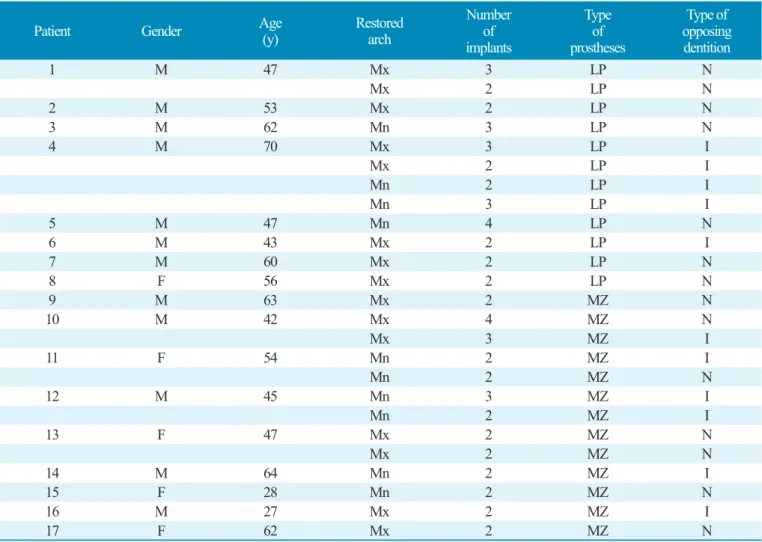
From 2015 to 2017, a total of 17 patients (male: 12, female: 5) were recruited at Pusan National
University Dental Hospital (Table 1). This study was conducted with the consent of the patient under
consideration of the National Bioethics Committee (IRB No. PNUDH- 2014-001-MD). 60 implant-
supported prostheses were planned and followed up for 12 months after placement; 30 lithium disilicate
pressed zirconia prostheses and 30 monolithic zirconia prostheses in the number of missing tooth. The subjects of this clinical study were patients over 21 years of age who were systemically healthy. Patients with missing tooth in the maxillary or mandibular posterior were included. They have sufficient bone width (at least 6 mm) and height (at least 10 mm) so that bone grafting is not necessary. Patients with one or more of the following diagnoses were excluded from subjects: alcohol or drug abuse, smoking, severe bruxism or other destructive habits, medical or psychological conditions preventing implant placement.
2. Clinical procedure
External type implants (USII, Osstem Co., Seoul, Korea) were placed as the number of missing tooth.
Table 1. Datum of patients and implants
Kyoung-Woo Roh et al. : Clinical Evaluation of Lithium Disilicate Pressed Zirconia and Monolithic Zirconia in Posterior Implant-supported Prostheses. Implantology 2019
Type of prostheses: LP. Lithium disilicate pressed zirconia, MZ. Monolithic zirconia, Type of opposing dentition: N. Natural teeth, I. Implant prostheses.
Patient Gender Age
(y) Restored
arch
Number implants of
Type of prostheses
Type of opposing dentition
1 M 47 Mx 3 LP N
Mx 2 LP N
2 M 53 Mx 2 LP N
3 M 62 Mn 3 LP N
4 M 70 Mx 3 LP I
Mx 2 LP I
Mn 2 LP I
Mn 3 LP I
5 M 47 Mn 4 LP N
6 M 43 Mx 2 LP I
7 M 60 Mx 2 LP N
8 F 56 Mx 2 LP N
9 M 63 Mx 2 MZ N
10 M 42 Mx 4 MZ N
Mx 3 MZ I
11 F 54 Mn 2 MZ I
Mn 2 MZ N
12 M 45 Mn 3 MZ I
Mn 2 MZ I
13 F 47 Mx 2 MZ N
Mx 2 MZ N
14 M 64 Mn 2 MZ I
15 F 28 Mn 2 MZ N
16 M 27 Mx 2 MZ I
17 F 62 Mx 2 MZ N
The surgical treatment was performed by specialists in periodontology, oral and maxillofacial surgery.
Two-stage surgical procedure was performed according to the surgical instructions of the manufacturer.
After completion of the second surgery, final impressions at fixture level were made with poly vinyl siloxane (Imprint II Garant regular/light body, 3M ESPE, Minnesota, USA) using the individual tray for open impression technique. The impressions of the opposing arch were made with alginate (aroma fine plus, GC, Tokyo, Japan). Titanium customized abutment (Pre-milled bar, Dio, Busan, Korea) that is set to the same margin level as gingiva were fabricated by CAD software (Exocad DentalCAD 2.2, Exocad, Darmstadt, Germany) and Milling machine (Trione Z, Dio, Busan, Korea). Provisional restorations were used to confirm the patient's discomfort for a month. A final impression was made at the abutment level for the final prostheses. The master casts were scanned by the AutoScan 3D Dental Scanner (Hangzhou Shining 3D Tech Co., Ltd., Hangzhou, China). The implant-supported prostheses are fabricated into two groups: the lithium disilicate pressed on zirconia-based prostheses (LP group, n=30) and the monolithic zirconia prostheses (MZ group, n=30). All the ceramic materials are presented in Table 2. To fabricate the LP group, 0.5 mm-thick zirconia coping (Zirtooth Fulluster, HASS, Gangneung, Korea) was made first. After fabricating the zirconia coping, liner powder (Rosetta Ceram Liner, HASS, Gangneung, Korea) was applied to the surface of the zirconia coping in order to improve the bond strength and wettability between the zirconia and the lithium disilicate glass ceramic veneer. And heat treatment was done. A wax veneer structure was fabricated by milling a wax block (TOTEM, Qingdao Totem Candle Industry, Shandong, China) using the Exocad software. The veneer was fixed to the coping by applying heat to its margin. And after investing, it was burned out at 880°C for 30 minutes (Burnout Furnace L 1/12, Nabertherm, Bremen, Germany). After that, glass ingot (Rosetta UltraPress, HASS, Gangneung, Korea) was put in to the investment ring, and the latter was pressed with the pressing furnace (Horizon Press, Shenpaz Dental Ltd., Migdal HaEmek, Israel) to bond the zirconia and the lithium disilicate glass Table 2. Materials used and properties of each group
Kyoung-Woo Roh et al. : Clinical Evaluation of Lithium Disilicate Pressed Zirconia and Monolithic Zirconia in Posterior Implant-supported Prostheses. Implantology 2019
*
CTE: Coefficient of Thermal Expansion.
Group Component System CTE
*(×10
6/℃) Flexural strength (MPa)
LP
Heat-pressed lithium disilicate glass ceramic veneer Rosetta UltraPress 9.7 450
Liner Rosetta Ceram Liner - -
CAD/CAM zirconia coping Zirtooth Fulluster 10.8 1,250
MZ CAD/CAM zirconia bridge Zirtooth Fulluster 10.8 1,250
ceramic. According to the instruction of the manufacturer, sandblasting (50 μm glass beads at 1 bar pressure) and glazing (IPS e.max Ceram glaze paste, Ivoclar Vivadent, Schaan, Leichtenstein) were performed. To fabricate the MZ group, a zirconia block (ZirtoothFulluster, HASS, gangneung, Korea) at green stage was milled. The occlusal surface is designed to have a uniform thickness of 1.5-2 mm.
Milled zirconia prostheses was gone through the sintering and glazing. All implant-supported prostheses were cemented using self-adhesive resin cement (G-CEM LinkAce, GC America, Alsip, IL, USA). For the purpose of standardization, the same dental technician executed the manufacturing process.
3. Clinical examination
The following criteria were evaluated with reference to clinical examination and radiographs. Clinical examinations and radiographs were performed at 12 months after placement of the implant-supported prostheses, and complications were examined.
1) Implant survival rate
Implant survival rate was evaluated according to the criteria presented by Cochran et al.
12. The evaluation criteria were: (1) no persistent discomfort such as pain, foreign body sensation, and abnormal sensation, (2) no persistent symptoms of peri-implant infection, such as pus discharge, and no relapse of such symptoms (3) no clinical mobility of the implants (4) no radiographic lucency around the implant, and no rapidly progressing bone loss.
2) Implant marginal bone resorption
Radiographs were taken using the parallel technique with a portable radiographic device (Port II, Genoray Co., Sungnam, Korea). The implant length and marginal bone level (distance from the implant platform to the top of the marginal bone) were measured using i-Solution (Olympus B × 51; Olympus Inc., Tokyo, Japan) and then the amount of marginal bone resorption compared with original bone level was calculated by comparing the implant length
13.
3) Probing depth
The probing depth was measured at one point on each mesial, distal, buccal and lingual side around
the implant in parallel with the long axis of the implant with Merritt-B periodontal probe, and then mean
value was calculated
14.
4) Plaque Index
According to the criteria of Mombelli et al.
15, the plaque attached to the surface of the implant was measured and a score from 0 to 3 was assigned.
5) Bleeding Index
The bleeding tendency was assessed using a Merrit-B periodontal probe according to the criteria proposed by Mombelli et al.
156) Calculus
Depending on the presence or absence of calculus, a score of 0 or 1 was given.
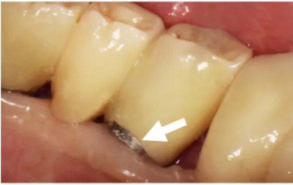
7) Complications
Complications were classified as (1) technically related, such as fracture of framework, connector fracture, chipping, crack, loss of retention, fixture fracture, screw fracture, screw loosening, (2) biologically related, such as peri-implantitis, crestal bone loss, gingival swelling, suppuration, food impaction. And frequency of complications was investigated.
4. Statistical analysis
To compare the marginal bone resorption and probing depth averages of the two groups, the independent T-test was used. Chi-square test was used to verify the significance of plaque index, calculus, bleeding index, and complications according to prostheses materials. All statistics were performed using SPSS (ver. 21.0, SPSS Inc., Chicago, IL, USA) at a significance level of 5%.
Ⅲ. Results
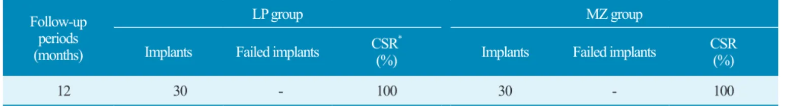
1. Implant survival rate
Of 17 patients with implant-supported prostheses, 60 implants were placed (Fig. 1): 30 implants in
the LP group and 30 implants in the MZ group. All of 60 implants were loaded for 12 months after the
definitive prostheses was placed. There were no failed implants and all implants were normal in function
without clinical mobility (Table 3).
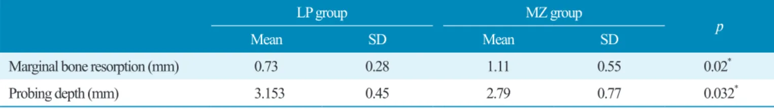
2. Marginal bone resorption
The mean values and standard deviations of implant marginal bone resorption at 1,3,12 months are shown in Table 4. The LP and MZ group showed 0.73±0.28 mm and 1.11±0.55 mm respectively at 12 months. The LP group had a significantly lower implant marginal bone resorption than the MZ group ( p
< 0.05).
3. Probing depth
The mean values and standard deviations of probing depth at 12 months are shown in Table 4. The LP group showed 3.15±0.45 mm and the MZ group showed 2.79±0.77 mm at 12 months. The LP group showed a significantly greater probing depth than the MZ group ( p < 0.05).
Fig. 1. Distribution of implant-supported prostheses.
Kyoung-Woo Roh et al. : Clinical Evaluation of Lithium Disilicate Pressed Zirconia and Monolithic Zirconia in Posterior Implant-supported Prostheses. Implantology 2019
Table 3. Cumulative survival rate of the implants
Kyoung-Woo Roh et al. : Clinical Evaluation of Lithium Disilicate Pressed Zirconia and Monolithic Zirconia in Posterior Implant-supported Prostheses. Implantology 2019
*
CSR: Cumulative survival rate of implants.
Follow-up periods (months)
LP group MZ group
Implants Failed implants CSR
*(%) Implants Failed implants CSR
(%)
12 30 - 100 30 - 100
4. Plaque Index and calculus
No plaque index was the most frequently observed in the MZ group (87%) and the score of 1 was the most frequently observed in the LP group (63%). The difference in the plaque index was significant ( p
< 0.05). The calculus in the LP group (47%) was significantly higher ( p < 0.05) than in the MZ group (0%) ( p < 0.05, Table 5).
5. Bleeding Index
No Bleeding index was the most frequently observed in the MZ group (70%). Relatively more bleeding was observed in the LP group and the difference in the bleeding index was significant ( p < 0.05, Table 5).
Table 4. The average value of marginal bone resorption and probing depth (12 months)
Kyoung-Woo Roh et al. : Clinical Evaluation of Lithium Disilicate Pressed Zirconia and Monolithic Zirconia in Posterior Implant-supported Prostheses. Implantology 2019
*
Mean values showed significant difference based on independent T-test ( p < 0.05).
LP group MZ group
Mean SD Mean SD p
Marginal bone resorption (mm) 0.73 0.28 1.11 0.55 0.02
*Probing depth (mm) 3.153 0.45 2.79 0.77 0.032
*Table 5. Plaque index and calculus, bleeding index
Kyoung-Woo Roh et al. : Clinical Evaluation of Lithium Disilicate Pressed Zirconia and Monolithic Zirconia in Posterior Implant-supported Prostheses. Implantology 2019
†
Frequency distribution of plaque index, calculus, bleeding index.
*