서 론
팽대부주위암에 의한 폐쇄성 황달이 있는 환자에 있어, 수 술 전에 담도배액술을 시행하는 것이 환자의 수술 후 경과 에 도움이 되는지에 대해서는 아직 논란이 많다.(1-7) 그러 나 담관염이 동반되거나 간기능의 이상이나 심한 영양결핍 상태가 있는 경우, 수술 전 평가 및 수술대기시간의 연장 등으로 인해 수술의 지연이 예상되는 경우들에서는 수술 전 담도배액술이 시행되고 있다.(8,9)
수술 전 담도배액술은 크게 경피경간 담도배액술(per- cutaneous transhepatic biliary drainage, PTBD)과 내시경적 담 도배액술(endoscopic biliary drainage)로 나누어 볼 수 있고, 내시경적 담도배액술은 다시 내시경적 경비담도배액술(endo- scopic nasogastric biliary drainage, ENBD)과 내시경적 역행 성담도배액술(endoscopic retrograde biliary drainage, ERBD) 로 구분할 수 있다. 경피경간 담도배액술은 수술 전 담도조
Preoperative Biliary Drainage for Periampullary Cancer: A comparison between endoscopic drainage and percutaneous transhepatic drai- nage
Dae-Wook Hwang, M.D., Sun-Whe Kim, M.D., Yoo-Seok Yoon, M.D., Ji-Hoon Kim, M.D.1, Jin-Young Jang, M.D.
and Yong-Hyun Park, M.D.
Purpose: A preoperative biliary drainage procedure (endo- scopic nasogastric biliary drainage, ENBD; endoscopic retrograde biliary drainage, ERBD; or percutaneous trans- hepatic biliary drainage, PTBD) is infrequently performed in periampullary cancer patients with obstructive jaundice.
Among these different biliary drainage procedures, a safer and more informative procedure should be performed in the indicated cases. However, no comparative study has been done between the two biliary drainage methods (endoscopic vs. percutaneous). The aim of this study is to compare the clinical outcome of these two biliary drainage methods in periampullary cancer and to suggest guidelines for selecting the appropriate preoperative biliary drainage procedure.
Methods: Between January 1996 and June 2002, 25 pa- tients underwent pancreaticoduodenectomy (Whipples' opera- tion or pylorus preserving pancreaticoduodenectomy) after ENBD/ERBD (Group A) due to periampullary cancer. Twenty- five patients who ubderwent PTBD preoperatively were matched with Group A, according to age group, sex, diagnosis, and type of operation during the same period (Group B).
Results: There were no differences in operative time, intra- operative/postoperative transfusion, total/postoperative length of hospital stay, incidence of postoperative complication, TNM staging, or perineural/endovascular/endolymphatic inva-
팽대부주위암에서 수술 전 내시경적 담도배액술과 경피경간 담도배액술의 임상적 비교
서울대학교 의과대학 외과학교실 및 1병리학교실
황대욱․김선회․윤유석․김지훈1․장진영․박용현
책임저자:김선회, 서울특별시 종로구 연건동 28 ꂕ 110-744, 서울대학교병원 외과 Tel: 02-760-2315, Fax: 02-745-2282 E-mail: [email protected]
접수일:2003년 7월 5일, 게재승인일:2003년 9월 1일
sion. However, the thickness of CBD wall (Group A:Group B=1.78±0.55 mm:1.14±0.37 mm, P<0.001) and degree of inflammation of the CBD wall (Group A> Group B, P<
0.001) were significantly different between the two groups.
Conclusion: Although a significant difference of clinical outcome between the two preoperative biliary drainage methods could not be identified in this study, the inflamma- tion of operative field resulting from ENBD/ERBD is ex- pected to cause surgical difficulties and ultimately affect postoperative complications. (J Korean Surg Soc 2003;65:
413-419)
Key Words: Preoperative biliary drainage, Obstructive Jaun- dice, Periampullary cancer, Pancreaticoduode- nectomy
중심 단어: 수술 전 담도배액술, 팽대부주위암, 폐쇄성 황달, 췌십이지장절제술
ꠏꠏꠏꠏꠏꠏꠏꠏꠏꠏꠏꠏꠏꠏꠏꠏꠏꠏꠏꠏꠏꠏꠏꠏꠏꠏꠏꠏꠏꠏꠏꠏꠏꠏꠏꠏꠏꠏꠏꠏꠏꠏꠏꠏꠏꠏꠏꠏ Department of Surgery and 1Pathology, Seoul National University College of Medicine
ꠏꠏꠏꠏꠏꠏꠏꠏꠏꠏꠏꠏꠏꠏꠏꠏꠏꠏꠏꠏꠏꠏꠏꠏꠏꠏꠏꠏꠏꠏꠏꠏꠏꠏꠏꠏꠏꠏꠏꠏꠏꠏꠏꠏꠏꠏꠏꠏꠏꠏꠏꠏꠏꠏꠏꠏꠏꠏꠏꠏꠏꠏꠏꠏꠏꠏꠏꠏꠏꠏꠏꠏꠏꠏꠏꠏꠏꠏꠏꠏꠏꠏꠏꠏꠏꠏꠏꠏꠏꠏꠏꠏꠏꠏꠏꠏꠏꠏꠏꠏꠏꠏꠏꠏꠏꠏꠏꠏꠏꠏꠏꠏꠏꠏꠏ 영술을 통한 병변부의 확인 및 진단에 도움이 되고, 수술
후에도 도관이 유지되므로 지속적으로 담도감압이 가능하며, 수술 후 담도조영술을 시행할 수 있다는 장점이 있다.(10-13) 그러나 담관염, 담즙누출 및 이로 인한 복막염, 출혈, 삽입 부 동통, 도관폐쇄 및 이탈, 체액 및 전해질 손실 등의 단점 도 있는 것으로 알려져 있다.(6,10-15) 이에 반해 내시경적 담도배액술은 내독소혈증 및 사망률의 감소, T 세포기능 이 상의 정상화, 장간순환의 회복, 체액 및 전해질의 손실 방 지, 시술과 동시에 조직검사가 가능하다는 등의 장점이 있 는 반면에, 담관염, 췌장염, 십이지장 천공, 출혈, 도관폐쇄, 삽관부위 주변의 염증성 반응을 초래하는 등의 단점을 가 지고 있다.(8-13,16-18)
임상적으로 외과의의 입장에서 보면 팽대부주위암에 대 한 수술 시 내시경적 담도배액술을 시행한 환자의 경우 경 피경간담도배액술을 시행한 환자보다 도관이 병변부위를 지나게 됨으로써 발생하는 병변주위의 염증으로 인해, 박 리 등의 수술 과정상 기술적 어려움이 있고, 절제연의 확인 이 불명확한 점 등을 경험하게 된다. 따라서 팽대부주위암 에 대한 수술적 치료를 고려할 때 어떠한 배액술이 유리할 지가 고려되어야 할 것이지만, 현재까지 수술과 연관하여 두 가지 배액술을 비교한 연구는 거의 없는 실정이다.(19-21) 본 연구에서는 팽대부주위암으로 인해 발생한 폐쇄성 황 달이 있는 환자에서 상술한 두 담도배액술이 수술 전후의 임상경과에 미치는 영향을 비교 분석하여, 수술 전 담도배 액술이 필요한 경우 이의 선택에 대한 지침을 마련해보고 자 한다.
방 법
1) 대상
1996년 1월 1일부터 2002년 6월 30일까지 서울대학교병 원 외과에서 팽대부주위암에 대해 근치적으로 휘플씨 수술 이나 유문보존췌십이지장절제술을 시행받은 273명의 환자 에 대한 의무기록을 후향적으로 검토한 결과, 수술 전 담도 배액술을 시행한 환자는 총 221명으로, 내시경적 담도배액 술을 시행한 환자는 25명, 경피경간 담도배액술을 시행한 환자는 196명이었다.
2) 방법
경피경간 담도배액술은 돼지꼬리형 카테터를 사용하여 폐쇄가 있는 병변부의 근위부까지 도관을 삽입함으로써 병 변부의 내강을 통과하지 않았다. 내시경적 담도배액술은 경비담도배액술과 역행성담도배액술을 포함한 것으로 폐 쇄가 있는 병변부의 내강을 통과하는 카테터나 스텐트를 사용하였다.
이 환자들을 성별, 연령군, 수술명, 수술 후 진단명이 일 치하도록 각각 1:1로 무작위 짝짓기를 시행하여, 수술 전
내시경적 담도배액술을 시행한 군(Group A)과 경피경간 담 도배액술을 시행한 군(Group B)에서 각각 25명의 환자들이 짝 짓기 되었다. 이 환자들에 대해서 연령, 성별, 수술 후 진단명, 수술명 이외에, 과거력, 발현증상, 담도배액술 및 수술 전후의 간기능검사, 수술시간, 수술 중 및 수술 후의 수혈량, 전체 및 수술 후 재원기간, 담도배액술 후 합병증, 수술 후 합병증 등을 조사하였다.
병리학적 검사는 종양세포의 침윤 정도, 염증세포의 침 윤정도, 총담관의 비후 여부를 광학현미경의 40배, 100배, 200배 배율로 한명의 병리의에 의해서 재검되었다. 총담관 비후에 대해서는 병변 근위부의 총담관검체를 사용하여, 병변에 의한 종양세포의 침윤이 없는 부분에서 시행하였 고, 총담관 비후에 대해서는 대안렌즈의 미세측정기를 이 용하였다.
3) 통계
Windows SPSS 10.0 k를 사용하여 시술 및 수술 전후의 임상 지표 및 검사결과들에 대하여 Student t-test를 사용하 여 평균비교 및 빈도분석을 하였고, Fisher's exact test를 사 용하여 교차분석을 시행하였다. 각각의 경우에 있어서 통 계학적 유의수준은 P값이 0.05 미만으로 하였다.
결 과
1) 임상적 결과
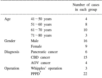
대상군의 짝짓기된 각 25명의 환자 중에서 남자는 16명 (64%), 여자는 9명(36%)였고, 평균연령은 Group A에서
Table 1. Distributions of age group, sex, postoperative diagnosis in patients (Group A and B)*
ꠚꠚꠚꠚꠚꠚꠚꠚꠚꠚꠚꠚꠚꠚꠚꠚꠚꠚꠚꠚꠚꠚꠚꠚꠚꠚꠚꠚꠚꠚꠚꠚꠚꠚꠚꠚꠚꠚꠚꠚꠚꠚꠚꠚꠚꠚꠚꠚꠚꠚꠚꠚꠚꠚꠚ Number of cases
in each group ꠏꠏꠏꠏꠏꠏꠏꠏꠏꠏꠏꠏꠏꠏꠏꠏꠏꠏꠏꠏꠏꠏꠏꠏꠏꠏꠏꠏꠏꠏꠏꠏꠏꠏꠏꠏꠏꠏꠏꠏꠏꠏꠏꠏꠏꠏꠏꠏꠏꠏꠏꠏꠏꠏꠏ
Age 41∼50 years 4
51∼60 years 8
61∼70 years 10
71∼80 years 3
Gender Male 16
Female 9
Diagnosis Pancreatic cancer 6
CBD cancer 15
AOV cancer 4
Operation Whipples’ operation 3
PPPD† 22
ꠏꠏꠏꠏꠏꠏꠏꠏꠏꠏꠏꠏꠏꠏꠏꠏꠏꠏꠏꠏꠏꠏꠏꠏꠏꠏꠏꠏꠏꠏꠏꠏꠏꠏꠏꠏꠏꠏꠏꠏꠏꠏꠏꠏꠏꠏꠏꠏꠏꠏꠏꠏꠏꠏꠏ
* The total number of patients in this table is 25 (in each group), Group A (n=25) and Group B (n=25) has equal distribution; †PPPD
= pylorus preserving pancreaticoduodenectomy.
ꠏꠏꠏꠏꠏꠏꠏꠏꠏꠏꠏꠏꠏꠏꠏꠏꠏꠏꠏꠏꠏꠏꠏꠏꠏꠏꠏꠏꠏꠏꠏꠏꠏꠏꠏꠏꠏꠏꠏꠏꠏꠏꠏꠏꠏꠏꠏꠏꠏꠏꠏꠏꠏꠏꠏꠏꠏꠏꠏꠏꠏꠏꠏꠏꠏꠏꠏꠏꠏꠏꠏꠏꠏꠏꠏꠏꠏꠏꠏꠏꠏꠏꠏꠏꠏꠏꠏꠏꠏꠏꠏꠏꠏꠏꠏꠏꠏꠏꠏꠏꠏꠏꠏꠏꠏꠏꠏꠏꠏꠏꠏꠏꠏꠏꠏ 59.84±8.47세, Group B에서 59.48±8.80세였다. 각 군에서
의 연령군별 분포를 보면 41∼50세가 4명, 51∼60세가 8명, 61∼70세가 10명, 71∼80세가 3명이었다. 진단은 각 군에서 췌장암 6예, 총담관암 15예, 바터씨팽대부암 4예였으며, 고 전적 휘플씨 수술은 두 군에서 각각 3예, 유문보존췌십이지 장절제술은 22예씩 시행되었으며, 췌공장 문합은 총 50예 모두에서 duct-to-mucosa 방법을 사용하였다(Table 1).
과거력상 당뇨, 고혈압, 심장질환, 간질환, 호흡기질환 등 이 있었던 환자는 Group A와 Group B에서 유의한 차이는 없었다(P=NS) (Table 2). 담도배액술전 황달, 발열, 우상복 부 동통 등의 담도염의 증상을 보인 환자는 각 군에서 6명 (24%)과 11명(44%)로 두 군 사이에 유의한 차이는 없었다 (P=0.21).
수술 전 총빌리루빈치의 최대값 및 배액술 후의 최소값은 Group A와 Group B에서 각각 12.774±5.44 mg/dl와 15.419
±10.13 mg/dl로 통계적으로 유의한 차이는 없었다(P=
0.313). 총빌리루빈치의 하강정도, 수술시간, 수술 중 및 수 술 후의 수혈량, 수술 후 재원기간 등에서도 유의한 차이는 없었다. 배액관의 제거는 Group A의 경우 수술 중에, Group B의 경우 수술 후 7일에 PTBD를 통한 담도조영술을 시행
하여 누출이나 폐쇄 등의 이상소견이 없는 경우에 시행하 였으며, 이를 기준으로 한 배액관 거치기간에도 두 군 사이 에 유의한 차이는 보이지 않았다(Table 3).
배액술과 관련하여 발생한 합병증은 출혈, 췌장염, 담관염 등이 있었으며, Group A와 B에서 각각 14예와 7예씩 발생 하여, Group A에서 배액술 관련 합병증 발생률이 통계적인 유의성에는 이르지 못하였지만 높은 경향을 보였다(P=
0.058). 수술 후 합병증으로는 창상감염, 위배출지연(기준:
10일 이상 위배액관을 삽관하여야 했던 경우), 문합부누출 등이 있었고 두 군에서 각각 4예, 3예, 3예와 3예, 1예, 1예가
Table 3. Perioperative variables associated with operation and biliary drainage
ꠚꠚꠚꠚꠚꠚꠚꠚꠚꠚꠚꠚꠚꠚꠚꠚꠚꠚꠚꠚꠚꠚꠚꠚꠚꠚꠚꠚꠚꠚꠚꠚꠚꠚꠚꠚꠚꠚꠚꠚꠚꠚꠚꠚꠚꠚꠚꠚꠚꠚꠚꠚꠚꠚꠚꠚꠚꠚꠚꠚꠚꠚꠚꠚꠚꠚꠚꠚꠚꠚꠚꠚꠚꠚꠚꠚꠚꠚꠚꠚꠚꠚꠚꠚꠚꠚꠚꠚꠚꠚꠚꠚꠚꠚꠚꠚꠚꠚꠚꠚꠚꠚꠚꠚꠚꠚꠚꠚꠚꠚꠚꠚꠚꠚꠚ
Group A Group B P
ꠏꠏꠏꠏꠏꠏꠏꠏꠏꠏꠏꠏꠏꠏꠏꠏꠏꠏꠏꠏꠏꠏꠏꠏꠏꠏꠏꠏꠏꠏꠏꠏꠏꠏꠏꠏꠏꠏꠏꠏꠏꠏꠏꠏꠏꠏꠏꠏꠏꠏꠏꠏꠏꠏꠏꠏꠏꠏꠏꠏꠏꠏꠏꠏꠏꠏꠏꠏꠏꠏꠏꠏꠏꠏꠏꠏꠏꠏꠏꠏꠏꠏꠏꠏꠏꠏꠏꠏꠏꠏꠏꠏꠏꠏꠏꠏꠏꠏꠏꠏꠏꠏꠏꠏꠏꠏꠏꠏꠏꠏꠏꠏꠏꠏꠏ
Total bilirubin, max. (mg/dl) 12.774±5.44 15.419±10.13 0.313
Total bilirubin, min. (mg/dl) 4.56±3.34 5.83±3.99 0.232
Daily diminution of total bil. (mg/dl/day)* 0.495±0.399 0.896±1.18 0.168
Operative time (mim) 408.2±48.58 395.6±78.58 0.499
Intraop. transfusion (units) 0.56±1.04 0.44±0.91 0.668
Postoperative transfusion (units)† 0.2±0.5 0.76±1.39 0.065
Length of stay, postop. (days) 27.12±10.77 25.48±12.26 0.618
Duration of drain (days) 14.96±7.65 16.73±10.29 0.498
ꠏꠏꠏꠏꠏꠏꠏꠏꠏꠏꠏꠏꠏꠏꠏꠏꠏꠏꠏꠏꠏꠏꠏꠏꠏꠏꠏꠏꠏꠏꠏꠏꠏꠏꠏꠏꠏꠏꠏꠏꠏꠏꠏꠏꠏꠏꠏꠏꠏꠏꠏꠏꠏꠏꠏꠏꠏꠏꠏꠏꠏꠏꠏꠏꠏꠏꠏꠏꠏꠏꠏꠏꠏꠏꠏꠏꠏꠏꠏꠏꠏꠏꠏꠏꠏꠏꠏꠏꠏꠏꠏꠏꠏꠏꠏꠏꠏꠏꠏꠏꠏꠏꠏꠏꠏꠏꠏꠏꠏꠏꠏꠏꠏꠏꠏ
*Maximum total bilirubin minimum total bilirubin/(date of drainage insertion - date of operation); †Packed RBC or whole blood, till postoperative 3rd day.
Table 2. Comorbid conditions
ꠚꠚꠚꠚꠚꠚꠚꠚꠚꠚꠚꠚꠚꠚꠚꠚꠚꠚꠚꠚꠚꠚꠚꠚꠚꠚꠚꠚꠚꠚꠚꠚꠚꠚꠚꠚꠚꠚꠚꠚꠚꠚꠚꠚꠚꠚꠚꠚꠚꠚꠚꠚꠚꠚꠚ
Group A Group B P
ꠏꠏꠏꠏꠏꠏꠏꠏꠏꠏꠏꠏꠏꠏꠏꠏꠏꠏꠏꠏꠏꠏꠏꠏꠏꠏꠏꠏꠏꠏꠏꠏꠏꠏꠏꠏꠏꠏꠏꠏꠏꠏꠏꠏꠏꠏꠏꠏꠏꠏꠏꠏꠏꠏꠏ
Diabetes militus 5 3 NS
Hypertension 4 5 NS
Cardiologic diseases 1 2 NS
Pulmonologic diseases 2 3 NS
Hepatologic diseases 1 1 NS
ꠏꠏꠏꠏꠏꠏꠏꠏꠏꠏꠏꠏꠏꠏꠏꠏꠏꠏꠏꠏꠏꠏꠏꠏꠏꠏꠏꠏꠏꠏꠏꠏꠏꠏꠏꠏꠏꠏꠏꠏꠏꠏꠏꠏꠏꠏꠏꠏꠏꠏꠏꠏꠏꠏꠏ
Total 13 (52%) 14 (56%) NS
ꠏꠏꠏꠏꠏꠏꠏꠏꠏꠏꠏꠏꠏꠏꠏꠏꠏꠏꠏꠏꠏꠏꠏꠏꠏꠏꠏꠏꠏꠏꠏꠏꠏꠏꠏꠏꠏꠏꠏꠏꠏꠏꠏꠏꠏꠏꠏꠏꠏꠏꠏꠏꠏꠏꠏ
Table 4. Profiles of complications related to biliary drainage and operation
ꠚꠚꠚꠚꠚꠚꠚꠚꠚꠚꠚꠚꠚꠚꠚꠚꠚꠚꠚꠚꠚꠚꠚꠚꠚꠚꠚꠚꠚꠚꠚꠚꠚꠚꠚꠚꠚꠚꠚꠚꠚꠚꠚꠚꠚꠚꠚꠚꠚꠚꠚꠚꠚꠚꠚ
Group A Group B p
ꠏꠏꠏꠏꠏꠏꠏꠏꠏꠏꠏꠏꠏꠏꠏꠏꠏꠏꠏꠏꠏꠏꠏꠏꠏꠏꠏꠏꠏꠏꠏꠏꠏꠏꠏꠏꠏꠏꠏꠏꠏꠏꠏꠏꠏꠏꠏꠏꠏꠏꠏꠏꠏꠏꠏ
Procedure-related complications 0.058
Bleeding 2 0
Pancreatitis 3 0
Cholangitis 9 7
Total 14 (56%) 7 (28%)
ꠏꠏꠏꠏꠏꠏꠏꠏꠏꠏꠏꠏꠏꠏꠏꠏꠏꠏꠏꠏꠏꠏꠏꠏꠏꠏꠏꠏꠏꠏꠏꠏꠏꠏꠏꠏꠏꠏꠏꠏꠏꠏꠏꠏꠏꠏꠏꠏꠏꠏꠏꠏꠏꠏꠏ
Postoperative complications 0.254
Wound problems 4 3
Delayed gastric emptying 3 1
Anastomosis leakage 3* 1†
Others 3‡ 4§
Total 13 (52%) 9 (36%)
ꠏꠏꠏꠏꠏꠏꠏꠏꠏꠏꠏꠏꠏꠏꠏꠏꠏꠏꠏꠏꠏꠏꠏꠏꠏꠏꠏꠏꠏꠏꠏꠏꠏꠏꠏꠏꠏꠏꠏꠏꠏꠏꠏꠏꠏꠏꠏꠏꠏꠏꠏꠏꠏꠏꠏ
*Pancreaticojejunostomy site leakage in 2 cases, choledochojejuno- stomy site leakage in 1 case; †Pancreaticojejunostomy site leakage in 1 case; ‡Postoperative bleeding in 1 case, pulmonary embolism 1 case, pancreatitis in 1 case; §Postoperative bleeding in 1 case, hepatic failure in 1 case, pleural effusion in 1 case, abdominal fluid collection in 1 case.
ꠏꠏꠏꠏꠏꠏꠏꠏꠏꠏꠏꠏꠏꠏꠏꠏꠏꠏꠏꠏꠏꠏꠏꠏꠏꠏꠏꠏꠏꠏꠏꠏꠏꠏꠏꠏꠏꠏꠏꠏꠏꠏꠏꠏꠏꠏꠏꠏꠏꠏꠏꠏꠏꠏꠏꠏꠏꠏꠏꠏꠏꠏꠏꠏꠏꠏꠏꠏꠏꠏꠏꠏꠏꠏꠏꠏꠏꠏꠏꠏꠏꠏꠏꠏꠏꠏꠏꠏꠏꠏꠏꠏꠏꠏꠏꠏꠏꠏꠏꠏꠏꠏꠏꠏꠏꠏꠏꠏꠏꠏꠏꠏꠏꠏꠏ 발생하였다.(22) 문합부 누출은 Group A에서는 췌공장문합
부누출(pancreaticojejunostomy leakage, 기준: 1주일 이상 50 ml/day 이상의 췌액이 누출되거나 경피적 천자에 의해 복강
내 췌액저류가 확인된 경우) 2예, 총담관공장문합부누출 (choledochojejunostomy leakage) 1예, Group B에서는 췌공장 문합부누출 1예가 있었다. 기타 수술 후 합병증으로는 Group A에서 출혈 1예, 폐색전 1예, 췌장염 1예가, Group B에서는 출혈 1예, 흉막삼출 1예, 복강내 액체저류 1예가 있었다. 이상과 같은 수술 후 발생한 합병증을 종합하였을 때 발생률에서 두 군 사이에 유의한 차이는 없었다(Table 4).
2) 병리학적 검사결과
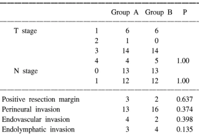
두 군 사이에서의 T stage 및 N stage는 통계적으로 유의 한 차이를 보이지 않았으며, 절제연 양성여부 및 신경주위/
혈관내/림프관내 암세포 침윤여부에서도 두 군 사이에 유 의한 차이는 없었다(Table 5). 병리 조직 중 직접적인 암세 포의 침윤이 없는 부분의 총담관에 대한 염증세포의 침윤 정도 및 총담관벽의 비후소견에 대하여 염증세포의 침윤 정도에 따라 염증소견이 없거나 미약한 경우를 Grade I, 중 등도 이하의 염증소견이 점막면에 국한되어 있거나 경한
Fig. 1. Microscopic finding of the CBD specimens (H & E stain, ×40). (A) Grade I, scanty or absent inflammation, (B) Grade II, mild to moderate inflammation confined to surface or diffusely mild inflammation, (C) Grade III, marked inflammation confined to surface or diffusely moderate inflammation, (D) Grade IV, diffusely severe inflammation.
Table 5. Assessments of pathological results
ꠚꠚꠚꠚꠚꠚꠚꠚꠚꠚꠚꠚꠚꠚꠚꠚꠚꠚꠚꠚꠚꠚꠚꠚꠚꠚꠚꠚꠚꠚꠚꠚꠚꠚꠚꠚꠚꠚꠚꠚꠚꠚꠚꠚꠚꠚꠚꠚꠚꠚꠚꠚꠚꠚꠚ Group A Group B P ꠏꠏꠏꠏꠏꠏꠏꠏꠏꠏꠏꠏꠏꠏꠏꠏꠏꠏꠏꠏꠏꠏꠏꠏꠏꠏꠏꠏꠏꠏꠏꠏꠏꠏꠏꠏꠏꠏꠏꠏꠏꠏꠏꠏꠏꠏꠏꠏꠏꠏꠏꠏꠏꠏꠏ
T stage 1 6 6
2 1 0
3 14 14
4 4 5 1.00
N stage 0 13 13
1 12 12 1.00
ꠏꠏꠏꠏꠏꠏꠏꠏꠏꠏꠏꠏꠏꠏꠏꠏꠏꠏꠏꠏꠏꠏꠏꠏꠏꠏꠏꠏꠏꠏꠏꠏꠏꠏꠏꠏꠏꠏꠏꠏꠏꠏꠏꠏꠏꠏꠏꠏꠏꠏꠏꠏꠏꠏꠏ
Positive resection margin 3 2 0.637
Perineural invasion 13 16 0.374
Endovascular invasion 4 2 0.398
Endolymphatic invasion 3 4 0.135
ꠏꠏꠏꠏꠏꠏꠏꠏꠏꠏꠏꠏꠏꠏꠏꠏꠏꠏꠏꠏꠏꠏꠏꠏꠏꠏꠏꠏꠏꠏꠏꠏꠏꠏꠏꠏꠏꠏꠏꠏꠏꠏꠏꠏꠏꠏꠏꠏꠏꠏꠏꠏꠏꠏꠏ
ꠏꠏꠏꠏꠏꠏꠏꠏꠏꠏꠏꠏꠏꠏꠏꠏꠏꠏꠏꠏꠏꠏꠏꠏꠏꠏꠏꠏꠏꠏꠏꠏꠏꠏꠏꠏꠏꠏꠏꠏꠏꠏꠏꠏꠏꠏꠏꠏꠏꠏꠏꠏꠏꠏꠏꠏꠏꠏꠏꠏꠏꠏꠏꠏꠏꠏꠏꠏꠏꠏꠏꠏꠏꠏꠏꠏꠏꠏꠏꠏꠏꠏꠏꠏꠏꠏꠏꠏꠏꠏꠏꠏꠏꠏꠏꠏꠏꠏꠏꠏꠏꠏꠏꠏꠏꠏꠏꠏꠏꠏꠏꠏꠏꠏꠏ
염증소견이 미만성으로 있는 경우를 Grade II, 심한 염증소 견이 점막면에 국한되어 있거나 중등도의 염증소견이 미만 성으로 관찰되는 경우를 Grade III, 미만성의 심한 염증소견 이 있는 경우를 Grade IV로 구분하였다(Fig. 1). 이러한 기준 하에서 Group A에 속한 환자들의 검체에서 통계적으로 유 의하게 더 심한 염증소견을 볼 수 있었다(P=0.001). 총담관 벽에서 관찰되는 비후의 정도를 비교하였을 때, 두 군은 각각 1.78±0.55 mm와 1.14±0.37 mm로 유의한 결과를 나타내었 다(P<0.001)(Table 6).
고 찰
팽대부 주위암에 의한 폐쇄성 황달이 있는 환자에서 수 술 전 담도배액술에 대해서는 많은 논란이 있다. 여기에 대 한 대부분의 연구들은 수술 전 담도배액술을 시행하는 것 자체가 수술 후의 유병률 및 사망률에 어떠한 영향을 미치 는가에 초점을 두고 시행되었다.(1,2,4,6,8,15) 그러나 서론 에서 언급한 바와 같이 많은 경우에서 수술 전 담도배액술 을 시행하지 않을 수 없는 환자들을 실제로 접하게 되며, 이런 경우 어떤 담도배액술을 택할 것인지에 대한 참고할 만한 연구는 아직 없는 실정이다.(1) 지금까지 담도배액술 의 종류에 따른 임상경과를 비교한 연구들은 대개 수술보 다는 시술 자체와 관련된 합병증 및 사망률을 거론하고 있 거나, 고식적 처치로서의 담도배액술과 관련한 비교들을 보고하고 있다.(1,6,18)
본 연구는 동일한 진단명과 수술명을 가진 환자를 짝짓 기를 통해 무작위로 선택함으로써, 수술 방법 또는 서로 다 른 진단명에 의한 오류를 배제하였다. 이를 바탕으로 저자 들은 팽대부 주위암으로 진단되었고 근치적 절제가 가능하 였던 환자들 중에서, 수술 전 담도배액술이 필요했던 환자
들을 대상으로, 수술 전 담도배액술을 시행할 수 밖에 없는 경우에 어떤 담도배액술이 수술 후의 임상경과에 더 유리 한가를 판단하고자 하였다.
결과에서 내시경적 담도배액술을 시행한 군과 경피적 담 도배액술을 시행한 군 사이의 임상경과 및 수술 중/후 변수 들에 통계적으로 유의한 차이는 없었으나, 내시경적 담도 배액술을 시행한 군에서 경피적 담도배액술을 시행한 군보 다 더 심한 총담관의 염증반응이 유발됨을 확인할 수 있었 다. 이는 내시경적 담도배액술이 가지는 특징인 폐쇄부위 를 가로질러 도관이 거치됨으로써 발생하는 담도계내로의 장내용물의 역류에 의한 세균 오염, 도관자체에 의한 폐쇄 로 유발되는 담도계의 세균집락형성, 그리고 담도폐쇄에 의해 만성 염증반응과 혈관울혈이 발생한 담관에 도관이 거치됨으로 인해 광범위한 점막손상과 담관섬유화가 유발 되는 것 등이 원인으로 생각된다.(1,16,23)
Karsten 등(16)은 총담관의 폐쇄를 유발하는 악성종양으 로 수술을 시행한 30명의 환자 중, 수술 전 총담관내 스텐트 를 삽입한 13명에서 총담관의 심한 염증성 변화와 섬유화, 궤양성 변화가 유발되며, 이로 인해 현저한 총담관의 비후 가 발생하고, 담즙배양시 수술 전 스텐트를 시행하지 않은 군에 비해 더 높은 세균배양 양성률(stent group:nonstent group=82%:24%)이 나타남을 보고하고 있어, 본 연구의 결 과를 뒷받침하고 있다. 또한 Taylor 등(24)은 췌십이지장절 제술을 시행한 597명의 환자를 대상으로 하여, 수술 전 스 텐트 삽입술을 시행한 군에서의 췌공장문합부누출의 빈도 가 스텐트삽입술을 시행하지 않은 군에 비해 더 높았다고 (10% vs. 4%: P=0.02) 보고하여, 스텐트 삽입에 의해 유발되 는 염증반응으로 인한 임상적 결과의 차이를 확인바 있다.
이런 보고들과 저자들의 경험을 바탕으로 하였을 때 내 시경적 담도배액술의 총담관에 대한 염증반응 유발은 수술 을 시행함에 있어 기술적 어려움을 초래하고, 절제연의 침 범여부를 판단하기 어려워 수술범위 결정이 힘들어지며, 이로 인한 수술시간의 연장과 수술 중 및 수술 후의 출혈을 포함한 합병증의 발생 가능성이 높아지는 등의 문제점과 연 관이 있을 것이라고 생각된다.
하지만 본 연구에서는 내시경적 담도배액술을 시행한 군 과 경피적 담도배액술을 시행한 군사이에 임상적 결과들의 통계적으로 유의한 차이는 나타나지 않았다. 이는 본 연구 에서 내시경적 담도배액술을 시행한 환자군의 크기가 경피 적 담도배액술을 시행한 환자 군에 비해 상대적으로 작아, 연구에 포함된 대상환자군의 크기가 크지 않았기 때문일 것이라 추측되고, 대상 환자군의 크기가 충분히 확보될 경 우, 통계적 유의성이 나타날 것으로 생각된다. 그러나 담도 배액술의 시술과 관련된 합병증 발생률을 살펴보면 내시경 적 담도배액술을 시행한 군에서 통계적 유의성은 없었으나 더 높은 경향을 보였는데, 이는 담관염의 발생은 비슷하였 지만 출혈과 췌장염이 내시경적 담도배액술을 시행한 군에 Table 6. Comparison of microscopic findings
ꠚꠚꠚꠚꠚꠚꠚꠚꠚꠚꠚꠚꠚꠚꠚꠚꠚꠚꠚꠚꠚꠚꠚꠚꠚꠚꠚꠚꠚꠚꠚꠚꠚꠚꠚꠚꠚꠚꠚꠚꠚꠚꠚꠚꠚꠚꠚꠚꠚꠚꠚꠚꠚꠚꠚ Group A Group B P-value ꠏꠏꠏꠏꠏꠏꠏꠏꠏꠏꠏꠏꠏꠏꠏꠏꠏꠏꠏꠏꠏꠏꠏꠏꠏꠏꠏꠏꠏꠏꠏꠏꠏꠏꠏꠏꠏꠏꠏꠏꠏꠏꠏꠏꠏꠏꠏꠏꠏꠏꠏꠏꠏꠏꠏ
Severity of inflammation, CBD wall*
I 12
II 7 12
III 14 1
IV 4 <0.001
ꠏꠏꠏꠏꠏꠏꠏꠏꠏꠏꠏꠏꠏꠏꠏꠏꠏꠏꠏꠏꠏꠏꠏꠏꠏꠏꠏꠏꠏꠏꠏꠏꠏꠏꠏꠏꠏꠏꠏꠏꠏꠏꠏꠏꠏꠏꠏꠏꠏꠏꠏꠏꠏꠏꠏ Thickness of CBD
1.78±0.55 1.14±0.37 <0.001 wall (mean±SD, mm)
ꠏꠏꠏꠏꠏꠏꠏꠏꠏꠏꠏꠏꠏꠏꠏꠏꠏꠏꠏꠏꠏꠏꠏꠏꠏꠏꠏꠏꠏꠏꠏꠏꠏꠏꠏꠏꠏꠏꠏꠏꠏꠏꠏꠏꠏꠏꠏꠏꠏꠏꠏꠏꠏꠏꠏ SD = standard deviation. *Severity of inflammation. Grade I = scanty or absent inflammation; Grade II = mild to moderate inflammation confined to surface or diffusely mild inflammation;
Grade III = marked inflammation confined to surface or diffusely moderate inflammation; Grade IV = diffusely severe inflammation.
ꠏꠏꠏꠏꠏꠏꠏꠏꠏꠏꠏꠏꠏꠏꠏꠏꠏꠏꠏꠏꠏꠏꠏꠏꠏꠏꠏꠏꠏꠏꠏꠏꠏꠏꠏꠏꠏꠏꠏꠏꠏꠏꠏꠏꠏꠏꠏꠏꠏꠏꠏꠏꠏꠏꠏꠏꠏꠏꠏꠏꠏꠏꠏꠏꠏꠏꠏꠏꠏꠏꠏꠏꠏꠏꠏꠏꠏꠏꠏꠏꠏꠏꠏꠏꠏꠏꠏꠏꠏꠏꠏꠏꠏꠏꠏꠏꠏꠏꠏꠏꠏꠏꠏꠏꠏꠏꠏꠏꠏꠏꠏꠏꠏꠏꠏ 서 많이 발생하였기 때문이다. 이러한 측면에서 내시경적
담도배액술이 경피경간 담도배액술에 비해 보다 침습적인 시술이라고 볼 수도 있지만 시술자의 숙련도에 따라 차이 가 있을 것으로 생각된다.
따라서 수술 전에 담도배액술을 시행할 경우에는 해당 병원에 어떤 형태의 배액술에 숙련된 시술자가 있는지를 충분히 고려해야 하겠고, 내시경적 배액술의 경우 출혈, 췌 장염과 같은 시술에 관련된 합병증들이 그 자체가 위험할 수 있을 뿐만 아니라 적절한 수술 시기를 지연시킬 수 있고, 수술해야 하는 췌장자체에 염증을 일으켜 수술을 어렵게 만들 수 있다는 것을 고려해야 하겠다.
결 론
팽대부 주위암에 대해 수술적 치료가 필요한 환자에서 선택적으로 수술 전 담도배액술을 시행하는 경우, 수술부 위의 염증성 변화를 유발함으로써 수술 중 및 수술 후의 합병증에 영향을 줄 수 있다고 보여지는 내시경적 담도배 액술 보다는, 경피경간 담도배액술이 보다 우선적으로 고 려되어야 할 것이고, 보다 객관적인 근거를 위해 추후 보다 많은 환자들을 대상으로 한 비교 연구가 필요할 것으로 생 각된다.
REFERENCES
1) Miguel ES, Thomas MK, Martin HP, Erik JAR, Huug O, Dirk JG. A meta-analysis on the efficacy of preoperative biliary drainage for tumors causing obstructive jaundice. Ann Surg 2002;236:17-27.
2) Armstrong CP, Dixon JM, Taylor TV, Davies GC. Surgical experience of deeply jaundiced patients with bile duct obstruction. Br J Surg 1984;71:234-8.
3) Greig JD, Krukowski ZH, Matheson NA. Surgical morbidity and mortality in one hundred and twenty-nine patients with obstructive jaundice. Br J Surg 1988;75:216-9.
4) Pitt HA, Gomes AS, Lois JF, Mann LL, Deutsch LS, Longmire WP Jr. Does preoperative percutaneous biliary drainage reduce operative risk or increast hospital cost? Ann Surg 1985;201:
545-53.
5) Pitt HA, Cameron JI, Postier RG, Gadacz TR. Factors affect- ing mortality in biliary tract surgery. Am J Surg 1981;141:
66-72.
6) Antony G S, Peter BC, R. Christopher G R, Richard RM, Adrian RW, Joseph WCL, et al. Randomized trial of endo- scopic versus percutaneous stent insertion in malignant obstr- uctive jaundice. Lancet 1987;2:57-62.
7) Stephen PP, Martin SK Jr., Kevin CC, Leslie HB, Murray FB.
Association of preoperative biliary drainage with postoperative
outcome following pancreaticoduodenectomy. Ann Surg 1999;
230:131-42.
8) Miguel ES, Rakesh SB, Erik AJR, Kees H, Huug O, Dirk JG.
The effect of preoperative biliary drainage on postoperative complications after pancreaticoduodenectomy. J Am Coll Surg 2001;192:726-34.
9) Derek JR, Philip S. External and internal-external biliary drainage in children with malignant obstructive jaundice.
Pediatr Radiol 2000;30:659-64.
10) Born P, Rosch T, Triptrap A, Bruhl K, Sandschin W, Allescher HD, et al. Long-term results of percutaneous transhepatic biliary drainage for benign and malignant bile duct strictures.
Scand J Gastroenterol 1998;33:544-9.
11) Lokich JJ, Kane RA, Harrison DA, McDermott WV. Biliary tract obstruction secondary to cancer: management guide- lines and selected literature review. J Clin Oncol 1987;
5:969-81.
12) Baijal SS, Dhiman RK, Gupta S. Percutaneous transhepatic biliary drainage in the management of obstructive jaundice.
Trop Gastroenterol 1997;18:167-71.
13) Kawarada Y, Higashiguchi T, Yokoi H, Vaidya P, Mizumoto R. Preoperative biliary drainage in obstructive jaundice. Hepa- togastroenterolory 1995;42:300-7.
14) Joseph PK, Bizer LS, Sprayrefen SS, Gliedman ML. Perc- utaneous transhepatic biliary drainage. Results and compli- cations in 81 patients. JAMA 1986;255:2763-7.
15) Gundry SR, Strodel WE, Knol JA, Eckhauser FE, Thompson NW. Efficacy of preoperative biliary tract decompression in patients with obstructive jaundice. Arch Surg 1984;119:
703-8.
16) Karsten TM, Coene PP, van Gulik TM, Bosma A, van Marle J, James J, et al. Morphologic changes of extrahepatic bile ducts during obstruction and subsequent decompression by endoprosthesis. Surgery 1992;111:562-8.
17) Karsten TM, Davids PH, van Gulik TM, Bosma A, Tytgat GN, Klopper PJ, et al. Effects of biliary endoprostheses on the extrahepatic bile ducts in relation to subsequent operation of the biliary tract. J Am Coll Surg 1994;178;343-52.
18) John S, Rolf PG, John C, Joseph A. Biliary decompression:
An institutional comparison of percutaneous and endoscopic methods. Radiology 1986;158:195-7.
19) Hatfield AR, Murray RS. Pre-operative biliary drainage in patients with obstructive jaundice. A comparison of the percutaneous transhepatic and endoscopic transpapillary routes. S Afr Med J 1981;60;737-42.
20) Yoshida S, Kiyota K, Mukai H, Nishimura K, Cho E, Kobayashi M, et al. Clinical evaluation of endoscopic retro- grade biliary drainage (ERBD) in comparison with that of precutaneous transhepatic biliary drainage (PTBD). Nippon Shokakibyo Gakkai Zasshi 1985;82:638-47.
21) Jeremy A, Tibble and Stuart R. Cairns. Role of endoscopic endoprostheses in proximal malignant biliary obstruction. J
ꠏꠏꠏꠏꠏꠏꠏꠏꠏꠏꠏꠏꠏꠏꠏꠏꠏꠏꠏꠏꠏꠏꠏꠏꠏꠏꠏꠏꠏꠏꠏꠏꠏꠏꠏꠏꠏꠏꠏꠏꠏꠏꠏꠏꠏꠏꠏꠏꠏꠏꠏꠏꠏꠏꠏꠏꠏꠏꠏꠏꠏꠏꠏꠏꠏꠏꠏꠏꠏꠏꠏꠏꠏꠏꠏꠏꠏꠏꠏꠏꠏꠏꠏꠏꠏꠏꠏꠏꠏꠏꠏꠏꠏꠏꠏꠏꠏꠏꠏꠏꠏꠏꠏꠏꠏꠏꠏꠏꠏꠏꠏꠏꠏꠏꠏ Hepatobiliary Pancreat Surg 2001;8:118-23.
22) Kim SH, Park SJ, Jang JY, Park YC, Lee KU, Choe KJ, et al. Forty-year experience with the pancreatoduodenectomy. J Korean Surg Soc 2000;59:643-50.
23) Mark GC, Andreas NA. Internal stenting in malignant biliary
obstruction. World J Surg 2001;25:355-61.
24) Taylor AS, Charles JY, John LC, Henry AP, Keith DL. Do preoperative biliary stents increase postpancreaticoduodenec- tomy complications? J Gastrointest Surg 2000;4:258-68.