Percutaneous transhepatic biliary drainage (PTBD) performed in obstructive biliary disease is an effective interventional procedure for pallia- tive treatment that alleviates jaundice or chol- angitis through bile duct decompression and bile drainage. Recently, with the increase of invasive procedures such as PTBD and liver biopsy in the diagnosis and treatment of hepatobiliary and pan- creatic diseases, the prevalence of complications like hemorrhage or sputum sanguineum is also increasing.1,2 The authors have experienced the case of endoscopic removal of a remained draw- string after PTBD on jaundice due to choledocho- lithiasis.
CASE
An 85-year-old male patient visited an external hospital due to nausea and high fever that had been present since 3 days before the visit, where he received abdominal ultrasonography and showed distal choledocholithiasis and bile duct dilatation. Accordingly, acute cholangitis was sus- pected, and he was transferred to the current hospital. Two years ago, he had received neph- rectomy on the right side due to kidney cancer, and has been on endocrinotherapy for prostatic cancer since a year ago. He had no other medical history such as diabetes, hepatitis or tuberculosis.
Case Report
Endoscopic Removal of Remained Drawstring After Percutaneous Transhepatic Biliary Drainage
Tae Wook Yoon, Geun Yong Jung, Young Jun Park, Jun Young Choi, Jee Hwan Jung, Tae Gyoon Kim Department of Internal Medicine, VHM Medical center, Seoul, Korea
The percutaneous transhepatic biliary drainage (PTBD) is an effective intervention as a palliative therapy for relieving a jaundice and cholangitis. It may be used in place of Endoscopic retrograde cholangiopancreatography (ERCP) in the obstructive biliary disease. Recently, by developing invasive procedures, the incidence of the complications such as bleeding and perforation has been increasing in the diagnosis and treatment of hepatobiliary disease. We report here on a case of remained drawstring after PTBD in a 85-year-old man. The patient was conducted PTBD for relieving a jaundice and cholangitis. And then the patient had complained of abdominal pain constantly. A few days later, we removed PTBD and attempted ERCP for removal of CBD stone.
The ERCP showed remained drawstring around ampulla of vater and we removed it by IT knife. The drawstring was successfully removed.
Key Words: Acute cholangitis, Endoscopic retrograde cholangiopancreatography (ERCP), Percutaneous transhepatic biliary drainage (PTBD)
Corresponding Author: Tae Gyoon Kim, Department of Internal Medicine, VHM Medical center, 53, Jihwangdo-ro 61-gil, Gangdong-gu, Seoul 05368, Korea
Tel: +82-2-2225-1114 Fax: +82-2-2225-4374 Email: [email protected]
Received:
Revised:
Accepted:
Jun. 23, 2015 Sep. 02, 2015 Sep. 16, 2015
Kosin Medical Journal 2016;31:173-178.
The vital signs at the time he was hospitalized were as follows: blood pressure 104/71 ㎜Hg, pulse rate 108 times per minute, respiration rate 18 times per minute, and temperature 36.5℃. There was nothing abnormal in auscultation. There was also no hepatosplenomegaly in abdominal pal- pation, and no oppressive pain or rebound tenderness. The complete blood cell count was as follows: white blood cell count 8290/㎣, hemo- globin 11.2 g/dL, platelet count 152000/㎣. In se- rum biochemistry test, total protein was 6.1 g/dL, Albumin 3.8 g/dL, AST 128 U/dL, ALT 248 U/dL, total bilirubin 3.0 ㎎/dL, alkine phosphatase 401 U/dL, glutamyl transpeptidase 971 U/dL, amylase 99 U/L, lipase 29 U/L, procalcitonin 2.6 ng/mL, hs CRP 39.5 ㎎/L, and PT was INR 1.03. Serum electrolyte was Na 141 mEq/L, K 4.2 mEq/L, and
Cl 89 mEq/L.
In abdominal CT scan, choledochal duct and intrahepatic duct were dilated, and 9㎜ gallstones were observed in the distal choledochal duct. (Fig.
1) The gallstones in the patient’s bile duct were about to be removed through endoscopic retro- grade cholangiopancreatography (ERCP), but the ampulla of Vater were swollen and cannulation failed; thus, PTBD was performed. The results of the blood test performed 4 days after visiting the hospital were AST 26 U/dL, ALT 53 U/dL, total bilirubin 0.6 mg/dL, alkine phosphatase 258 U/dL, glutamyl transpeptidase 610 U/dL, hs CRP 21.2
㎎/L, and there was no fever. The symptoms of nausea were improved, but the patient showed mild right upper quadrant abdominal pain. There were no other clinical symptoms and the vital
Fig. 1. Black arrow in abdomen CT demonstrating 9mm sized biliary stone in the distal common bile duct.
signs were normal, and indexes of liver function and inflammation improved in the blood test.
Thus, ERCP was scheduled for 2 weeks later while observing the progress of the right upper quad- rant abdominal pain and performing antibiotic treatment.
The patient constantly suffered from right up- per quadrant abdominal pain, which was so sig- nificantly worsened that he could barely move in the morning on the 14th day of hospitalization;
thus, PTBD was removed. Only the catheter was removed while the drawstring remained, so the tip of the remaining drawstring was fixed with a mosquito forceps, and ERCP was performed to
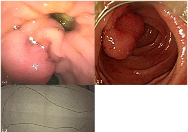
check the remaining gallstones and swollen am- pulla of Vater in the afternoon. ERCP showed the drawstring that tightens the ampulla of Vater on the duodenum (Fig. 2-1). The drawstring remain- ing in the body was deep inside the mucous mem- brane around the ampulla of Vater, which could be taken out with the IT knife used in endoscopic sphincterotomy (Fig. 2-2, 2-3). Iatrogenic ulcer was observed around the ampulla of Vater, which may have been due to the tightening of the draw- string, and ERCP would be performed later to check the remaining gallstones due to the risk of tresis.
The patient no longer complained of abdominal Fig. 2-1. Endoscopy showed an ampulla of vater tightened by a remained drawstring after removing percutaneous transhepatic biliary drainage.
Fig. 2-2. Endoscopy showed an ampulla of vater after removing a remained drawstring.
Fig. 2-3. The remained drawstring to prevent a drainage tube from falling into the body.
Kosin Medical Journal 2016;31:173-178.
pain, and ERCP was performed again on the 21st day of hospitalization. He was discharged from the hospital after verifying that there were no gall- stones in the choledochal duct. Since then, he has been under longitudinal tracking as an outpatient.
DISCUSSION
ERCP was originally adopted as a diagnostic procedure, but it is currently being used as an alternative to surgery for removing gallstones in the choledochal duct. ERCP, sphincterotomy and gallstone removal are safe and effective proce- dures to remove gallstones in the extrahepatic bile duct, and multiple retrospective studies have shown that there is only a 5 ~ 10% of possibility of complications such as enterobrosia, hemor- rhage, pancreatitis and cholangitis.3 Moreover, in about 5 ~ 10% of cases, for reasons such as post- operative anatomy or inevitable difficulty in en- tering the ampulla of Vater, PTBD is a highly use- ful interventional procedure.3-5 However, less in- vasive methods are preferred in treatment, and thus the use of PTBD is decreasing compared to ERCP, and the incidence of complications from PTBD is reported as 9 ~ 61%.1,6,7 In this case, the ampulla of Vater was swollen and entry to the am- pulla of Vater using ERCP was failed, and thus PTBD was used.
Nennstiel et al.1 pointed out that the major complications of PTBD include obstruction, ecto- py, cholangitis, defluvium, hemorrhage, and pain,
and risk factors include malignant diseases, anamnesis of experiencing complications before, bilateral drainage, and stricture of proximal bili- ary tree; they thus encouraged the consideration of other procedures when there is at least one of these risk factors. This case did not have the aforementioned risk factors, but the drawstring was tightening the ampulla of Vater when the PTBD was removed, and thus was endoscopically removed. The cholangiography taken during PTBD reveals that the drawstring was entangled and fixated around the ampulla of Vater, and thus it may have not been removed when removing PTBD (Fig. 3).
When performing and removing PTBD based on this case, there are two things to note. First, when performing PTBD after failing to have access to the ampulla of Vater and thus failing ERCP, there are cases in which the catheter is drawn out to the duodenum8 and fixed by expecting it to play the role of a guide to expand the choledochal duct around the ampulla of Vater and approach the choledochal duct. But if the ampulla of Vater is swollen, as shown in this case, it is necessary to note that the drawstring may tighten the ampulla of Vater. Second, when removing the catheter, unlock the locking device or cut the catheter at the top of the locking device to untie the fixed drawstring to take out the catheter. Here, it is necessary to check whether the drawstring in the catheter is also removed.
Recently, methods that are less painful to the patients and less invasive are preferred over surgi-
cal methods.9,10 This report describes an endo- scopic removal of drawstring, as there have been no reports of cases in which the drawstring re- maining in the body was removed after removing PTBD was performed after a failure of ERCP.
REFERENCES
1. Nennstiel S, Weber A, Frick G, Haller B, Meining A, Schmid RM, et al. Drainage-related Complications in Percutaneous Transhepatic Biliary Drainage: An Analysis Over 10 Years. J Clin Gastroenterol 2014.
2. Oh HC, Lee SK, Lee TY, Kwon S, Lee SS, Seo DW, et al. Analysis of percutaneous transhepatic cholangioscopy-related complications and the risk factors for those complications. Endoscopy 2007;39:731-6.
3. Enochsson L, Swahn F, Arnelo U, Nilsson M, Löhr M, Persson G. Nationwide, population-based da- ta from 11,074 ERCP procedures from the Swedish Registry for Gallstone Surgery and ERCP.
Gastrointestinal Endoscopy 2010;72:1175-84.
4. Eickhoff A, Schilling D, Jakobs R, Weickert U, Hartmann D, Eickhoff JC, et al. Long-term out- come of percutaneous transhepatic drainage for Fig. 3. The twisted biliary tube on the duodenum.
Kosin Medical Journal 2016;31:173-178.
benign bile duct stenoses. Rocz Akad Med Bialymst 2005;50:155-60.
5. Nikola Y. Kolev, Valentin L, Ignatov, Anton Y.
Tonev, Aleksanmdar K. Zlatorov, Elitsa P. Encheva, Tanya N. Kirilova, et al. BILIARY DRAINAGE.
Journal of IMAB–Annual Proceeding (Scientific Papers) 2013;19:465-9.
6. Günther R, Schild H, Thelen M. Review article:
percutaneous transhepatic biliary drainage: ex- perience with 311 procedures. Cardiovasc In- tervent Radiol 1988;11:65-71.
7. Mayumi T, Someya K, Ootubo H, Takama T, Kido T, Kamezaki F, et al. Progression of Tokyo Guidelines and Japanese Guidelines for manage- ment of acute cholangitis and cholecystitis. J
UOEH 2013;35:249-57.
8. Lee TH, Park SH, Lee SH, Lee CK, Lee SH, Chung IK, et al. Modified rendezvous intrahepatic bile duct cannulation technique to pass a PTBD cathe- ter in ERCP. World J Gastroenterol 2010;16:5388.
9. Kühn JP, Busemann A, Lerch MM, Heidecke CD, Hosten N, Puls R. Percutaneous biliary drainage in patients with nondilated intrahepatic bile ducts compared with patients with dilated intrahepatic bile ducts. AJR Am J Roentgenol 2010;195:851-7.
10. Oberholzer K, Pitton M, Mildenberger P, Lechner C, Düber C, Thelen M. [The current value of percutaneous transhepatic biliary drainage].
RoFo 2002;174:1081-8.