Correspondence:Kwang-Woong Lee, Department of Sur- gery, Seoul National University College of Medi- cine, 101 Daehak-ro, Jongno-gu, Seoul 110-744, Korea
Tel: +82-2-760-2511, Fax: +82-2-766-3975 E-mail: [email protected]
Received : November 29, 2011, Revised : August 11, 2012, Accepted : August 13, 2012
Sang Myung Woo and Hyun Beom Kim contributed equally to this work as first authors.
New Strategy in Cases of Failed Endoscopic Intervention of Biliary Strictures after Living Donor Liver Transplantation:
Percutaneous Transhepatic Biliary Stent Insertion and Subsequent Endoscopic Treatment
Sang Myung Woo, M.D.
1, Hyun Beom Kim, M.D.
1, Kwang-Woong Lee, M.D.
1,2, Woo Jin Lee, M.D.
1, Young-Kyu Kim, M.D.
1, Sung-Sik Han, M.D.
1and Sang-Jae Park, M.D.
1Center for Liver Cancer, National Cancer Center
1, Goyang,
Department of Surgery, Seoul National University College of Medicine
2, Seoul, Korea
Background: In cases of endoscopic intervention treatment for biliary stricture which fail, a percutaneous approach can be subsequently attempted. However, the quality of life is lower for those patients with percutaneous transhepatic biliary drainage (PTBD) tubes than those with endoscopic retrograde biliary drainage tubes. In this study, we report the outcome of the application of percutaneous transhepatic biliary stenting (PTBS) for use in subsequent endoscopic treatment of biliary stricture after living donor liver transplantation (LDLT).
Methods: Of 165 patients who underwent LDLT, 40 (24.2%) were diagnosed with anastomotic biliary strictures. Of these patients, seven agreed to treatment using PTBS using a plastic stent with endoscopic follow-up instead of treatment by insertion of a PTBD tube, and were enrolled in this study.
Results: In all seven patients, the use of this technique enabled effective advancement of a guide wire and successful placement of one or two plastic stents (7 or 10 Fr) into the PTBD tract. There were no PTBS-related complications associated with the procedure. The median duration for stent use was 40.3 weeks (range; 27.6~65.0). Upon final removal of all stents, the stricture had been resolved in four (57%) of the seven patients.
Conclusions: Our study data suggested that, after failed use of ERCP in the treatment of biliary stricture after LDLT, the use of PTBS and ERCP may be an effective and safe treatment.
Key Words: Bile ducts, Complication, Bile duct stent 중심 단어: 담도, 합병증, 담도 스텐트
Introduction
Liver transplantation (LT) is a widely accepted treat- ment for end-stage liver disease and early staged hep- atocellular carcinoma. Despite great improvements in the surgical techniques and standardization of the
method of biliary reconstruction, the biliary tract is still
the most common site for postoperative complica-
tions(1). Biliary complications are more common in
living donor LT (LDLT) patients than in deceased do-
nor LT (DDLT) patients, occurring in up to 32% of
LDLT patients compared to 10∼15% of patients who
undergo DDLT(2,3). The non-operative management
of biliary complications following LT has become
standard practice with primarily endoscopic techniques
as the preferred diagnostic and therapeutic modalities,
obviating the need for surgery in a majority of
patients. However, technically, LDLT presents a chal-
lenge, with the most common reason for failure being
the inability to traverse the stricture and complex pe-
ripheral anastomosis, rendering plastic stent placement
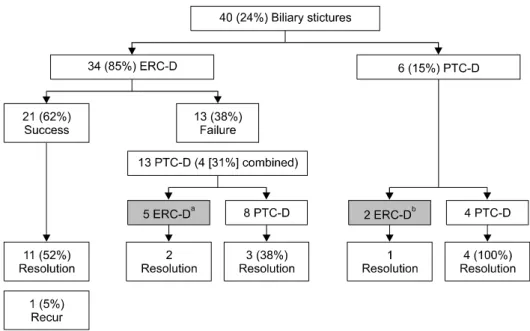
Fig. 1. Among 13 patients who had failure in initial endoscopic ap- proach and received PTBD. Five pa- tients
awho agreed with endo- scopic follow-up instead of PTBD tubes were enrolled this study;
Among 6 patients for whom PTBD was done as an initial approach,
b
two patients who wanted endo- scopic follow-up was also enrolled in this study. Abbreviations: ERC- D, endoscopic retrograde chol- angiography with dilation; PTC-D, percutaneous transhepatic chol- angiography with dilation.
difficult(4).
The success rates of endoscopic treatment for anas- tomotic strictures in LDLT are significantly lower than in DDLT at 60∼75%(3-6). In failed cases, a percuta- neous approach can be attempted. However, an ex- ternal catheter has a major potential for infection and dislodgement, and it is associated with catheter main- tenance problems as well as with discomfort for the patient. In addition, the number of interventions (including tube management) was reported to be high- er in the percutaneous intervention group than in en- doscopic treatment group(7).
Percutaneous transhepatic biliary stenting (PTBS) is a well-established interventional radiology procedure used in patients with biliary obstruction for decom- pression of intra- and extrahepatic bile ducts. Subse- quent endoscopic treatment after PTBS will also have a positive impact on quality of life in failed cases with initial endoscopic treatment. Therefore, we adopted a strategy of plastic stent insertion using percutaneous transhepatic biliary drainage (PTBD) and subsequent balloon dilatation and plastic stent change using endo- scopic retrograde cholangiopancreatography (ERCP).
The aim of this study was to evaluate both the feasi- bility of this new strategy in treatment of biliary stric- ture after LDLT and the clinical outcomes of this protocol.
Materials and Methods 1) Patients
Of 165 patients who underwent LDLT with duct-to- duct biliary reconstruction between February 2005 and September 2009 at the National Cancer Center, Goyang, Korea, 40 (24.2%) were diagnosed with anas- tomotic biliary strictures. Of these 40 patients, 35 pa- tients (87%) had undergone ERCP as an initial ap- proach (Fig. 1). Among them, 14 patients had failure in initial endoscopic approach and received PTBD.
Among them, five patients who agreed with endo- scopic follow-up instead of PTBD tubes were enrolled this study. Among six patients for whom PTBD was done as an initial approach, two patients who wanted endoscopic follow-up were also enrolled in this study.
Therefore, PTBS insertion for subsequent endoscopic treatment was performed in seven patients. All pa- tients had intolerable pain or recurrent bile leakage around the PTBD tube, and informed consents were obtained before all procedures.
2) Study definition
The first clue that a biliary complication has arisen
may be an asymptomatic increase in serum trans-
aminase or bilirubin levels. Symptoms are often
non-specific and include itching and fever. The initial
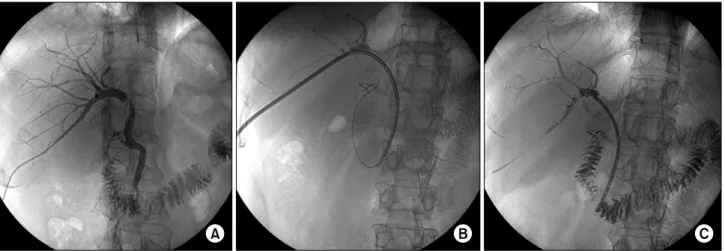
Fig. 2. Percutaneous transhepatic biliary stent insertion. (A) The anastomotic stricture before stent insertion. (B) The placement of the guidewires and the insertion of the plastic stent at the anastomotic stricture. (C) The plastic stent left in place after treatment.
evaluation should include ultrasonography with a Doppler examination of the hepatic vessels, followed by a liver biopsy or biliary imaging, depending on the pattern of liver test abnormalities. Anastomotic biliary stricture was defined as a dominant narrowing at the anastomotic site, without effective passage of contrast material, as identified by cholangiography. Ischemic biliary strictures, defined as strictures that extend more than 0.5 cm proximal to the anastomosis, were ex- cluded from this study. Minimal narrowing at the site of the anastomosis that did not impede the flow of contrast material was not considered a clinically sig- nificant stricture and hence was also excluded.
A successful outcome after completion of therapy was defined as the sustained improvement in liver en- zymes shown by the results of laboratory studies and by the patency of the anastomotic site shown on cholangiography. The biliary anastomosis was consid- ered patent when an 8∼10 mm retrieval balloon could be withdrawn through the anastomosis, followed by prompt emptying of contrast material observed with fluoroscopy during ERCP.
3) PTBS insertion
All PTBD procedures were conducted throughout the study by the same experienced interventional radi- ologist (H.B. Kim). Under the guidance of ultrasound or a C-arm for fluoroscopy, the postero-inferior or
postero-superior segmental duct of the transplanted liver was targeted with a 21-gauge puncture needle.
After obtaining a cholangiogram, a guidewire (Terumo Medical Co., Tokyo, Japan) was passed through the stricture segment of the bile duct (anastomosis site).
Then, an 8.5 Fr drainage catheter with multiple side holes (Cook Medical Inc., Bloomington, IN, USA) was placed across the stricture. A two-stage procedure for transhepatic stent placement was used. An 8.5 Fr drainage catheter was placed on the first day. Five to 7 days later, the transhepatic tract and the anastomotic stricture were dilated up to 12 Fr, and a 7 or 10 Fr plastic stent (Wilson-Cook Medical Inc., Winston- Salem, NC, USA) was inserted (Fig. 2). The distal ends of the stents were placed across the ampulla into the duodenum to make subsequent endoscopic treatments easy.
Procedure-related cholangitis was defined as the on- set of fever (>38.2
oC) and/or leukocytosis (white blood cell count >10,000/mm
3) along with abdominal pain or tenderness in the right upper quadrant.
Pancreatitis was diagnosed when serum amylase levels
rose to more than 3 times the normal limit (60∼180
U/L) with notable persistent abdominal pain for more
than 24 hours after the procedure. Significant bleeding
was defined as a requirement either for a blood trans-
fusion of more than 2 units or for hemostatic proce-
dures after PTBS.
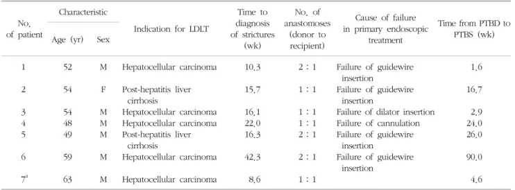
Table 1. Patient characteristics
No.
of patient
Characteristic
Indication for LDLT
Time to diagnosis of strictures
(wk)
No. of anastomoses
(donor to recipient)
Cause of failure in primary endoscopic
treatment
Time from PTBD to PTBS (wk) Age (yr) Sex
1 2 3 4 5 6 7
a52 54 54 48 49 59 63
M F M M M M M
Hepatocellular carcinoma Post-hepatitis liver
cirrhosis
Hepatocellular carcinoma Hepatocellular carcinoma Post-hepatitis liver
cirrhosis
Hepatocellular carcinoma Hepatocellular carcinoma
10.3 15.7 16.1 22.0 16.3 42.3 8.6
2:1 1:1 1:1 1:1 2:1 2:1 1:1
Failure of guidewire insertion
Failure of guidewire insertion
Failure of dilator insertion Failure of cannulation Failure of guidewire
insertion
Failure of guidewire insertion
1.6 16.7 2.9 24.0 26.0 90.0 4.6 Abbreviations: LDLT, living donor liver transplantation; PTBD, percutaneous transhepatic biliary drainage; PTBS, percutanous trans- hepatic biliary stenting.
a
PTBD was used as initial approach for biliary stricture.
4) Subsequent ERCP
The ERCP was performed by one of two experi- enced endoscopists (S.M. Woo and W.J. Lee). ERCP was scheduled electively at intervals of 2∼3 months for evaluation of the stricture and for stent exchange after PTBS. ERCP was performed earlier in cases of cholangitis or worsening liver function indicated by test results. At subsequent ERCP, the stents were ex- tracted using the polypectomy snare or a large forceps. After selective biliary cannulation, a chol- angiogram was obtained to evaluate the biliary anastomosis. When the stricture was determined to be clinically significant (as defined above), a biliary sphincterotomy was performed to allow placement of multiple stents. The anastomotic stricture was then di- lated by using high-pressure pneumatic biliary dilation balloons that ranged in size 4∼10 mm (Hurricane RX Dilation Balloons; Microvasive Endoscopy, Boston Scientific Co., Natick, MA, USA). The balloon diameter was limited by the diameter of the intrahepatic bile ducts. The maximum number of polyethylene straight stents that could be accommodated within the stricture was inserted. A cholangiogram was then obtained by occluding the distal common bile duct with a dis- tended retrieval balloon (8.5∼15 mm). If the chol-
angiogram showed a patent anastomotic stricture, then treatment was discontinued. If the stricture persisted, balloon dilation and maximal stent placement were repeated. The patient then returned at intervals of 2∼
3 months for further courses of endoscopic treatment until the stricture was patent or until the completion of 12 months of therapy.
5) Immunosuppression and peri-procedural mana- gement
All the patients were initially on a triple-drug im- munosuppressive regimen that consisted of cyclo- sporine A or tacrolimus, corticosteroids, and mycophe- nolate mofetil, with the latter two medications being tapered off at approximately 3 months and 1 year af- ter LT, respectively. When taking the immunocompro- mised status into consideration, antibiotics (1 g cefo- taxime, or 200 mg ciprofloxacin intravenously for pa- tients with allergies) were routinely administered be- fore and after an endoscopy or PTBD for the initial and subsequent procedures.
Results
The seven patients were six males and one female
and the median age of the recipients was 54 years
Table 2. Outcomes of percutanous transhepatic biliary stenting (PTBS) insertion, liver chemistries in individual patients No. of
patient
Success of PTBS
PTBS-related complication
Total bilirubin (mg/dL) Alkaline phosphatase (IU/L) Alanine aminotransferase (IU/L)
Pre Post Pre Post Pre Post
1 2 3 4 5 6 7
Yes Yes Yes Yes Yes Yes Yes
No No No No No No No
1.8 5.6 2.1 0.7 0.8 6.1 1.8
1.4 5.5 1.0 0.8 0.7 5.5 1.4
750 820 72 325 95 278 144
478 717 63 278 116 261 332
45 251 155 16 15 121 18
732 169 9 13 22 105 12
Table 3. Outcomes of percutanous transhepatic biliary stent insertion and subsequent endoscopic retrograde cholangiopancreatog- raphy (ERCP), balloon size, maximum number of stents in individual patients
No. of patient Stent duration (wk) Total No. of subsequent ERCP
Maximum balloon size (mm)
Maximum No. of
stents Current state 1
2 3 4 5 6 7
64.9 37.3 40.3 27.6 65.0 32.6 54.0
5 1 3 2 3 2 4
6 6 8 6 6 6 6
2 1 2 2 1 2 2
Success Failure
aSuccess Failure
bSuccess Ongoing Success
a