외상성 췌장 손상에서 내시경적 담췌관 조영술의 역할
울산대학교 의과대학 서울아산병원 외과학교실 외상 및 중환자외과
인제대학교 의과대학 해운대백병원 외과학교실1
울산대학교 의과대학 서울아산병원 내과학교실 위장관내과2
정민영∙김영환∙경규혁1∙이성구2∙홍석경
─ Abstract ─
The Role of Endoscopic Retrograde Cholangiopancreatography (ERCP) in the Treatment of Traumatic Pancreas Injury
Min-young Jeong, M.D., Young-hwan Kim, M.D., Kyu-hyouck Kyoung, M.D.1, Sung Koo Lee, M.D.2, Suk-kyung Hong, M,D., Ph.D.
Division of Trauma and Surgical Critical Care, Department of General Surgery, University of Ulsan College of Medicine, Asan Medical Center,
Department of General Surgery, Inje University, Haeundae Paik Hospital1 Division of Gastroenterology, Department of Internal Medicine, University of Ulsan College of Medicine, Asan Medical Center2
Purpose: Blunt pancreatic injury has a high mortality rate, especially if adequate management is delayed.
Although many guidelines exist for diagnosis and treatment, there is no consensus to date. Therefore, we ana- lyzed the role of endoscopic retrograde cholangiopancreatography (ERCP) as a diagnostic and therapeutic tool for the treatment of traumatic pancreatic injury.
Methods: We retrospectively reviewed the electronic medical records (EMR) database at Asan Medical Center (Seoul, South Korea) to identify all patients diagnosed with trauma to the pancreas between June 2003 and December 2010. Clinical and operative findings, CT (computed tomography) images, and ERCP findings were assessed.
Results: A total of 40 patients were evaluated in this study. Of these, 14 patients underwent diagnostic ERCP, and 26 did not. Of the 14 patients who underwent diagnostic ERCP, 5 were found to have normal pan- creatic ducts, thereby preventing a needless laparotomy in these patients. Of the patients diagnosed with ductal injury, four were treated with endoscopic intervention, and four underwent an exploratory laparotomy. The remaining patient was treated with radiologic intervention (percutaneous drainage) to manage pancreatic pseudocyst formation.
Conclusion: Our findings suggest that ERCP is a beneficial diagnostic and therapeutic modality for the treat- ment of traumatic pancreatic injury. (J Korean Soc Traumatol 2011;24:136-142)
Key Words: Pancreatic duct, Endoscopic retrograde cholangiopancreatography
� Address for Correspondence : Suk-kyung Hong, M.D., Ph.D.
Division of Trauma and Surgical Critical Care,Department of Surgery, Ulsan University College of Medicine, Asan Medical Center, 388-1 Pungnap-2dong, Songpa-gu, Seoul 138-736, Korea
Tel : 82-2-3010-3510, Fax : 82-2-3010-6701, E-mail : [email protected]
접수일: 2011년 9월 15일, 심사일: 2011년 9월 20일, 수정일: 2011년 10월 13일, 승인일: 2011년 10월 19일
I. BACKGROUND
Pancreatic injury is not common in blunt trauma, but, when it occurs, has a high mortality rate, especially if ade- quate management is delayed.(1,2) The reported morbidity rate in patients that suffer traumatic pancreatic injury is 45%, and can increase to as high as 60% if treatment is delayed.(3~6) Complications arise following surgical treat- ment of injuries to the pancreas in 26% to 86% of patients.(7) Thus, trauma surgeons find it challenging to diagnose and treat traumatic injury to the pancreas, and are cautious in recommending surgery.
The primary factor associated with mortality and mor- bidity in patients with traumatic injury to the pancreas is integrity of the main pancreatic duct.(3,5,7,8) Although many guidelines exist for diagnosis and treatment, there is no widely accepted treatment protocol to date.(9~11) Endoscopic retrograde cholangio pancreatography (ERCP) is regarded as the most accurate tool for evaluating injury to the main pancreatic duct, with subtotal or total pancreatec- tomy recommended in patients with injury to the main pancreatic duct (Fig. 1).(7) However, this type of surgery is itself associated with high rates of morbidity, including bleeding, intra-abdominal infection, anastomosis site leakage, and pancreatico-duodenal fistula in the acute phase, and the development of pseudocysts, diabetes, and chronic pan-
creatitis in the chronic phase.(7) Therefore, we analyzed the functionality of ERCP in traumatic pancreatic injury as a diagnostic and therapeutic tool.
II. MATERIALS AND METHODS
A retrospective review of the electronic medical records (EMR) database at Asan Medical Center (Seoul, South Korea) was performed to identify all patients diagnosed with traumatic injury to the pancreas between June 2003 and December 2010. A total of 59 patients were initially identified using the search term “pancreas injury”. After excluding those who underwent surgery at another hospital before admission and those who had no suspected pancreas injury on initial CT, 40 patients with pancreatic trauma were selected for study.
Clinical and operative findings, CT (computed tomogra- phy) images, and ERCP findings were assessed. ERCP was performed on patients with stable vital signs and no injuries to other abdominal organs. The severity of injury to the pancreas was graded according to the American Association for the Surgery of Trauma (AAST) Organ Injury Scale (OIS) I.(12) Patient age, Injury Severity Score (ISS),(13) number of days in hospital, NPO (nil per os) time and the occurrence of early and late complications were also evaluated.
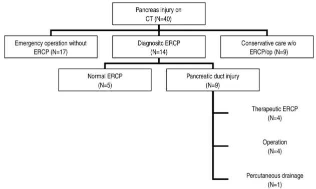
Fig. 1. Flow chart illustrating the patients reviewed in this study
III. RESULTS
Over a seven year period, 40 patients were treated at Asan Medical Center for traumatic pancreatic injury. Of these, 14 patients underwent diagnostic ERCP. During the study period, only one patient (2.5%) died. Of the 14 patients who underwent diagnostic ERCP, five had normal pancreatic ducts, whereas ductal injury was diagnosed in nine of the patients. In the patients diagnosed with ductal injury, four were treated by endoscopic intervention and four underwent exploratory laparotomy. Only one patient received radiologic intervention (percutaneous drainage) to
manage complications arising from pancreatic pseudocyst formation (Fig. 1).
Identification of normal pancreatic ducts by diagnostic ERCP in five patients prevented unnecessary laparotomy in these cases. Of the nine patients in whom ductal injury was revealed by ECRP, four received surgical treatment;
two cases of distal pancreatectomy, one case of pylorus preserving pancreaticoduodenectomy and one case of abscess drainage. Of the remaining patients, three were treated by endoscopic pancreatic stent insertion and one underwent endoscopic pancreatic drainage (Table 1). The remaining case was treated by radiologic intervention
Table 1. Characteristics of patients with ductal injury diagnosed by ERCP
Treatment options Patient number Age (y/o) NPO time / Management Complications Hospital days
Surgical treatment (N=4) 1 50 35/51 *DP Wound dehiscence
2 49 56/73 DP Diabetes mellitus
3 29 07/16 �PPPD Diabetes mellitus
4 32 125 Abscess drainage Necrotizing pancreatitis
Endoscopic intervention (N=4) 1 30 14/24 Pancreatic None
2 25 18/23 stent Pancreatic ductal stenosis
3 54 14/19 Pancreatic None
4 62 46/52 stent None
�ENBD Pancreatic
stent
Radiologic intervention (N=1) 1 47 07/14 Percutaneous None
drainage
* DP: distal pancreatectomy
�PPPD: Pylorus preserving pancreaticoduodenectomy
�ENBD: endoscopic nasobiliary drainage
Fig. 2. (A) Initial CT scan of a 30-year old patient with main pancreatic ductal leakage and pancreatic pseudocyst. (B) Follow-up ERCP image after endoscopic intervention shows a normal pancreatic duct.
A B
(Table 1).
The grade of pancreatic injury was similar between the two groups, as were the mean age of the patients in each group (diagnostic ERCP followed by therapeutic ERCP=43
±18 years old, diagnostic ECRP followed by surgery=43±
12 years old). NPO time, ISS and hospital stay were lower in the endoscopic intervention group than the surgical treat- ment group (23±15 vs. 33±26, 12±9.1 vs. 26±10, and 30
±15 vs. 47±29, respectively) but due to the small number of patients, the differences were not significant (p>0.05) (Table 2). Two patients developed diabetes, and wound dehiscence was a problem in the surgical intervention group. In the cases of other organ damage and unstable vital sign, we performed emergent operation without delay.
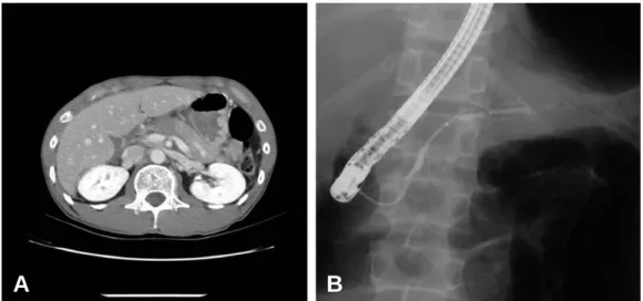
Of the four patients who underwent endoscopic interven- tion (Fig. 1), the first was 30 years old, with main pancre- atic ductal leakage and a pancreatic pseudocyst visible on initial CT scan and ERCP images (Fig. 2A). Endoscopic nasopancreatic drainage (ENPD) was performed one week
after trauma, but follow-up ERCP continued to show leak- age of the main pancreatic duct, which was treated by stent insertion. The patient was discharged from the hospi- tal after 24 days without complications. ERCP performed two months after stent insertion showed no evidence of ductal leakage or pseudocyst; therefore, the stent was removed (Fig. 2B).
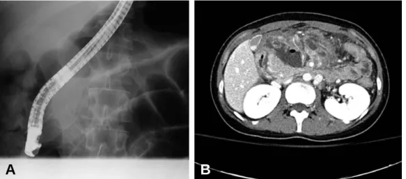
The second patient who underwent therapeutic ERCP was 25 years old, with leakage of the main pancreatic duct on initial ERCP (Fig. 3A). A 3Fr ERPD (endoscopic ret- rograde pancreatic drainage) stent (Cook, Bloomington, USA) was inserted, but CT scan six days later showed acute pancreatitis and the presence of a pseudocyst (Fig.
3B). Therapeutic NPO was continued, and a stent exchange to 5Fr (Cook, Bloomington, USA) was per- formed. CT performed 10 days later showed that the size of the pseudocyst had decreased and pancreatitis had improved. The patient was discharged at 2 months after the trauma, at which time a follow up ERCP showed a
Fig. 3. (A) Initial ERCP image of a 25-year old patient showing leakage of the main pancreatic duct. (B) Follow up CT scan 6 days after the trauma. Acute pancreatitis and a pseudocyst are still evident.
A B
Table 2. Clinical characteristics of patients who underwent therapeutic ERCP or surgery after diagnostic ERCP Endoscopic intervention group Surgical treatment group
(N=4) (N=4)
Age 43±18 43±12
Sex (Male:Female) 3:1 4:0
NPO time (days) 23±15 33±26
*AAST (CT score) 3.2±1.0 3.7±0.6
�ISS .12±9.1 26±10
Hospital days 30±15 47±29
* AAST: American Association for the Surgery of Trauma
�ISS: Injury severity score
stricture at the neck of the pancreatic duct and proximal dilatation. A 7Fr stent (Cook, Bloomington, USA) was therefore reinserted and exchanged for a 10Fr stent (Cook, Bloomington, USA) after 3 months. Two months later, the stent was removed without any complications.
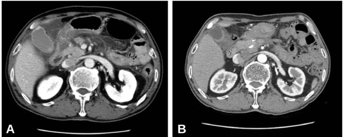
The third patient who underwent therapeutic ERCP was a 54-year old male with a large pancreatic pseudocyst and ductal disruption (Fig. 4A). ENBD was performed and the patient was discharged after 19 days. The follow up CT scan is shown in Fig. 4B.
The fourth patient that underwent therapeutic ERCP was a 62-year old male who was treated by endoscopic nasopancreatic drainage after injury to the main pancreatic duct (5A). Two weeks later, the endoscopic nasopancreatic drainage (ENPD) was removed and an endoscopic retro- grade pancreatic duct (ERPD) stent was inserted. At 46 days after stent insertion, clinical prognosis was improved,
and the patient was discharged from the hospital. Two months after discharge, the stent was removed without any complications (Fig. 5B). Fig. 5A is the first CT image obtained after trauma and Fig. 5B shows the healed pan- creatic duct after treatement.
IV. DISCUSSION
We evaluated the role of ERCP in determining the severity of traumatic pancreas injury. Proper evaluation may prevent unnecessary treatments, such as surgery, which is accompanied by high rates of mortality and morbidity.
Patients with major ductal injury revealed by ERCP should be treated by therapeutic ERCP procedures, such as endo- scopic retrograde pancreatic drainage (ERPD) or endoscopic stent insertion, before surgical options are considered. In addition, delayed diagnosis of major pancreatic ductal injury
Fig. 4. (A) CT scan of a 54-year old patient who underwent therapeutic ERCP for a large pancreatic pseudocyst and ductal disrup- tion. The patient was discharged 19 days after ENBD. A follow up CT scan is shown in (B).
A B
Fig. 5. (A) Initial CT image of a patient who underwent therapeutic ERCP after trauma. (B) The pancreatic duct is restored after treatement.
A B
increases mortality and morbidity rates. Initial CT may be negative in 15% to 40% of patients with an MPD injury.(10,14~16) Indeed, a study of CT and ERCP in 23 patients with pancreatic injury found that CT predicted main pancreatic ductal injury in only 6 of 11 patients (55%).(10)
1. Diagnostic ERCP
Diagnostic ERCP is considered the most accurate diag- nostic modality for detecting damage to the pancreatic duct.(17) Patients with any evidence of pancreatic ductal injury on CT should be assessed by magnetic resonance cholangiopancreatography (MRCP). Patients with intact pancreatic ducts may be treated conservatively, whereas those with ductal disruptions should undergo ERCP to evaluate the severity of the injury more precisely.
Undiagnosed disruption of the pancreatic duct may result in significant complications, including prolonged periods of intensive care.(6) In this study, patients who were suspect- ed to have traumatic pancreatic injury were examined by diagnostic ERCP. Diagnostic ERCP revealed normal ducts in five of these patients, thereby preventing needless laparo- tomy in these cases. Surgery presents high risks in patients with traumatic pancreatic injury. The identification of five patients with normal ducts by ERCP spared them from the risks associated with surgery and recovery, and empha- sizes the importance of diagnostic ERCP when deciding how to manage of the pancreatic injury patients.
2. Therapeutic ERCP
Percutaneous aspiration and drainage to treat pancreatic fluid collection was first suggested in 1976,(18) and thera- peutic ERCP has since been used to manage patients with main pancreatic duct injury without the need for surgery.
In addition, one patient with incomplete ductal disruption was successfully treated without surgery,(17) and stent insertion has been used for definitive management of isolat- ed injuries within the proximal pancreatic duct.(3) Another study described the use of endoscopic transpapillary pancre- atic duct stenting in 11 children with pancreatic injury after blunt abdominal trauma.(19)
In this study, four patients with main pancreatic ductal injury were successfully managed by therapeutic ERCP.
Due to the small number of patients, a significant differ-
ence in outcomes between therapeutic ERCP and surgery was not achieved. However, late complication rates between these two groups differed considerably. Of the three patients who underwent surgery, two developed diabetes and one required a second operation due to wound dehis- cence. In contrast, none of the four patients who underwent therapeutic ERCP developed diabetes or required repetition of the procedure. Moreover, hospital stay and NPO times were shorter in the therapeutic ERCP group.
Therapeutic ERCP can also result in complications, including acute pancreatitis, bacterial infection, and aspira- tion pneumonia. Several patients developed minor ductal stenosis after ERCP, but this was not considered to be a serious complication. The decision to perform therapeutic ERCP or surgery depends on whether there is damage to other organs and whether the patient is stable.
Our study had several limitations, including its retrospec- tive design and the small number of patients. In addition, patient management was at the discretion of individual clinicians. The difference in ISS between the two groups may suggest selection bias, indicating that the incidence of injury to other organs was greater in the non-ERCP group.
Prospective studies, with larger numbers of patients and more careful analysis of the CT findings, are required to confirm our findings.
Patients with severe bleeding and peritonitis accompanied by injuries in other organs should receive emergency surgery. However, in patients who are hemodynamically stable but suspected of having sustained pancreatic injury through abdominal trauma, ERCP should be considered due to its diagnostic and therapeutic merits.
This study reviews our experience with patients at AMC. Based on our findings, we believe that ERCP repre- sents an important diagnostic and therapeutic modality in the treatement of traumatic pancreas injury. Further studies focusing on the role of ERCP as a therapeutic tool in patients who have sustained blunt pancreatic trauma will be needed.
V. CONCLUSION
Needless laparotomy was prevented by revealing the nor- mal pancreatic duct through the diagnostic ERCP proce- dure, and the patients got recovered without complications, in our study. So we once again emphasize the importance of ERCP as diagnostic modality.
The patients with main pancreatic duct injury got well without serious complications after the ERCP intervention, so ERCP intervention is a good treatment option for the patient with the pancretic duct injury.
REFERENCES
01) Wolf A, Bernhardt J, Patrzyk M, Heidecke CD. The value of endoscopic diagnosis and the treatment of pancreas injuries following blunt abdominal trauma.
Surg Endosc. 2005;19:665-9.
02) Jones RC. Management of pancreatic trauma. Am J Surg 1985;150:698-704.
03) Subramanian A, Dente CJ, Feliciano DV. The manage- ment of pancreatic trauma in the modern era. Surg Clin North Am 2007;87:1515-32, x.
04) Cogbill TH, Moore EE, Feliciano DV, Hoyt DB, Jurkovich GJ, Morris JA, et al. Conservative manage- ment of duodenal trauma: a multicenter perspective. J Trauma 1990;30:1469-75.
05) Akhrass R, Yaffe MB, Brandt CP, Reigle M, Fallon WF, Jr., Malangoni MA. Pancreatic trauma: a ten- year multi-institutional experience. Am Surg 1997;63:
598-604.
06) Wind P, Tiret E, Cunningham C, Frileux P, Cugnenc PH, Parc R. Contribution of endoscopic retrograde pan- creatography in management of complications following distal pancreatic trauma. Am Surg 1999;65:777-83.
07) Patton JH, Jr., Fabian TC. Complex pancreatic injuries.
Surg Clin North Am 1996;76:783-95.
08) Bhasin DK, Rana SS, Rawal P. Endoscopic retrograde pancreatography in pancreatic trauma: need to break the mental barrier. J Gastroenterol Hepatol 2009;24:
720-8.
09) Lin BC, Chen RJ, Fang JF, Hsu YP, Kao YC, Kao JL. Management of blunt major pancreatic injury. J Trauma 2004;56:774-8.
10) Kim HS, Lee DK, Kim IW, Baik SK, Kwon SO, Park
JW, et al. The role of endoscopic retrograde pancre- atography in the treatment of traumatic pancreatic duct injury. Gastrointest Endosc 2001;54:49-55.
11) Rescorla FJ, Plumley DA, Sherman S, Scherer LR, 3rd, West KW, Grosfeld JL. The efficacy of early ERCP in pediatric pancreatic trauma. J Pediatr Surg 1995;30:
336-40.
12) Moore EE, Cogbill TH, Malangoni MA, Jurkovich GJ, Champion HR, Gennarelli TA, et al. Organ injury scal- ing, II: Pancreas, duodenum, small bowel, colon, and rectum. J Trauma 1990;30:1427-9.
13) Osler T, Baker SP, Long W. A modification of the injury severity score that both improves accuracy and simplifies scoring. J Trauma 1997;43:922-5; discussion 5-6.
14) Lin BC, Liu NJ, Fang JF, Kao YC. Long-term results of endoscopic stent in the management of blunt major pancreatic duct injury. Surg Endosc 2006;20:1551-5.
15) Fennema EM, Nellensteijn DR, Nieuwenhuijs VB, van Rheenen PF, Ten Duis HJ, Hulscher JB. [Pancreatic injury in abdominal trauma in children: difficult to diagnose and treat.]. Ned Tijdschr Geneeskd 2011;155:
A2406.
16) Valentino M, Ansaloni L, Catena F, Pavlica P, Pinna AD, Barozzi L. Contrast-enhanced ultrasonography in blunt abdominal trauma: considerations after 5 years of experience. Radiol Med 2009;114:1080-93.
17) Takishima T, Hirata M, Kataoka Y, Asari Y, Sato K, Ohwada T, et al. Pancreatographic classification of pancreatic ductal injuries caused by blunt injury to the pancreas. J Trauma 2000;48:745-51; discussion 51-2.
18) Hancke S, Pedersen JF. Percutaneous puncture of pan- creatic cysts guided by ultrasound. Surg Gynecol Obstet 1976;142:551-2.
19) Houben CH, Ade-Ajayi N, Patel S, Kane P, Karani J, Devlin J, et al. Traumatic pancreatic duct injury in children: minimally invasive approach to management.
J Pediatr Surg 2007;42:629-35.