Percutaneous transhepatic biliary drainage catheter fracture:
A case report
Jia Rui Kwan1, Keith Sheng Hng Low2, Rahul Lohan3, and Vishal G Shelat4
1Lee Kong Chian School of Medicine, Nanyang Technological University,
2Yong Loo Lin School of Medicine, National University of Singapore,
Departments of 3Radiology and 4General Surgery, Khoo Teck Puat Hospital, Singapore
Percutaneous transhepatic biliary drainage (PTBD) is safe treatment for biliary decompression given certain indications.
However, this is temporary until definitive drainage is established. We report on a 76-year-old lady with recurrent pyo- genic cholangitis and PTBD catheter fracture. She had hepatitis B virus-related Child-Pugh class A liver cirrhosis, hypo- thyroidism, hyperlipidaemia, and previous atrial fibrillation with a background of mild mitral, tricuspid and aortic valvular regurgitation. She had history of laparoscopic cholecystectomy in the past. She was deemed to be a high operative risk and declined hepatic resection. She had undergone multiple endoscopic and percutaneous biliary interventions to control sepsis and stone burden. A bilateral PTBD catheter was left in situ with plans for 3-monthly change. However, she defaulted follow-up and presented 11 months later with complaints of pain over the drain site and inability to flush the right catheter. Abdominal X-ray and computed tomography scans detected right catheter fracture at two places, making three fragments. She underwent percutaneous removal of the proximal fragment by an interventional radiology team. A temporary 4 Fr catheter was inserted to maintain biliary access. Endoscopic removal of the intra-biliary frag- ments was done the next day. Complete removal was confirmed on fluoroscopy. Finally, the 4 Fr catheter was replaced by a new 12 Fr catheter. The patient was discharged well. (Ann Hepatobiliary Pancreat Surg 2018;22:282-286) Key Words: Biliary drainage; Catheter fracture; Cholangitis
Received: November 19, 2017; Revised: February 11, 2018; Accepted: February 16, 2018 Corresponding author: Vishal G Shelat
Department of General Surgery, Khoo Teck Puat Hospital, Level 6, Surgery Office, 90, Yishun Central 768828, Singapore Tel: +65-6357-7807, Fax: +65-6357-7809, E-mail: [email protected]
Copyright Ⓒ 2018 by The Korean Association of Hepato-Biliary-Pancreatic Surgery
This is an Open Access article distributed under the terms of the Creative Commons Attribution Non-Commercial License (http://creativecommons.org/
licenses/by-nc/4.0) which permits unrestricted non-commercial use, distribution, and reproduction in any medium, provided the original work is properly cited.
Annals of Hepato-Biliary-Pancreatic Surgery ∙ pISSN: 2508-5778ㆍeISSN: 2508-5859
INTRODUCTION
Percutaneous transhepatic biliary drainage (PTBD) is safe treatment for biliary decompression. It is usually done to control acute hepatobiliary sepsis or as a pallia- tion for malignant jaundice. In most instances, PTBD is a temporary procedure prior to definitive intervention.
However, PTBD can be left life-long in patients with pre- dicted short life expectancy or prohibitive operative risk.
We report on an elderly female patient with multiple co- morbidities and recurrent pyogenic cholangitis (RPC) where bilateral PTBD catheters were inserted for sepsis.
Thereafter the patient elected to have this left in situ until the catheter fracture warranted admission.
CASE
A 76-year-old lady with a history of RPC was admitted with hepatobiliary sepsis. She was deemed to be a high operative risk because of hepatitis B virus-induced Child A liver cirrhosis, hypothyroidism, hyperlipidaemia, and previous atrial fibrillation with a background of mild mi- tral, tricuspid, and aortic valve regurgitation. She had un- dergone laparoscopic cholecystectomy previously. She had also undergone multiple prior endoscopic and percuta- neous biliary interventions to control sepsis and stone burden. The brushings and cytology from the intrahepatic ducts did not find malignancy. She was offered hepatic resection, but declined because of the high operative risk.
She opted for indwelling percutaneous internal-external biliary drains with 3-monthly changes. In the last visit pri- or to this admission, she was discharged with a 12 Fr in-
Fig. 1. Abdominal X-ray showing intrabiliary fracture of a right percutaneous transhepatic biliary drainage (PTBD) cath- eter (arrow A) and normal left-side PTBD catheter.
Fig. 2. Computed tomography scan showing fractured right biliary drainage catheter at liver capsule with gap between fragments (arrow B).
ternal–external biliary drain (Navarre®; Bard Biopsy Systems, Tempe, AZ, USA) in the right hepatic duct and a 16 Fr drainage catheter (Cook Medical, Bloomington, IN, USA) in the left hepatic duct. The catheters were spi- gotted on discharge and the patient was taught to flush the catheters once a day with normal saline. However, she defaulted the follow up and was readmitted after 11 months with complaints of pain over the drain site and inability to flush the right catheter. The left drain site was normal. A plain abdominal x-ray revealed fractures of the right PTBD catheter with a normal left PTBD catheter (Fig. 1).
This result was further confirmed by a computed to- mography (CT) scan, which showed a break in the right PTBD catheter near the liver capsule, with a 4 cm gap between the ends (Fig. 2), as well as a break at the distal segment. The left PTBD catheter was intact.
The catheter was fractured at two places with three fragments needing removal. The management of the cath- eter fracture was performed in stages. Firstly, radio- logically guided percutaneous removal of the proximal fragment was performed. Further percutaneous attempt at removal of the middle and distal fragments was un- successful, because the strings of the catheter could not be pulled. A temporary 4 Fr Cobra angiographic catheter (Cook Medical, Bloomington, IN, USA) was inserted over the strings of the fractured catheter into the duodenum to
drain the right hepatic duct and retain right biliary access.
Second, endoscopic retrieval of the remaining fractured fragments (Fig. 3) was done the next day. The in- tra-biliary catheter fragments were still tied to the duode- nal components by the interlocking suture. Thus, all the catheter fragments could be successfully removed by the forceps. At this point, two catheters remained in place:
the 4 Fr temporary right biliary access catheter and the 16Fr left biliary access catheter. Last, interventional radi- ology-guided exchange of the 4 Fr catheter to a 12F in- ternal–external biliary drain (Navarre®; Bard Biopsy Systems, Tempe, AZ, USA) was then done the following day. The successful removal and insertion was confirmed by fluoroscopy (Fig. 4).
DISCUSSION
RPC is a triad of stones, sepsis, and stricture.1 RPC pa- tients have either definitive surgery performed or perma- nent metallic biliary endoprosthesis inserted depending on the clinical state. Placement of a PTBD catheter intended to be permanent is a rare clinical need, and for the cathe- ter to fracture is even rarer. Clinical presentation depends on the site of fracture. If left in situ, the retained catheter fragments in the duodenum behave like migrated endo- scopically placed biliary stents, and can migrate further into the small bowel, leading to intestinal obstruction.
Fig. 3. Endoscopic view of distal fracture fragment (arrow A), intact left PTBD catheter (Arrow B), and temporarily inserted 4 Fr right biliary catheter (arrow C).
Fig. 4. Fluoroscopic image after removal of fractured right PTBD catheter with 12 Fr replacement catheter.
Besides intestinal obstruction, other reported complica- tions of the migrated biliary stents include intestinal per- foration, intra-abdominal abscess, and fistula formation.2 We performed a literature search on PubMed with search terms “(percutaneous transhepatic biliary drainage OR biliary catheter OR biliary drainage catheter OR bili- ary endoprosthesis OR biliary drainage tube) AND (breakage OR catheter fracture OR tube fracture OR frag- ment OR segment) AND (liver OR bile duct OR biliary system OR bile)” from January 01, 1980 to December 14, 2017. No restriction on language was applied, an ad refer- ence list of all the articles was screened to include addi- tional case reports. A total of 132 reports was generated.
Title and abstracts of the reports were screened by first author Kwan JR, and any doubt was clarified by senior author Shelat VG. The findings are presented in Table 1.3-10
In our patient, the fracture was at two sites: (a) at the liver capsule and (b) at the intra-biliary portion. Our pa- tient presented with mild right upper quadrant pain and inability to flush the catheter. It is likely that, because of the prolonged duration, a fistulous track had already ma- tured, and she hence did not develop biliary peritonitis.
We believe that the fracture could have been caused by the catheter becoming brittle over time, leading to sponta- neous fracture. In our experience, if there is no catheter fracture, blockage is almost certain after 3 months; so we
advocate changing the catheter every 3 months. To pre- vent early blockage, we advocate daily flushing of the catheter. Plain abdominal radiography is adequate to diag- nose catheter migration or fracture, as in our patient.
However, a CT scan is warranted to evaluate the under- lying pathology, disease progression, or secondary com- plications that result from the fracture. In our patient, an abdominal film showed a large gap between the two frag- ments of the catheter.
Management of fractured PTBD includes both pre- ventive and corrective measures for fractured segments and management of the underlying pathology for which the PTBD is inserted. As with our patient, a multi- disciplinary approach involving the surgical, medical, and radiological teams is integral to achieving good outcomes.
Removal of catheter fragments percutaneously via radio- logical guidance or via endoscopic retrograde cholangiog- raphy should first be considered. Surgical intervention should be the last option if all else fails or if the patient develops biliary peritonitis. In patients requiring a long-term catheter, options of metallic biliary endopros- thesis should be explored. Biodegradable biliary stents are also reportedly used in benign biliary strictures.11 In pa- tients wishing to leave the PTBD for the long term, they should be instructed about the possibility of fracture and to reinforce the need to change the PTBD catheter every 3 months.
In conclusion, this case highlights the importance of close follow-up. Catheters and plastic stents are not de- signed for long-term use. Therefore it is our institutional
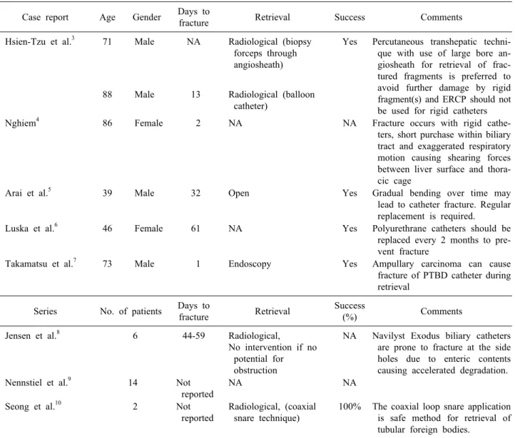
Table 1. Reported cases on percutaneous biliary catheter fracture Case report Age Gender Days to
fracture Retrieval Success Comments
Hsien-Tzu et al.3 71 Male NA Radiological (biopsy forceps through angiosheath)
Yes Percutaneous transhepatic techni- que with use of large bore an- giosheath for retrieval of frac- tured fragments is preferred to avoid further damage by rigid fragment(s) and ERCP should not be used for rigid catheters
88 Male 13 Radiological (balloon
catheter)
Nghiem4 86 Female 2 NA NA Fracture occurs with rigid cathe-
ters, short purchase within biliary tract and exaggerated respiratory motion causing shearing forces between liver surface and thora- cic cage
Arai et al.5 39 Male 32 Open Yes Gradual bending over time may
lead to catheter fracture. Regular replacement is required.
Luska et al.6 46 Female 61 NA Yes Polyurethrane catheters should be
replaced every 2 months to pre- vent fracture
Takamatsu et al.7 73 Male 1 Endoscopy Yes Ampullary carcinoma can cause
fracture of PTBD catheter during retrieval
Series No. of patients Days to
fracture Retrieval Success
(%) Comments
Jensen et al.8 6 44-59 Radiological,
No intervention if no potential for obstruction
NA Navilyst Exodus biliary catheters are prone to fracture at the side holes due to enteric contents causing accelerated degradation.
Nennstiel et al.9 14 Not
reported
NA NA
Seong et al.10 2 Not
reported
Radiological, (coaxial snare technique)
100% The coaxial loop snare application is safe method for retrieval of tubular foreign bodies.
NA, not available; ERCP, Endoscopic retrograde cholangiopancreatography; PTBD, Percutaneous transhepatic biliary drainage
recommendation to change catheters every 3 months.
Close follow-up of patients who are on PTBD and patient education on regular flushing and catheter care is im- portant to prevent catheter malfunction.
REFERENCES
1. Kwan KEL, Shelat VG, Tan CH. Recurrent pyogenic cholangitis:
a review of imaging findings and clinical management. Abdom Radiol (NY) 2017;42:46-56.
2. Strode MA, Bandera BC, Deveaux P, Rice RD. Migrated biliary stent complicated by small bowel obstruction. Am Surg 2013;
79:E253-E254.
3. Hsien-Tzu L, Hsiuo Shan T, Nai Chi C, Yi Yang L, Yi You C, Chien An L. Percutaneous transhepatic techniques for retriev- ing fractured and intrahepatically dislodged percutaneous trans- hepatic biliary drainage catheters. Diagn Interv Radiol 2017;23:
461-464.
4. Nghiem DD. Bile leakage after fracture of a percutaneous trans- hepatic biliary drainage catheter. JAMA 1984;251:892.
5. Arai M, Furuya M, Shimizu Y, Okino T, Yokomuro S, Arima Y, et al. A piece of catheter for liver abscess drainage was left in a liver due to fracture: report a case. Tando 2010;24:209-212.
6. Luska G, Elgeti H, Lux M. Repeated breaks of polyurethane catheters as a complication of internal bile duct drainage. Rofo 1981;135:731-733.
7. Takamatsu S, Nagano H, Ootsukasa S, Kawachi Y, Maruyama H. Ampullary carcinoma associated with removing fractured per- cutaneous transhepatic biliary drainage catheter for hilar cholangiocarcinoma. HPB 2016;18:e472.
8. Jensen LE, Collins Z, Lemons S, Johnson PL. Fracture of Navilyst Exodus biliary catheters. J Vasc Interv Radiol 2011;22:
261-262.
9. Nennstiel S, Weber A, Frick G, Haller B, Meining A, Schmid RM, et al. Drainage-related complications in percutaneous trans- hepatic biliary drainage: an analysis over 10 years. J Clin Gastroenterol 2015;49:764-770.
10. Seong CK, Kim YJ, Chung JW, Kim SH, Han JK, Kim HB, et al. Tubular foreign body or stent: safe retrieval or reposition- ing using the coaxial snare technique. Korean J Radiol 2002;3:
30-37.
11. Siiki A, Rinta-Kiikka I, Sand J, Laukkarinen J. Endoscopic bio- degradable biliary stents in the treatment of benign biliary stric- tures: First report of clinical use in patients. Dig Endosc 2017;
29:118-121.