Korean Circulation Journal
Introduction
Early generation drug-eluting stents (DES), releasing Sirolimus or Paclitaxel, have reduced the occurrence of restenosis and the need for repeat revascularization, compared with bare metal stents (BMS).
1-4)Stent thrombosis can be exaggerated by basic DES prop- erties that inhibit intimal regeneration and healing processes. There
Print ISSN 1738-5520 • On-line ISSN 1738-5555
Long-Term Safety and Efficacy of Sirolimus- and Paclitaxel-Eluting Stents in Patients With Acute Myocardial Infarction:
Four-Year Observational Study
Gye-Sik Min, CRC, Jae-Hwan Lee, MD, Jae-Ho Park, MD, Ung-Lim Choi, MD, Young-Dal Lee, MD, Seok-Woo Seong, MD, Seon-Ah Jin, MD, Soo-Jin Park, MD, Jun-Hyeong Kim, MD,
Jae-Hyeong Park, MD, Si Wan Choi, MD, Jin-Ok Jeong, MD, and In-Whan Seong, MD
Department of Cardiology, Chungnam National University School of Medicine, Daejeon, Korea
Background and Objectives: The comparison of long-term clinical effects between Sirolimus-eluting stent (SES) and Paclitaxel-eluting stents (PES) for treatment of acute myocardial infarction (AMI) remains unclear. Seeking to clarify this issue, we performed a retrospective analysis to evaluate four-year clinical outcomes of SES compared to PES treated AMI patients.
Subjects and Methods: From January 2004 to August 2006, all patients with acute ST-segment elevation myocardial infarction and acute non-ST segment elevation myocardial infarction who underwent percutaneous coronary intervention (PCI) by implantation of ei- ther SES or PES were enrolled. The occurrences of cardiac and non-cardiac deaths, recurrent infarction, target vessel revascularization (TVR) and stent thrombosis were analyzed. The composite end points of these major adverse cardiac events (MACE) were also analyzed.
Results: During the study period, a total of 668 AMI patients had visited, of which 522 patients (299 with SES and 223 with PES) were en- rolled. During the four-year clinical follow-up, both groups showed similar occurrences of non-cardiac death (14.6±2.2% vs. 18.3±3.0%, p=0.26); cardiac death (6.8±1.52% vs. 11.2±2.6%, p=0.39); re-infarction (3.3±1.1% vs. 6.4±1.8%, p=0.31); and stent thrombosis (3.2±1.1%
vs. 5.4±1.7%, p=0.53). However, occurrences of TVR {4.0±1.2% vs. 10.0±3.0%, hazard ratio (HR)=0.498, 95% confidence interval (CI)=
0.257-0.967, p=0.039} and MACE (19.4±2.5% vs. 29.4±3.5%, HR=0.645, 95% CI=0.443-0.940, p=0.021) were significantly lower in the SES population.
Conclusion: In AMI patients treated with either SES or PES implantation, the former had a significantly lower risk of TVR and MACE during four-year clinical follow-up. Rates of death, cardiac death or recurrent infarction, and stent thrombosis were similar. (Korean Circ J 2012;
42:266-273)
KEY WORDS: Acute myocardial infarction; Percutaneous coronary intervention; Stents.
Received: August 31, 2011 / Revision Received: October 10, 2011 / Accepted: October 27, 2011
Correspondence: Jae-Hwan Lee, MD, Department of Cardiology, Chungnam National University School of Medicine, 282 Munhwa-ro, Jung-gu, Daejeon 301-721, Korea
Tel: 82-42-208-8237, Fax: 82-42-280-8238, E-mail: [email protected]
• The authors have no financial conflicts of interest.
This is an Open Access article distributed under the terms of the Creative Commons Attribution Non-Commercial License (http://creativecommons.org/licenses/
by-nc/3.0) which permits unrestricted non-commercial use, distribution, and reproduction in any medium, provided the original work is properly cited.
has been concern about the use of DES in highly thrombogenic situ- ations, such as acute myocardial infarction (AMI). However, based on the favorable results of clinical studies, DES have been widely used in more complex clinical and anatomic situations, including AMI.
5-7)Two early generation DES: the Sirolimus-eluting stent (SES) (Cor-
dis, Johnson and Johnson, Miami Lakes, FL, USA) and the Paclitaxel-
eluting stent (PES) (Boston Scientific, Natick, MA, USA) have revealed
improvements in clinical and angiographic outcomes in the treat- ment of many coronary lesions, compared with BMS.
2-4)However, to date there is limited long term clinical data directly comparing outcomes of SES with PES implantation, in the treatment of AMI patients. In this retrospective study we compared four-year clinical outcomes of SES versus PES implantation in patients with AMI.
Subjects and Methods
Study patients
From January 2004 to August 2006, we evaluated all ST-segment elevation myocardial infarction (STEMI) and non-ST segment eleva- tion myocardial infarction (NSTEMI) patients who had been treated with either SES or PES implantation, at the Chungnam National University Hospital. They were retrospectively analyzed for four years after the index percutaneous coronary intervention (PCI).
Treatment methods
All patients were treated according to standardized guidelines.
8)Patients with acute STEMI all underwent a primary PCI. The infarct- related lesions were assessed by electrocardiogram, echocardio- gram and coronary angiogram by the attending physician. All pro- cedures were performed according to standard techniques, and the final interventional strategy was left to the discretion of the operators. The culprit lesions were fully covered with a single or multiple stents. Direct implantation of a stent without prior balloon dilatation was also allowed. Adjunctive balloon dilatation within the stent was performed where necessary. The final inflation was performed using either a stent balloon or another short balloon wi- thin the stent. Intervention in non-infarct-related arteries during the initial procedure was discouraged, especially in STEMI patients. The removal of thrombi by aspiration catheter was performed at the operator’s discretion. Glycoprotein IIb/IIIa receptor antagonists were selectively used according to the operator’s judgment.
Study methods
We included consecutive patients with AMI who underwent PCI.
We collected initial and follow-up clinical outcomes from their me- dical records and analyzed angiographic findings. Of the patients lost-to-follow-up, clinical data were acquired by means of tele- phone interview. The occurrences and causes of death were assessed by the medical records of our own or from other clinics, telephone in- terview, or from the data of Statistics Korea.
Quantitative coronary angiography
The coronary angiographies were performed after administra- tion of intracoronary nitroglycerin, when possible. Quantitative cor-
onary analysis was performed by an experienced investigator, who was not aware of treatment assignment, using the guiding catheter for magnification calibration with an automated edge-detection sys- tem (CAAS V, Pie Medical Imaging). The quantitative measurements included: reference diameter; lesion length; and the minimal luminal diameter before the procedure, after the procedure and at follow- up. The in-segment or target lesion was defined as the in-stent segment plus the adjacent proximal and distal 5 mm segments.
The in-segment minimal lumen diameter was determined both af- ter the procedure and at follow-up. An acute gain was defined as a change in minimal luminal diameter between pre- and post-in- tervention measurements. A late loss was defined as a change in minimal luminal diameter between post-intervention and follow- up. A recurrent restenosis was defined as an in-segment diameter stenosis ≥50% according to follow-up angiography.
Definition of clinical event
Procedural success was defined as no laboratory death, no em- ergency bypass surgery, and thrombolysis in myocardial infarction (TIMI) grade 2 flow in the distal part of the infarct related artery with a residual stenosis less than 30%. Reinfarction was diagnosed based on recurrent symptoms and/or new electrocardiographic changes in association with a re-elevation of creatine kinase-MB levels >1.5 times the previous value if within 48 hours, or >3 times the upper normal limit if longer than 48 hours from the index infarction. We applied the Academic Research Consortium definitions for stent thrombosis.
9)With respect to timing, stent thrombosis is classified as acute, subacute, late, and very late. By the level of certainty, it is defined as definite, probable, or possible. Definite and probable stent thromboses were included in major adverse cardiac events (MACE).
A target lesion revascularization (TLR) was considered if the target lesion stenosis was at least 50% of the diameter, in the presence of ischemic signs or symptoms or when target lesion stenosis was at least 70%. A TLR was defined as a repeat intervention or bypass sur- gery of the target lesion owing to restenosis or reocclusion of the target lesion.
A target vessel revascularization (TVR) was defined as a repeat
revascularization of an infarct-related artery. The occurrence of
MACE including death, reinfarction, stent thrombosis (definite and
probable), and TVR at 48 months were evaluated. Death from car-
diac causes included: death from recurrent myocardial infarction,
cardiac perforation, pericardial tamponade, arrhythmia or conduc-
tion abnormality, complications of the index procedure, and heart
failure or stroke during follow-up. All deaths that could not be clearly
attributed to a non-cardiac cause were also considered to be car-
diac deaths.
Statistical analysis
Data are expressed as mean±SD or median (range) for continu- ous variables and as frequencies (percentages) for the categorical variables. Differences between groups were assessed using Chi- square (χ
2) or Fisher’s exact tests for categorical variables, and un- paired t-tests or Mann-Whitney U tests for continuous variables.
The relative risk and its 95% confidence interval (CI) were computed for outcome measures. Event-free survival (events: death, reinfarc- tion, TVR, stent thrombosis and MACE) during four years was ana- lyzed using the Kaplan-Meier method, and the differences between groups were assessed by a log-rank test.
Multivariate analyses involved a backwards elimination tech- nique, variables with a p of <0.20 and clinically relevant predictors were used in the final model. All p of were two-sided and a proba- bility value of p<0.05 was considered significant. Statistical analy- sis was performed using commercially available software {Statistical Package for the Social Sciences (SPSS) 17.0 for Windows, SPSS Inc., Chicago, IL, USA}.
Results
From January 2004 to August 2006, a total of 668 AMI patients were admitted at the Chungnam National University Hospital. Of them, 176 patients were excluded from this study; 59 patients were treated with balloon angioplasty alone, 25 were treated with BMS implantation and two with other types of DES, 27 patients deferr- ed from angioplasty, eleven were treated with bypass surgery, nine were diagnosed with variant angina, five were treated with thrombi suction alone, and eight could not be recanalized due to poor gen- eral condition. Finally, 522 patients treated with either SES (n=299, 57.3%) or PES (n=223, 46.7%) implantation were included in this study (Fig. 1).
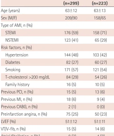
Baseline clinical characteristics
Baseline clinical characteristics are shown in Table 1. Mean age
(63±12 years) and sex distribution were identical for both groups.
The proportion of STEMI was significantly lower in the SES popula- tion compared to the PES population (59% vs. 71%, p<0.001). The risk factors for ischemic heart disease, underlying heart disease, and the frequency of arrhythmia were equal in both groups. The left ven- tricular ejection fraction of 51% was also equal in the two groups.
Fig. 1. Study population. STEMI: ST-elevation myocardial infarction, NSTE- MI: non-ST elevation myocardial infarction, BMS: bare-metal stent, DES:
drug-eluting stent, PCI: percutaneous coronary intervention, SES: Sirolim- us-eluting stent, PES: Paclitaxel-eluting stent.
All acute STEMI/NSTEMI patients (January 2004-August 2006, n=668)
N=522
N=146 Excluded
SES (n=299) STEMI (n=176) NSTEMI (n=123)
PES (n=223) STEMI (n=168) NSTEMI (n=65)
Balloon angioplasty (59) BMS (25)
Other DES (2) Medical treatment (27) Bypass surgery (11) Variant angina (9) Suction or thrombolysis (5) Failed PCI (8)
Table 1. Baseline clinical characteristics SES (n=299)
PES
(n=223) p
Age (years) 63±12 63±13 0.52
Sex (M/F) 209/90 158/65 0.85
Type of AMI, n (%)
STEMI 176 (59) 158 (71) 0.006
NSTEMI 123 (41) 65 (29)
Risk factors, n (%)
Hypertension 144 (48) 103 (42) 0.66
Diabetes 82 (27) 60 (27) 0.92
Smoking 171 (57) 121 (54) 0.53
T-cholesterol >200 mg/dL 84 (29) 54 (26) 0.48
Family history 16 (5) 10 (5) 0.69
Previous PCI, n (%) 15 (5) 13 (6) 0.70
Previous MI, n (%) 18 (6) 9 (4) 0.43
Previous CABG, n (%) 2 (1) 0 (0) 0.24
Preinfarction angina, n (%) 75 (25) 50 (23) 0.53
LVEF (%) 51±12 51±11 0.77
VT/V-fib, n (%) 15 (5) 14 (6) 0.54
Atrial fibrillation, n (%) 8 (3) 4 (2) 0.38
Complete AV block, n (%) 25 (8) 25 (11) 0.30
SES: Sirolimus-eluting stent, PES: Paclitaxel-eluting stent, AMI: acute myo- cardial infarction, NSTEMI: non-ST elevation myocardial infarction, STEMI:
ST-elevation myocardial infarction, PCI: percutaneous coronary interven- tion, CABG: coronary artery bypass graft, LVEF: left ventricular ejection fraction, VT: ventricular tachycardia, V-fib: ventricular fibrillation, AV: atrio- ventricular
Table 2. Procedural data including pain-to-ER time and door-to-balloon time for patients with acute myocardial infarction
SES (n=299)
PES
(n=223) p
STEMI/NSTEMI 176/123 168/65
STEMI (minutes)
Pain-to-ER time 19-1380 (208) 30-1715 (180) 0.99 Door-to-Balloon time 20-1818 (68) 15-1690 (67) 0.31 NSTEMI (hours)
Pain-to-ER time 0.3-480 (10) 0.2-168 (11) 0.55
Door-to-Balloon time 0.5-187 (15) 0.6-232 (14) 0.21
SES: Sirolimus-eluting stent, PES: Paclitaxel-eluting stent, STEMI: ST-eleva-
tion myocardial infarction, NSTEMI: non-ST elevation myocardial infarction,
ER: emergency room
Reperfusion time
The durations from symptom onset to emergency room (ER) ar- rival (pain-to-ER time) and from ER arrival to reperfusion (door-to-
balloon time) are shown in Table 2. The median of pain-to-ER time was about 3 hours in STEMI and about 10 hours in NSTEMI patients.
The median of door-to-balloon time was less than 70 minutes in STEMI and about 15 hours in NSTEMI patients. Pain-to-ER and door- to-balloon times were the same for the two groups.
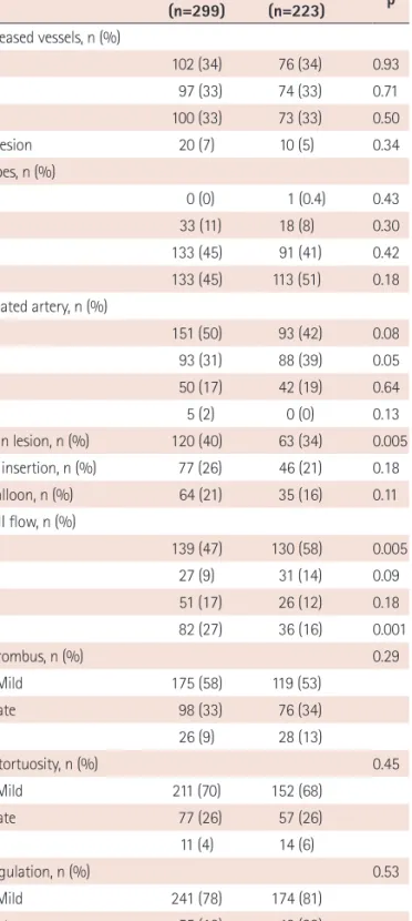
Coronary angiographic findings
Coronary angiographic findings are shown in Table 3. Angiogra- phic diagnoses and lesion types were the same between the two groups. In the SES group, the incidence of the infarct-related artery being the right coronary artery was significantly lower than the PES group (31% vs. 39%, p=0.05). A bifurcation lesion was more frequ- ent in the SES group compared with the PES group (40% vs. 34%, p=0.005). The frequency of initial TIMI 0 flow was lower in the SES group (47% vs. 58%, p=0.005) and TIMI 3 flow was higher (27% vs.
16%, p=0.001). There were no significant differences in the presence of intraluminal thrombi, proximal tortuosity, lesion angulation, or ostial location between the two groups.
Table 3. Coronary angiographic findings SES (n=299)
PES
(n=223) p
No. of diseased vessels, n (%)
1 VD 102 (34) 76 (34) 0.93
2 VD 97 (33) 74 (33) 0.71
3 VD 100 (33) 73 (33) 0.50
LMCA lesion 20 (7) 10 (5) 0.34
Lesion types, n (%)
A 0 (0) 1 (0.4) 0.43
B1 33 (11) 18 (8) 0.30
B2 133 (45) 91 (41) 0.42
C 133 (45) 113 (51) 0.18
Infarct related artery, n (%)
LAD 151 (50) 93 (42) 0.08
RCA 93 (31) 88 (39) 0.05
LCx 50 (17) 42 (19) 0.64
LMCA 5 (2) 0 (0) 0.13
Bifurcation lesion, n (%) 120 (40) 63 (34) 0.005
Two GWs insertion, n (%) 77 (26) 46 (21) 0.18
Kissing balloon, n (%) 64 (21) 35 (16) 0.11
Initial TIMI flow, n (%)
0 139 (47) 130 (58) 0.005
I 27 (9) 31 (14) 0.09
II 51 (17) 26 (12) 0.18
III 82 (27) 36 (16) 0.001
Visible thrombus, n (%) 0.29
None-Mild 175 (58) 119 (53)
Moderate 98 (33) 76 (34)
Heavy 26 (9) 28 (13)
Proximal tortuosity, n (%) 0.45
None-Mild 211 (70) 152 (68)
Moderate 77 (26) 57 (26)
Severe 11 (4) 14 (6)
Lesion angulation, n (%) 0.53
None-Mild 241 (78) 174 (81)
Moderate 55 (18) 48 (22)
Heavy 3 (1) 1 (0.1)
Ostial lesion, n (%) 48 (10) 28 (13) 0.32
SES: Sirolimus-eluting stent, PES: Paclitaxel-eluting stent, 1 VD: one vessel disease, 2 VD: two vessel disease, 3 VD: triple vessel disease, LAD: left ante- rior descending artery, RCA: right coronary artery, LCx: left circumflex ar- tery, LMCA: left main coronary artery, GW: guidewire, TIMI: Thrombolysis in Myocardial Infarction
Table 4. Procedural characteristics SES (n=299)
PES
(n=223) p
Temporary pacemaker, n (%) 13 (4) 19 (9) 0.06
IABP support, n (%) 6 (2) 7 (3) 0.41
Access site, n (%)
Femoral 42 (14) 25 (11) 0.36
Radial 257 (86) 198 (89)
GpIIb/IIIa inhibitor, n (%) 34 (11) 7 (3) 0.13
Thrombus aspiration, n (%) 71 (24) 80 (36) 0.09
Number of stents, n (%)
1 268 (90) 194 (87) 0.41
≥2 31 (10) 29 (13) 0.41
Stent diameter (mm) 3.28±0.29 3.29±0.30 0.50
Total stent length (mm) 30.7±3.9 30.8±4.4 0.86
Final balloon size (mm) 3.3±0.3 3.3±0.3 0.84
Final balloon pressure (atm) 15.3±3.8 15.1±4.1 0.50
Postdilation, n (%) 118 (40) 86 (39) 0.86
Nonculprit lesion PCI, n (%) 38 (9) 21 (13) 0.27 Post-TIMI flow, n (%)
0 2 (1) 4 (2) 0.66
1 3 (1) 3 (1) 0.66
2 31 (10) 26 (12) 0.23
3 263 (88) 190 (85) 0.12
Procedural success, n (%) 287 (96) 209 (94) 0.17
SES: Sirolimus-eluting stent, PES: Paclitaxel-eluting stent, IABP: intra-aortic
balloon pump, PCI: percutaneous coronary intervention, TIMI: Thrombolysis
in Myocardial Infarction
Procedural characteristics
Procedural characteristics are shown in Table 4. The frequencies of temporary pacemaker back-up, intra-aortic balloon pump support and glycoprotein IIb/IIIa inhibitor use were equivalent between the two groups. A transradial approach was performed in more than 85% of cases in both groups. Aspiration of thrombi using a suction catheter was performed in 24% of cases in the SES and 36% in the PES group (p=0.09). The majority of patients were treated with single stent implantation (90% in SES vs. 87% in PES, p=0.41). The length and the diameter of deployed stents were similar between the two groups. As were the size, length and the maximal inflation pressure of the final balloon used. Non-culprit lesion intervention was performed in 9% of the SES and 13% of the PES group (p=0.27).
The distribution of post-procedural TIMI flow and the procedural success rate were also similar.
Four-year clinical outcomes in ST-segment elevation myocardial infarction patients (n=334)
The occurrences of death (total and cardiac), recurrent infarction, stent thrombosis, TVR, and MACE (composite of death, re-infarction, stent thrombosis, and TVR) were the same between the SES and PES treated groups in 344 STEMI patients (Fig. 2).
Four-year clinical outcomes in non-ST segment elevation myocardial infarction patients (n=188)
The occurrences of death (total and cardiac), recurrent infarction,
Fig. 2. Four-year clinical outcomes in STEMI patients (n=334). A: total mortality. B: cardiac mortality. C: re-infarction. D: target vessel revascularization. E:
stent thrombosis (definite+probable). F: major adverse cardiac events. STEMI: ST-elevation myocardial infarction, PES: Paclitaxel-eluting stent, SES: Sirolim- us-eluting stent.
25 20 15 10 5 0
25 20 15 10 5 0
25 20 15 10 5 0
25 20 15 10 5 0
25 20 15 10 5 0
30 25 20 15 10 5 0
Incidence (%)Incidence (%)Incidence (%) Incidence (%)Incidence (%)Incidence (%)
0 12 24 36 48
0 12 24 36 48
0 12 24 36 48
Follow-up duration (months)
Follow-up duration (months)
Follow-up duration (months)
Follow-up duration (months)
Follow-up duration (months)
Follow-up duration (months)
0 12 24 36 48
0 12 24 36 48
0 12 24 36 48 12.5±2.8%
2.6±1.3%
1.9±1.1%
4.8±1.7%
7.1±2.1%
18.9±3.3%
Log-rank p=0.39
Log-rank p=0.41
Log-rank p=0.32
Log-rank p=0.54
Log-rank p=0.25
Log-rank p=0.171 PES (n=176)
SES (n=158)
PES (n=176) SES (n=158)
PES (n=176) SES (n=158)
PES (n=176) SES (n=158)
PES (n=176) SES (n=158)
PES (n=176) SES (n=158) 17.3±3.6%
5.4±2.0%
4.4±1.8%
10.1±3.2%
12.9±3.3%
27.1±4.2%
A
C
E
B
D
F
stent thrombosis or TVR were the same between the SES and PES treated groups in 168 NSTEMI patients (Fig. 3). However, the occurr- ence of MACE was significantly lower in the SES group {20.2±3.8%
vs. 35.9±6.5%, hazard ratio (HR)=0.512, 95% CI=0.281-0.933, p=
0.029} (Fig. 3F).
Four-year clinical outcomes in all patients (n=522)
The occurrences of death (total and cardiac), recurrent infarction, or stent thrombosis were not different between the two groups, in- cluding all 522 AMI patients (Fig. 4). However, the occurrence of TVR was significantly lower in the SES compared to the PES group (4.0±1.2% vs. 10.0±3.0%, HR=0.498, 95% CI=0.257-0.967, p=0.039) (Fig. 4D). The occurrence of MACE was also significantly lower in
the SES group (19.4±2.5% vs. 29.4±3.5%, HR=0.645, 95% CI=
0.443-0.940, p=0.021) (Fig. 4F).
Predictor of clinical outcomes
On multivariate analysis, all clinical and angiographic variables with p<0.2 in the univariate analysis and all clinically relevant pre- dictors were tested: age, sex, diabetes, cardiogenic shock, multives- sel disease, left main stem as the infarct related artery, pre-proce- dural TIMI 2/3 flow, post-procedural minimal lumen diameter, stent type, type of myocardial infarction, and the duration of dual antipla- telet therapy were tested. Independent predictors of 4-year MACE were age (HR: 1.026, 95% CI: 1.007 to 1.044, p=0.006) and diabetes mellitus (HR: 1.838, 95% CI: 1.170 to 2.886, p=0.006).
Fig. 3. Four-year clinical outcomes in NSTEMI patients (n=188). A: total mortality. B: cardiac mortality. C: re-infarction. D: target vessel revascularization. E:
stent thrombosis (definite+probable). F: major adverse cardiac events. NSTEMI: non-ST elevation myocardial infarction, PES: Paclitaxel-eluting stent, SES:
Sirolimus-eluting stent.
25 20 15 10 5 0
25 20 15 10 5 0
25 20 15 10 5 0
25 20 15 10 5 0
25 20 15 10 5 0
40 35 30 25 20 15 10 5 0
Incidence (%)Incidence (%)Incidence (%) Incidence (%)Incidence (%)Incidence (%)
0 12 24 36 48
0 12 24 36 48
0 12 24 36 48
Follow-up duration (months)
Follow-up duration (months)
Follow-up duration (months)
Follow-up duration (months)
Follow-up duration (months)
Follow-up duration (months)
0 12 24 36 48
0 12 24 36 48
0 12 24 36 48 17.6±3.6%
4.3±1.9%
5.1±2.0%
9.5±2.7%
6.2±2.3%
20.2±3.8%
Log-rank p=0.27
Log-rank p=0.40
Log-rank p=0.95
Log-rank p=0.32
Log-rank p=0.52
Log-rank p=0.029 PES (n=123)
SES (n=65)
PES (n=123) SES (n=65)
PES (n=123) SES (n=65)
PES (n=123) SES (n=65)
PES (n=123) SES (n=65)
PES (n=123) SES (n=65) 23.6±5.8%
8.8±3.8%
7.3±3.6%
14.9±4.9%
11.5±4.5%
35.9±6.5%
A
C
E
B
D
F
Discussion
This retrospective study compared four year clinical efficacy of SES and PES in patients with AMI who underwent PCI. The main findings of this study were: 1) Both SES and PES demonstrated a high procedural success rate and favorable clinical outcome, 2) In all AMI patients, the occurrence of TVR and MACE were significant- ly higher in the PES group. The occurrence of total death, cardiac dea- th, recurrent infarction, or stent thrombosis was the same.
Introduction of DES in the field of coronary intervention has mar- kedly reduced the occurrence of restenosis by reducing neointimal hyperplasia and it has demonstrated better clinical outcome for the last decade. In the early phase of the DES era many interven-
tionists hesitated to implant DES in highly thrombogenic situa- tions such as AMI, because of stent thrombosis concerns. Based on the results of favorable clinical studies, SES and PES have since been widely used in AMI patients.
5-7)10)However, there is limited long- term clinical data directly comparing SES and PES in AMI.
Although there have been several studies which reported that SES was superior to PES in terms of TVR,
4)10-12)no study has declared a superior long-term clinical efficacy of SES in terms of MACE. In our present study, however, the incidence of 4-year MACE as well as TVR was significantly lower in the SES group. It is hard to explain the statistical difference of MACE in this study even small sample size, the more difference of MACE after 2-year may partially explain this result.
Fig. 4. Four-year clinical outcomes in all patients (n=522). A: total mortality. B: cardiac mortality. C: re-infarction. D: target vessel revascularization. E:
stent thrombosis (definite+probable). F: major adverse cardiac events. PES: Paclitaxel-eluting stent, SES: Sirolimus-eluting stent.
25 20 15 10 5 0
25 20 15 10 5 0
25 20 15 10 5 0
25 20 15 10 5 0
25 20 15 10 5 0
30 25 20 15 10 5 0
Incidence (%)Incidence (%)Incidence (%) Incidence (%)Incidence (%)Incidence (%)
0 12 24 36 48
0 12 24 36 48
0 12 24 36 48
Follow-up duration (months)
Follow-up duration (months)
Follow-up duration (months)
Follow-up duration (months)
Follow-up duration (months)
Follow-up duration (months)
0 12 24 36 48
0 12 24 36 48
0 12 24 36 48 14.6±2.2%
3.3±1.1%
3.2±1.1%
6.8±1.5%
4.0±1.2%
19.4±2.5%
Log-rank p=0.26
Log-rank p=0.31
Log-rank p=0.53
Log-rank p=0.39
Log-rank p=0.039
Log-rank p=0.021 PES (n=223)
SES (n=299)
PES (n=223) SES (n=299)
PES (n=223) SES (n=299)
PES (n=223) SES (n=299)
PES (n=223) SES (n=299)
PES (n=223) SES (n=299) 18.3±3.0%
6.4±1.8%
5.4±1.7%
11.2±2.6%
10.0±3.0%
29.4±3.5%