Acute and Long-Term Angiographic Outcomes of Side Branch Stenosis after Randomized Treatment of Zotarolimus-, Sirolimus-, and Paclitaxel-Eluting Stent for Coronary Artery Stenosis
This was designed to assess the outcomes of side branch (SB) stenosis after implantation of three drug-eluting stents (DES). From 2,645 patients in the ZEST (Comparison of the Efficacy and Safety of Zotarolimus-Eluting Stent with Sirolimus-Eluting and PacliTaxel- Eluting Stent for Coronary Lesions) Trial, 788 patients had 923 bifurcation lesions with SB
≥ 1.5 mm were included. SB was treated in 150 lesions, including 35 (3.8%) receiving SB stenting. Of untreated SB with baseline stenosis < 50%, the incidences of periprocedural SB compromise was similar in the zotarolimus (15.8%), sirolimus (17.2%), and paclitaxel (16.6%) stent groups (P = 0.92). At follow-up angiography, delayed SB compromise occurred in 13.9%, 3.2%, and 9.4% (P = 0.010) of these groups. When classified into four groups (< 50%, 50%-70%, 70%-99%, and 100%), 9.0% of untreated SB were worsened, whereas improvement and stationary were observed in 9.6% and 81.4%. In a multivariable logistic regression model, main branch (MB) stenosis at follow-up (%) was the only independent predictor of SB stenosis worsening (odds ratio, 1.03; 95% confidence interval, 1.01-1.04; P < 0.001). After MB stenting in bifurcation lesions, a minority of SB appears to worsen. DES with strong anti-restenotic efficacy may help maintain SB patency.
Key Words: Drug-Eluting Stents; Bifurcation; Percutaneous Coronary Intervention;
Zotarolimus; Sirolimus; Paclitaxel Bong-Ki Lee1, Young-Hak Kim2,
Duk-Woo Park2, Sung-Cheol Yun3, Jung-Min Ahn2, Hae Geun Song2, Jong-Young Lee2, Won-Jang Kim2, Soo-Jin Kang2, Seung-Whan Lee2, Cheol Whan Lee2, Jae-Hwan Lee4, In-Whan Seong4, Seong-Wook Park2, and Seung-Jung Park2
1Division of Cardiology, Department of Internal Medicine, Kangwon National University School of Medicine, Kangwon National University Hospital, Chuncheon; 2Department of Cardiology, University of Ulsan College of Medicine, Asan Medical Center, Seoul; 3Division of Biostatistics, Center for Medical Research and Information, University of Ulsan College of Medicine, Asan Medical Center, Seoul;
4Department of Cardiology, Chungnam National University Hospital, Daejeon, Korea
Received: 5 May 2012 Accepted: 11 October 2012 Address for Correspondence:
Seung-Jung Park, MD
Department of Cardiology, University of Ulsan College of Medicine, Asan Medical Center, 88 Olympic-ro 43-gil, Songpa-gu, Seoul 138-736, Korea
Tel: +82.2-3010-4812, Fax: +82.2-475-6898 E-mail: [email protected]
This study was supported by funds from the CardioVascular Research Foundation, Seoul, Korea, and Medtronic Vascular, Santa Rosa, California.
http://dx.doi.org/10.3346/jkms.2012.27.12.1499 • J Korean Med Sci 2012; 27: 1499-1506
INTRODUCTION
Although the use of drug-eluting stent (DES) has improved the outcomes of percutaneous coronary intervention (PCI), proce- dures for treatment of bifurcation lesions remain challenging because of their technical complexity and unpredictable com- plications (1, 2). In particular, no optimal procedure for side branch (SB) treatment, using either balloon angioplasty or stent- ing, nor the timing thereof, has been determined. This issue is more clinically relevant when the bifurcation lesion is treated with a single-stent technique, in which stenting is performed for the main branch (MB) alone, leaving the SB untouched. For example, the benefits of kissing balloon angioplasty, which is
frequently applied in the single-stent technique, have not been completely evaluated (10, 11). Indeed, the long-term outcomes of SB stenosis are unclear because the natural course of SB after MB stenting has not been determined. Although studies using DES or bare-metal stent (BMS) have suggested spontaneous improvement of SB flow, the reports on the initial and follow- up angiographic outcomes of SB stenosis after DES implanta- tion are still limited (12-14). Therefore, the present study was designed to assess the frequency and outcomes of SB stenosis after implantation of three DESs, randomly evaluated in the Comparison of the Efficacy and Safety of Zotarolimus-Eluting Stent with Sirolimus-Eluting and PacliTaxel-Eluting Stent for Coronary Lesions (ZEST) trial (15).
MATERIALS AND METHODS Study populations
This study was a bifurcation substudy, which had been prespec- ified in the protocol of the ZEST trial. The ZEST trial was a pro- spective, randomized, single-blind, multicenter study compar- ing the safety and effectiveness of zotarolimus-eluting stents (ZES, Endeavor; Medtronic Vascular, Minneapolis, MN, USA), sirolimus-eluting stents (SES, Cypher select; Cordis, Johnson &
Johnson, Bridgewater, NJ, USA), and paclitaxel-eluting stents (PES, Taxus Liberte; Boston Scientific, Natick, MA, USA) in pa- tients randomized 1:1:1 (15). The study enrolled ‘all comers’ un- dergoing PCI except those with acute ST-segment elevation myo- cardial infarction necessitating primary PCI, severe left ventric- ular dysfunction with ejection fraction < 35%, cardiogenic shock, left main stenosis, in-stent restenosis of DES, or inability to re- ceive antiplatelet treatment. This bifurcation substudy retrospec- tively included patients enrolled in the ZEST trial who had bi- furcation coronary lesions with SB ≥ 1.5 mm in diameter with- in the stented segment of the MB.
Procedures and follow-up
All procedures were performed using standard techniques for PCI. As our protocol did not specify the methods to be used to treat bifurcation lesions, the choice of predilation, kissing bal- loon inflation, or stenting in SB was at the discretion of each phy- sician. All lesions in MB and SB were recommended to be treat- ed with the assigned DES type. Antithrombotic therapy consist- ed of standard dual antiplatelet therapy with 100 mg/day aspi- rin and 75 mg/day clopidogrel for at least 12 months after stent- ing.
Patients were followed-up at 30 days and 4, 9, and 12 months.
All patients were asked to receive angiographic follow-up 8 to 10 months after the procedure, or earlier if anginal symptoms occurred. Patient demographic, clinical, angiographic, proce- dural, and outcome characteristics were collected using dedi- cated electronic case report forms. All events, including death, myocardial infarction, and repeat revascularization, were cen- trally adjudicated by an independent clinical event committee based on the source documents collected at each hospital.
Angiographic analysis
Baseline, post-procedure, and follow-up angiograms were ana- lyzed using an automated edge-detection analysis system (CAAS- 5, Pie Medical Imaging, Maastricht, the Netherlands) in an an- giographic core laboratory of the CardioVascular Research Foun- dation (16, 17). Quantitative angiographic analysis of the MB was performed within the stented segment (in-stent) and over the entire segment, including the stent and margins 5 mm prox- imal and distal thereto (in-segment) (16). Measured variables included the reference diameter, minimal lumen diameter, per-
centage diameter stenosis, and late luminal loss. The reference diameter was determined by interpolation. Binary restenosis was defined as ≥ 50% percent stenosis on follow-up angiogra- phy. In SB analysis, all angiographic measurements were made by visual estimation. The degree of diameter stenosis was clas- sified as < 50%, 50%-70%, 70%-99%, and 100%, and vessel size was classified as < 1.5 mm, 1.5-2.0 mm, 2.0-2.5 mm, 2.5-3.0 mm, and ≥ 3.0 mm. Visually estimated bifurcation classifications were made according to the MEDINA classification (18). SB com- promise after a procedure (periprocedural) and at follow-up (delayed) was defined as ≥ 50% diameter stenosis at the ostial SB. Spontaneous recanalization was defined as the SB which had post-procedural flow of TIMI grade 0 but ≥ TIMI grade 1 at follow-up.
Study endpoints and definition
The primary endpoint of this study was SB diameter stenosis at follow-up angiography. All other angiographic parameters were considered to be secondary endpoints. Deaths were considered to be of cardiac origin unless a noncardiac cause could be iden- tified. Myocardial infarction was diagnosed based on the pres- ence of new Q waves in at least two contiguous leads on an elec- trocardiogram or an elevation of creatine kinase or its MB iso- enzyme to at least three times the upper limit of the normal range in at least two blood samples. Target lesion revasculariza- tion was defined as any revascularization with either PCI or by- pass surgery in the targeted segments and the adjacent 5 mm.
Stent thrombosis was defined as definite or probable according to the Academic Research Consortium definition (19).
Statistical analysis
Data for continuous and categorical variables are presented as means ± standard deviations and numbers (with percentages), respectively. Differences among treatment groups were evalu- ated by one-way ANOVA for continuous variables and by the chi-square or Fisher’s exact test for categorical variables, with the Bonferroni correction used for post-hoc comparisons. The variables were further compared using the Kruskal-Wallis test when they were not normally distributed by the Kolmogorov- Smirnov test. All analyses were based on the intention-to-treat principle.
Angiographic outcomes in SBs were estimated in the two co- horts, consisting of all SBs with or without treatment and naive SBs without any treatment during the principal procedure. In the naive SB group, independent predictors of worsening of SB stenosis were estimated using a multivariable logistic general- ized estimated equation model with robust standard errors that accounted for the clustering between lesions in the same sub- ject. The model for the outcome variable was reduced by using backward elimination until the model contained only factors with P values < 0.1. Because of the limited number of events,
the model used 15 covariates: age; gender; symptom presenta- tion; diabetes mellitus; chronic renal failure; hypercholesterol-
emia; MB stent length; MB stenosis at baseline, post-procedure and follow-up; MB in-stent late loss; SB size group; and SB ste- Table 1. Baseline clinical characteristics of patients
Characteristics ZES (N = 259) SES (N = 269) PES (N = 260) P value
Age (yr) 61.2 ± 9.1 61.3 ± 9.7 62.0 ± 9.4 0.55
Male gender 177 (68.3) 186 (69.1) 182 (70.0) 0.92
Diabetes mellitus 68 (26.3) 73 (27.1) 62 (23.8) 0.67
Hypertension 149 (57.5) 146 (54.3) 153 (58.8) 0.55
Hyperlipidemia 134 (51.7) 146 (54.3) 129 (49.6) 0.56
Current smoker 86 (33.2) 89 (33.1) 77 (29.6) 0.61
Family history of coronary disease 14 (5.4) 17 (6.3) 25 (9.6) 0.15
Previous coronary angioplasty*,† 10 (3.9) 25 (9.3) 26 (10.0) 0.016
Previous bypass surgery 1 (0.4) 2 (0.7) 3 (1.2) 0.60
Previous myocardial infarction† 3 (1.2) 11 (4.1) 14 (5.4) 0.029
Previous congestive heart failure 2 (0.8) 2 (0.7) 2 (0.8) > 0.99
Chronic renal failure 3 (1.2) 2 (0.7) 2 (0.9) 0.85
Peripheral vascular disease 5 (1.9) 7 (2.6) 7 (2.7) 0.83
Left ventricular ejection fraction 61.6 ± 8.0 61.3 ± 8.7 60.6 ± 7.8 0.35
Clinical indication Silent ischemia Chronic stable angina Unstable angina NSTEMI
12 (4.6) 96 (37.1) 128 (49.4) 23 (8.9)
16 (5.9) 116 (43.1) 118 (43.9) 19 (7.1)
10 (3.8) 92 (35.4) 133 (51.2) 25 (9.6)
0.40
*P = 0.05/3 for ZES vs SES, and †P = 0.05/3 for ZES vs PES, by post-hoc comparison. NSTEMI, non-ST elevation myocardial infarction; PES, paclitaxel-eluting stent; SES, siro- limus-eluting stent; ZES, zotarolimus-eluting stent.
Table 2. Lesion characteristics and procedures
Characteristics ZES (N = 305) SES (N = 324) PES (N = 294) P value
Main Vessel Location
Left anterior descending Left circumflex Right coronary De novo lesions Total occlusion*
No. of stents per lesion Length of stents per lesion, mm Mean stent size, mm Maximal device diameter, mm*,†
Direct stenting Debulking atherectomy Intravascular ultrasound guidance
231 (75.7) 46 (15.1) 28 (9.2) 305 (100)
11 (3.6) 1.3 ± 0.5 30.1 ± 14.2
3.2 ± 0.4 3.5 ± 0.5 21 (6.9) 9 (3.0) 158 (51.8)
241 (74.4) 47 (14.5) 36 (11.1) 323 (99.7) 28 (8.6) 1.3 ± 0.4 32.8 ± 16.9
3.2 ± 0.3 3.6 ± 0.4 25 (7.7) 15 (4.6) 169 (52.2)
223 (75.9) 40 (13.6) 31 (10.5) 289 (98.3) 21 (7.1) 1.2 ± 0.5 31.0 ± 13.9
3.3 ± 0.4 3.6 ± 0.5 20 (6.8) 6 (2.0) 159 (54.1)
0.93
0.022 0.033 0.47 0.070 0.060 0.028 0.89 0.18 0.84 Side Branch
Location
Left anterior descending Diagonal branch Septal branch
Ramus intermedius branch Left circumflex
Obtuse marginal branch Posterior descending artery Right posterolateral branch Right ventricular branch Left internal thoracic artery Any treatment
Predilation
Final kissing balloon inflation Stenting
Crush technique Culotte stenting T-stenting Kissing stenting
6 (2.0) 180 (59.0)
15 (4.9) 3 (1.0) 46 (15.1)
29 (9.5) 11 (3.6) 5 (1.6) 10 (3.3)
0 51 (16.7) 33 (10.8) 36 (11.8) 10 (3.3)
4 1 5 0
8 (2.5) 187 (57.7)
13 (4.0) 8 (2.5) 35 (10.8) 37 (11.4) 7 (2.2) 6 (1.9) 22 (6.8) 1 (0.3) 60 (18.5) 37 (11.4) 48 (14.8) 15 (4.6)
4 0 7 4
1 (0.3) 177 (60.2)
12 (4.1) 2 (0.7) 36 (12.2) 36 (12.2) 14 (4.8) 6 (2.0) 10 (3.4)
0 39 (13.3)
26 (8.8) 23 (7.8) 10 (3.4)
5 0 5 0
0.18
0.20 0.55 0.025 0.62
*P = 0.05/3 for ZES vs SES, and †P = 0.05/3 for SES vs PES, by post-hoc comparison.
nosis group at baseline and post-procedure. All P values were two-sided, and P values less than 0.05 were considered statisti- cally significant. SAS software, version 9.1 (SAS Institute, Cary, NC, USA) was used for statistical analysis.
Ethics statement
The study protocol was approved by the institutional review board at each participating center (IRB no. 2006-0295). All pa- tients provided written, informed consent for participation in this trial.
RESULTS
Clinical characteristics
Of the 2,645 patients with 3,613 lesions enrolled in the ZEST tri- al, 788 (29.8%) patients having 923 (25.5%) bifurcation lesions with SB ≥ 1.5 mm in diameter were included in this substudy.
Table 1 shows the baseline clinical characteristics of the ZES, SES, and PES patients. All variables were well matched except for the slightly lower incidence of previous PCI or myocardial infarction in the ZES group. Baseline characteristics of 3 DES groups were also well matched in patients with angiography follow-up except in the ZES group in the same manner.
Lesion characteristics and procedures
Lesion and procedural characteristics are shown in Table 2. In all three treatment groups, the left anterior descending artery of
the MB and the diagonal branch of the SB were most frequently involved. The prevalence of total occlusion was slightly lower in the ZES group, and the numbers and lengths of stents used in the MB were comparable in the three groups. However, the max- imal device diameter was slightly smaller in the ZES group. SB treatment was performed on 150 (16.3%) lesions, with a similar distribution across the three groups, whereas 35 (3.8%) lesions underwent stenting in both branches. Stents were used as the assigned randomization in all patients. Peri-procedural MI, de- fined as procedure-related increase of creatine kinase or its MB isoenzyme to at least three times the upper limit of the normal range, occurred in 24 (7.9%), 33 (10.2%) and 24 (8.2%) lesions after ZES, SES, and PES implantations, respectively (P = 0.53).
Angiographic analysis of the main branch
Table 3 shows the results of quantitative angiographic analysis of the MB. Follow-up angiography was obtained for 635 (68.8%) lesions, including 221 ZES (72.5%), 204 SES (63.0%), and 210 (71.4%) PES (P = 0.018) treated lesions. At baseline and imme- diately post-procedure, all angiographic parameters were almost identical among the three groups. However, at follow-up angi- ography, because of the lower late loss in the SES group, that group had a significantly higher minimal lumen diameter and a significantly smaller diameter stenosis than did the ZES or PES groups. When the differences were compared using the Krus- kal-Wallis test, the statistical differences were not changed in any variable. As a result, the restenosis rate was significantly
Table 3. Angiographic findings of main vessel
Characteristics ZES (N = 305) SES (N = 324) PES (N = 294) P value
Baseline
Proximal reference (mm) Distal reference (mm) Minimal lumen diameter (mm) Diameter stenosis (%) Lesion length (mm) Post-procedure
Minimal luminal diameter (mm) In stent
In segment Diameter stenosis (%) In stent In segment
3.48 ± 0.56 2.58 ± 0.49 1.00 ± 0.44 67.3 ± 13.3 26.1 ± 13.5
2.64 ± 0.47 2.31 ± 0.49 9.3 ± 9.0 16.5 ± 10.1
3.43 ± 0.56 2.55 ± 0.48 0.94 ± 0.51 69.3 ± 15.8 26.8 ± 14.4
2.62 ± 0.46 2.28 ± 0.49 9.5 ± 9.2 16.7 ± 9.3
3.45 ± 0.51 2.55 ± 0.45 0.95 ± 0.47 69.0 ± 15.0 26.2 ± 12.4
2.63 ± 0.41 2.29 ± 0.47 10.6 ± 8.2 17.6 ± 9.9
0.60 0.76 0.19 0.20 0.75
0.73 0.81 0.18 0.37 Follow-up
Minimal luminal diameter (mm) In stent*,†
In segment*,†
Diameter stenosis (%) In stent*,†
In segment*,†
Late loss, mm In stent*,†
In segment*,†
Restenosis In stent*,†
In segment*,†
2.07 ± 0.54 1.95 ± 0.50 29.7 ± 16.1 31.8 ± 16.5 0.59 ± 0.48 0.37 ± 0.49 18 (8.1) 24 (10.9)
2.53 ± 0.47 2.25 ± 0.46 14.5 ± 11.2 19.9 ± 11.3 0.16 ± 0.29 0.12 ± 0.28
1 (0.5) 2 (1.0)
2.15 ± 0.61 1.96 ± 0.57 27.3 ± 18.2 30.7 ± 18.5 0.51 ± 0.58 0.37 ± 0.57 20 (9.5) 25 (11.9)
< 0.001
< 0.001
< 0.001
< 0.001
< 0.001
< 0.001
< 0.001
< 0.001
*P = 0.05/3 for ZES vs SES, and †P = 0.05/3 for SES vs PES, by post-hoc comparison.
lower in the SES than in the ZES or PES groups. Angiographic outcomes at follow-up were, however, similar between the ZES and PES groups.
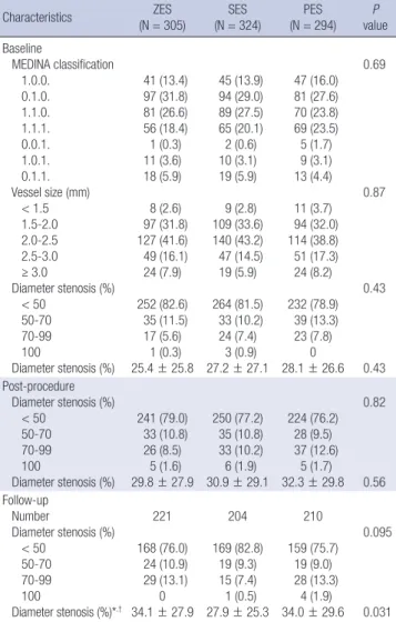
Angiographic analysis of all side branches
Table 4 shows angiographic analysis data of all SBs. True bifur- cation lesions involving both branches, defined as MEDINA classes 1.1.1., 1.0.1., and 0.1.1., were observed in 27.9% of the ZES, 29.0% of the SES, and 31.0% of the PES (P = 0.70) lesions.
SB vessel size was averaged between 2.0 and 2.5 mm. After the procedure, periprocedural SB compromise, defined as ≥ 50%
diameter stenosis, occurred in 21.0% of the ZES, 22.8% of the SES, and 23.8% of the PES (P = 0.70) lesions. At follow-up angi- ography, the mean value of diameter stenosis was lower in the SES than in the ZES and PES groups. Therefore, the prevalence of delayed SB compromise at follow-up was numerically lower in the SES (17.2%) than in the ZES (24.0%) and PES (24.3%) le- sions, but the difference did not attain statistical significance
(P = 0.14). However, when the diameter stenosis was compared using the Kruskal-Wallis test, the statistical significance was not significant at baseline (P = 0.61), post-procedure (P = 0.72) and follow-up (P = 0.29).
Angiographic analysis of side branches without treatment When we analyzed naive, untreated SBs, we observed true bifur- cation lesions in 21.7% of the ZES, 24.6% of the SES, and 27.5%
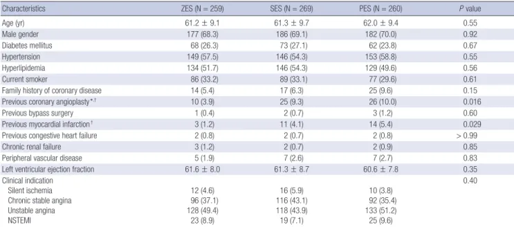
of the PES (P = 0.32) lesions (Table 5). After the procedure, peri- procedural SB compromise from non-diseased SBs occurred in 15.8% of the ZES, 17.2% of the SES, and 16.6% of the PES (P = 0.92) lesions (Fig. 1). However, delayed SB compromise at fol- low-up angiography was found in 13.9% of the ZES, 3.2% of the SES, and 9.4% of the PES (P = 0.010) lesions (Fig. 2). Although SB diameter stenosis at follow-up was numerically lower in the SES than in the ZES and PES groups, this did not attain statisti- cal significance by parametric and non-parametric analyses.
The incidence of SB total occlusion was 2.1% (16 lesions). Of the 10 occluded SBs having follow-up angiography, 6 showed spontaneous recanalization.
Table 5. Angiographic findings of side branches without treatment
Characteristics ZES
(N = 254)
SES (N = 264)
PES (N = 255)
P value Baseline
MEDINA classification 1.0.0.
0.1.0.
1.1.0.
1.1.1.
0.0.1.
1.0.1.
0.1.1.
Size, mm < 1.5 1.5-2.0 2.0-2.5 2.5-3.0 ≥ 3.0
Diameter stenosis (%) < 50
50-70 70-99 100
Diameter stenosis (%)
40 (15.7) 87 (34.3) 71 (28.0) 35 (13.8) 1 (0.4) 5 (2.0) 15 (5.9) 7 (2.8) 79 (31.1) 106 (41.7) 42 (16.5) 20 (7.9) 222 (87.4)
22 (8.7) 9 (3.5) 1 (0.4) 21.6 ± 23.8
40 (15.2) 81 (30.7) 76 (28.8) 43 (16.3) 2 (0.8) 8 (3.0) 14 (5.3) 9 (3.4) 88 (33.3) 119 (45.1) 36 (13.6) 12 (4.5) 227 (86.0)
20 (7.6) 16 (6.1) 1 (0.4) 24.4 ± 25.1
45 (17.6) 77 (30.2) 58 (22.7) 52 (20.4) 5 (2.0) 9 (3.5) 9 (3.5) 11 (4.3) 81 (31.8) 98 (38.4) 43 (16.9) 22 (8.6) 211 (82.7) 28 (11.0) 16 (6.3)
0 26.0 ± 25.3
0.38
0.53
0.51
0.13 Post-procedure
Diameter stenosis (%) < 50
50-70 70-99 100
Diameter stenosis (%) 199 (78.3) 26 (10.2) 24 (9.4) 5 (2.0) 30.2 ± 28.8
198 (75.0) 28 (10.6) 32 (12.1) 6 (2.3) 32.7 ± 29.9
187 (73.3) 26 (10.2) 37 (14.5) 5 (2.0) 34.1 ± 30.7
0.78
0.32 Follow-up
Number
Diameter stenosis (%) < 50
50-70 70-99 100
Diameter stenosis (%)
181 136 (75.1) 23 (12.7) 22 (12.2)
0 33.8 ± 27.7
169 137 (81.1) 17 (10.1) 14 (8.3) 1 (0.6) 29.4 ± 25.7
183 137 (74.9)
17 (9.3) 26 (14.2)
3 (1.6) 34.6 ± 29.5
0.26
0.17 Table 4. Angiographic findings of all side branches
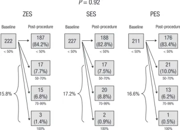
Characteristics ZES
(N = 305) SES
(N = 324) PES
(N = 294) P
value Baseline
MEDINA classification 1.0.0.
0.1.0.
1.1.0.
1.1.1.
0.0.1.
1.0.1.
0.1.1.
Vessel size (mm) < 1.5 1.5-2.0 2.0-2.5 2.5-3.0 ≥ 3.0
Diameter stenosis (%) < 50
50-70 70-99 100
Diameter stenosis (%)
41 (13.4) 97 (31.8) 81 (26.6) 56 (18.4) 1 (0.3) 11 (3.6) 18 (5.9) 8 (2.6) 97 (31.8) 127 (41.6) 49 (16.1) 24 (7.9) 252 (82.6) 35 (11.5) 17 (5.6) 1 (0.3) 25.4 ± 25.8
45 (13.9) 94 (29.0) 89 (27.5) 65 (20.1) 2 (0.6) 10 (3.1) 19 (5.9) 9 (2.8) 109 (33.6) 140 (43.2) 47 (14.5) 19 (5.9) 264 (81.5) 33 (10.2) 24 (7.4) 3 (0.9) 27.2 ± 27.1
47 (16.0) 81 (27.6) 70 (23.8) 69 (23.5) 5 (1.7) 9 (3.1) 13 (4.4) 11 (3.7) 94 (32.0) 114 (38.8) 51 (17.3) 24 (8.2) 232 (78.9) 39 (13.3) 23 (7.8)
0 28.1 ± 26.6
0.69
0.87
0.43
0.43 Post-procedure
Diameter stenosis (%) < 50
50-70 70-99 100
Diameter stenosis (%)
241 (79.0) 33 (10.8) 26 (8.5) 5 (1.6) 29.8 ± 27.9
250 (77.2) 35 (10.8) 33 (10.2) 6 (1.9) 30.9 ± 29.1
224 (76.2) 28 (9.5) 37 (12.6)
5 (1.7) 32.3 ± 29.8
0.82
0.56 Follow-up
Number
Diameter stenosis (%) < 50
50-70 70-99 100
Diameter stenosis (%)*,†
221 168 (76.0) 24 (10.9) 29 (13.1)
0 34.1 ± 27.9
204 169 (82.8)
19 (9.3) 15 (7.4) 1 (0.5) 27.9 ± 25.3
210 159 (75.7)
19 (9.0) 28 (13.3)
4 (1.9) 34.0 ± 29.6
0.095
0.031
*P = 0.05/3 for ZES vs SES, and †P = 0.05/3 for SES vs PES, by post-hoc compari- son.
When the degree of SB diameter stenosis was classified into four groups (< 50%, 50%-70%, 70%-99%, and 100%), worsening of SB stenosis occurred in 48 (9.0%) of lesions comprising 21 (11.6 %), 8 (4.7%), and 19 (10.4%) lesions of the ZES, SES and PES lesions (P = 0.059), respectively (Fig. 3), whereas improve- ment and stationary of SB stenosis were observed in 51 (9.6%) and 434 (81.4%) of lesions, respectively. In a multivariable logis- tic regression model, in-stent MB stenosis at follow-up (%) was the only independent predictor of worsening of SB stenosis (odds ratio, 1.03; 95% confidence interval, 1.01-1.04; P = 0.0003).
Clinical outcomes
Peri-procedural MI, defined as procedure-related increase of
creatine kinase or its MB isoenzyme to at least three times the upper limit of the normal range, occurred in 24 (7.9%), 33 (10.2
%), and 24 (8.2%) lesions after ZES, SES, and PES implantations, respectively (P = 0.53).
One-year clinical follow-up was completed for 97.2% of le- sions, and the clinical outcomes are shown in Table 6. There were no differences among the three groups in the incidence of death or myocardial infarction. However, the incidence of target lesion revascularization for MB, SB, and stent thrombosis, were higher in the PES than in the SES lesions. All target lesion revas- cularizations for SB restenoses were treated percutaneously.
One patient in the PES group received bypass graft surgery for MB restenosis. In naive SBs without treatment, target lesion re- vascularization was performed in 2 (0.7%) of the ZES, none of the SES, and 8 (2.8%) of the PES lesions (P = 0.002).
DISCUSSION
This was a substudy of the ZEST trial, a large randomized study comparing use of the three types of DESs for elective PCI in real
Baseline Post-procedure
ZES
P = 0.92
15.8%
< 50%
< 50%
50-70%
70-99%
100%
222 187
(84.2%)
17 (7.7%)
15 (6.8%)
3 (1.4%)
Baseline Post-procedure
SES
17.2%
< 50%
< 50%
50-70%
70-99%
100%
227 188
(82.8%)
17 (7.5%)
20 (8.8%)
2 (0.9%)
Baseline Post-procedure
PES
16.6%
< 50%
< 50%
50-70%
70-99%
100%
211 176
(83.4%)
21 (10.0%)
13 (6.2%)
1 (0.5%)
Fig. 1. Change in side branch stenosis between baseline and post-procedure for non- diseased side branches without treatment. Periprocedural side branch compromise, defined as ≥ 50% diameter stenosis from non-diseased (< 50%) side branches, oc- curred in 15.8% of the zotarolimus- (ZES), 17.2% of the sirolimus- (SES), and 16.6%
of the paclitaxel-eluting stent (PES) lesions (P = 0.92).
8-month
ZES
P = 0.010
13.9%
< 50%
< 50%
50-70%
70-99%
100%
137 118
(86.1%)
11 (8.0%)
8 (5.8%)
0
8-month
SES
3.2%
< 50%
< 50%
50-70%
70-99%
100%
125 121
(96.8%)
3 (2.4%)
1 (0.8%)
0
Post-procedure Post-procedure Post-procedure 8-month
PES
9.4%
< 50%
< 50%
50-70%
70-99%
100%
128 116
(90.6%)
7 (5.5%)
5 (3.9%)
0
Fig. 2. Change in side branch stenosis between post-procedure and 8-month follow- up from non- compromised side branches without treatment. Delayed side branch compromise, defined as ≥ 50% diameter stenosis at follow-up from non-compro- mised (< 50%) side branches, occurred in 13.9% of the zotarolimus- (ZES), 3.2% of the sirolimus- (SES), and 9.4% of the paclitaxel-eluting stent (PES) lesions (P = 0.010).
Table 6. One-year clinical outcomes per lesion
Characteristics ZES
(n = 299) SES
(n = 310) PES (n = 288) P
value Death
Cardiac Non-cardiac
1 (0.3) 1 0
1 (0.3) 1 0
3 (1.0) 2 1
0.41
Myocardial infarction Non-ST elevation ST elevation
26 (8.7) 24 2
36 (11.6) 34 2
25 (8.7) 25 0
0.37
Target lesion revascularization*
Main branch*
Side branch*
13 (4.3) 13 (4.3) 2 (0.7)
3 (1.0) 3 (0.9) 0
27 (9.4) 27 (9.4) 9 (3.1)
< 0.001
< 0.001 0.001 Stent thrombosis*
Definite Probable
1 (0.3%) 1 0
0 0 0
5 (1.7%) 4 1
0.023
*P = 0.05/3 for SES vs PES, by post-hoc comparison.
Fig. 3. Change in side branch stenosis between post-procedure and 8-month follow- up in all lesions without treatment. Changes in side branch stenosis were assessed according to stenosis group, classified as < 50%, 50%-70%, 70%-90%, and 100%.
The statistical difference of incidences was not significant (NS).
%
ZES (N = 181) SES (N = 169) PES (N = 183) 100
80
60
40
20
0
80.7
Improved No change Aggravation
85.2 78.7
7.7 11.6 10.1 10.9
4.7
P = 0.154
10.4
practice. The major finding of this study was that a minority of patients experienced a worsening of SB stenosis at follow-up angiography after single-stent implantation with DES for treat- ment of bifurcation lesions. In addition, because follow-up SB stenosis was mainly influenced by the severity of MB stenosis, DESs effectiveness to maintain small stenosis in MB at follow- up may help maintain the luminal patency of untreated SBs.
The ZEST trial was a study of ‘all-comers’ undergoing elective PCI with DES in real practice. Therefore, our subgroup analysis has the advantage of allowing us to assess outcomes patterns following current bifurcation treatments. In addition, we could compare the outcomes using the three current types of DESs to treat specific bifurcation lesion sets, with minimal selection bias.
Of all lesions treated with DESs, approximately one-fourth had SBs > 1.5 mm in diameter, requiring protection during PCI (10).
Of these SBs, approximately 20%, or 5% of all lesions, had signif- icant SB stenosis within true bifurcation segments. As a diseased SB is the most important predictor of SB compromise during MB stenting (8), special attention must be given to PCI lesions at risk for complications related to SB compromise. Because of the small proportion of true bifurcations, about one-fifth of bi- furcations underwent SB treatment, with less than 5% of all le- sions receiving stenting in both branches. Our conservative treat- ment pattern was indicated by previous results, showing a com- parable effectiveness of single- and two-stent techniques for treatment of bifurcation lesions (4, 6, 9).
The major purposes of this study were to assess the incidence of periprocedural SB compromise and the long-term angio- graphic outcomes. Few previous studies have described the long-term outcomes of untreated SBs after DES implantation in the MB (13). Furthermore, the insight on the ‘clinically signifi- cant SB stenosis’ has not been consistent across operators. The degree of SB compromise necessitating provisional SB stenting ranged from 50% to 100% in the diverse clinical trials (3, 5, 9). We found that significant periprocedural SB compromise after MB stenting occurred in 16.5% of non-diseased SBs. Although the three DESs had different stent platforms, the incidence of peri- procedural SB compromise were comparably low in all groups, indicating that the three current DES platforms are comparable in maintaining SB patency during the procedure. Consequent- ly, kissing balloon inflation with or without SB stenting was se- lectively performed in about 10% of patients.
The long-term differential effectiveness of the three DESs in maintaining SB patency was measured by analysis of follow-up SB stenosis. Overall, delayed SB compromise at follow-up was observed in only 9.3% of lesions, all of which had been free of stenosis just after the main procedure, in good agreement with the findings of a previous trial (the RAVEL trial), which com- pared follow-up angiographic outcomes of 118 SBs after SES and 124 SBs after BMS implantation (13). Of the 12 and 9 occlud- ed SBs, respectively, 11 (92%) and 6 (67%) spontaneously recan-
alized. Other studies using BMS also supported the phenome- non of natural recanalization of occluded SBs after stent implan- tation (12, 14, 20). In our present study, spontaneous recanali- zation occurred in 6 of the 10 occluded SBs. Therefore, our find- ings, together with previous results, suggest that routine treat- ment of compromised SBs (i.e., the ‘oculo-stenotic reflex’) dur- ing or after the procedure should be avoided, especially in le- sions receiving single-stent treatment for bifurcations. Recent results from the Nordic Bifurcation III trial also suggested that routine kissing-balloon inflation of bifurcation lesions may not provide superior clinical benefit compared with provisional SB treatment (11).
In previous studies, the predictors of delayed SB stenosis or occlusion were the preprocedural morphology or stenosis of SB (12, 13). In our multivariate analysis, however, the small MB stenosis at follow-up treated with SES compared to ZES or PES might translate to improved SB stenosis at follow-up, where an- giography showed that only 3% of lesions were delayed SB com- promise after SES implantation, compared with 14% for ZES and 9% for PES. This finding indicates that DES with higher an- ti-restenotic potencies may be effective even for preservation of untreated SB patency after single-stent bifurcation treatment.
Furthermore, the relative benefit of SES in reducing MB reste- nosis, compared with ZES or PES, was similarly observed as the major outcome of the ZEST trial (15). Nevertheless, all three DESs were associated with an improvement of stenosis in 10%
of SBs and a recanalization of 60% of occluded SBs, indicating that clinical outcomes in SB stenosis are relatively benign. Only 1.2% of SB compromises underwent target lesion revasculariza- tion when MB revascularizations were performed.
Our study had several limitations. First, this was a post-hoc analysis of a large clinical trial. Therefore, although the sub- group analyzed was extracted from a large randomized study, selection bias, resulting in a lack of statistical power, could not be completely avoided. In fact, although the delayed SB com- promise occurred less frequently after SES implantation for naive lesions without treatment, the numerical degree of steno- sis at follow-up was not statistically significant across the differ- ent DESs. Second, due to the limitation of current angiographic analysis system, quantitative angiographic analysis was not per- formed for SB (21). Therefore, because of visual estimation of SB stenosis, inter- or intra-observer variations might have oc- curred (21). A new dedicated quantitative analysis system for bifurcation lesions may improve the accuracy of measurement.
Third, because of the limited number of true bifurcation lesions, our results may not be extrapolated to treatment for complex bifurcation lesions. Fourth, a follow-up bias may occur due to the incomplete performance of angiographic follow-up. Finally, a lack of data on functional ischemia limits our understanding of the clinical implications of these angiographic results. As pre- viously reported, the correspondence rate between angiograph-
ic stenosis and flow impairment detected by fractional flow re- serve is low (7). Thus, future studies on bifurcation coronary le- sions are required. We found that the long-term incidence of aggravation of SB stenosis was low and may be associated with the degree of intimal hyperplasia in the MB after DES implanta- tion. Therefore, aggressive SB treatment of bifurcation lesions may not be necessary in real-world practice. Our results indi- cate the need for additional clinical studies to determine an ef- fective SB treatment, in view of the technical complexity, clini- cal prognosis, and costs of procedures addressing bifurcation coronary lesions.
In conclusion, after main-branch stenting in bifurcation le- sions, a minority of SB appeared to experience worsening of ste- nosis. DES with strong anti-restenotic efficacy may help main- tain SB patency.
REFERENCES
1. Kim YH, Park DW, Suh IW, Jang JS, Hwang ES, Jeong YH, Lee SW, Lee SW, Lee CW, Hong MK, et al. Long-term outcome of simultaneous kiss- ing stenting technique with sirolimus-eluting stent for large bifurcation coronary lesions. Catheter Cardiovasc Interv 2007; 70: 840-6.
2. Kim YH, Park SW, Hong MK, Park DW, Park KM, Lee BK, Song JM, Han KH, Lee CW, Kang DH, et al. Comparison of simple and complex stent- ing techniques in the treatment of unprotected left main coronary artery bifurcation stenosis. Am J Cardiol 2006; 97: 1597-601.
3. Pan M, de Lezo JS, Medina A, Romero M, Segura J, Pavlovic D, Delgado A, Ojeda S, Melián F, Herrador J, et al. Rapamycin-eluting stents for the treatment of bifurcated coronary lesions: a randomized comparison of a simple versus complex strategy. Am Heart J 2004; 148: 857-64.
4. Colombo A, Moses JW, Morice MC, Ludwig J, Holmes DR Jr, Spanos V, Louvard Y, Desmedt B, Di Mario C, Leon MB. Randomized study to eval- uate sirolimus-eluting stents implanted at coronary bifurcation lesions.
Circulation 2004; 109: 1244-9.
5. Steigen TK, Maeng M, Wiseth R, Erglis A, Kumsars I, Narbute I, Gunnes P, Mannsverk J, Meyerdierks O, Rotevatn S, et al. Randomized study on simple versus complex stenting of coronary artery bifurcation lesions: the Nordic bifurcation study. Circulation 2006; 114: 1955-61.
6. Ferenc M, Gick M, Kienzle RP, Bestehorn HP, Werner KD, Comberg T, Kuebler P, Büttner HJ, Neumann FJ. Randomized trial on routine vs.
provisional T-stenting in the treatment of de novo coronary bifurcation lesions. Eur Heart J 2008; 29: 2859-67.
7. Koo BK, Kang HJ, Youn TJ, Chae IH, Choi DJ, Kim HS, Sohn DW, Oh BH, Lee MM, Park YB, et al. Physiologic assessment of jailed side branch lesions using fractional flow reserve. J Am Coll Cardiol 2005; 46: 633-7.
8. Louvard Y, Thomas M, Dzavik V, Hildick-Smith D, Galassi AR, Pan M, Burzotta F, Zelizko M, Dudek D, Ludman P, et al. Classification of coro- nary artery bifurcation lesions and treatments: time for a consensus!
Catheter Cardiovasc Interv 2008; 71: 175-83.
9. Colombo A, Bramucci E, Saccà S, Violini R, Lettieri C, Zanini R, Sheiban I, Paloscia L, Grube E, Schofer J, et al. Randomized study of the crush technique versus provisional side-branch stenting in true coronary bifur-
cations: the CACTUS (Coronary Bifurcations: Application of the Crush- ing Technique Using Sirolimus-Eluting Stents) Study. Circulation 2009;
119: 71-8.
10. Lefèvre T, Louvard Y, Morice MC, Dumas P, Loubeyre C, Benslimane A, Premchand RK, Guillard N, Piéchaud JF. Stenting of bifurcation lesions:
classification, treatments, and results. Catheter Cardiovasc Interv 2000;
49: 274-83.
11. Niemelä M, Kervinen K, Erglis A, Holm NR, Maeng M, Christiansen EH, Kumsars I, Jegere S, Dombrovskis A, Gunnes P, et al. Randomized com- parison of final kissing balloon dilatation versus no final kissing balloon dilatation in patients with coronary bifurcation lesions treated with main vessel stenting: the Nordic-Baltic Bifurcation Study III. Circulation 2011;
123: 79-86.
12. Poerner TC, Kralev S, Voelker W, Sueselbeck T, Latsch A, Pfleger S, Schumacher B, Borggrefe M, Haase KK. Natural history of small and medium-sized side branches after coronary stent implantation. Am Heart J 2002; 143: 627-35.
13. Tanabe K, Serruys PW, Degertekin M, Regar E, van Domburg RT, Sousa JE, Wülfert E, Morice MC. Fate of side branches after coronary arterial sirolimus-eluting stent implantation. Am J Cardiol 2002; 90: 937-41.
14. Arora RR, Raymond RE, Dimas AP, Bhadwar K, Simpfendorfer C. Side branch occlusion during coronary angioplasty: incidence, angiographic characteristics, and outcome. Cathet Cardiovasc Diagn 1989; 18: 210-2.
15. Park DW, Kim YH, Yun SC, Kang SJ, Lee SW, Lee CW, Park SW, Seong IW, Lee JH, Tahk SJ, et al. Comparison of zotarolimus-eluting stents with sirolimus- and paclitaxel-eluting stents for coronary revascularization:
the ZEST (Comparison of the Efficacy and Safety of Zotarolimus-Eluting Stent with Sirolimus-Eluting and PacliTaxel-Eluting Stent for Coronary Lesions) randomized trial. J Am Coll Cardiol 2010; 56: 1187-95.
16. Popma JJ, Leon MB, Moses JW, Holmes DR Jr, Cox N, Fitzpatrick M, Douglas J, Lambert C, Mooney M, Yakubov S, et al. Quantitative assess- ment of angiographic restenosis after sirolimus-eluting stent implanta- tion in native coronary arteries. Circulation 2004; 110: 3773-80.
17. Mehran R, Dangas G, Abizaid AS, Mintz GS, Lansky AJ, Satler LF, Pich- ard AD, Kent KM, Stone GW, Leon MB. Angiographic patterns of in-stent restenosis: classification and implications for long-term outcome. Circu- lation 1999; 100: 1872-8.
18. Medina A, Suárez de Lezo J, Pan M. A new classification of coronary bifurcation lesions. Rev Esp Cardiol 2006; 59: 183.
19. Cutlip DE, Windecker S, Mehran R, Boam A, Cohen DJ, van Es GA, Steg PG, Morel MA, Mauri L, Vranckx P, et al. Clinical end points in coronary stent trials: a case for standardized definitions. Circulation 2007; 115:
2344-51.
20. Mazur W, Grinstead WC, Hakim AH, Dabaghi SF, Abukhalil JM, Ali NM, Joseph J, French BA, Raizner AE. Fate of side branches after intracoro- nary implantation of the Gianturco-Roubin flex-stent for acute or threat- ened closure after percutaneous transluminal coronary angioplasty. Am J Cardiol 1994; 74: 1207-10.
21. Goktekin O, Kaplan S, Dimopoulos K, Barlis P, Tanigawa J, Vatankulu MA, Koning G, Tuinenburg JC, Mario CD. A new quantitative analysis system for the evaluation of coronary bifurcation lesions: comparison with current conventional methods. Catheter Cardiovasc Interv 2007;
69: 172-80.