5
급성심근경색증 환자의 일차적 관동맥 스텐트 삽입술 시 삽입된 Sirolimus-eluting stent 와 Paclitaxel-eluting stent의
임상적 안정성 및 유용성 평가
민계식*, 한만석**
충남대병원 심혈관 조영실*, 충남대병원 영상의학과**
The evaluation for Clinical usefulness and Safety of Sirolimus-eluting stent and Paclitaxel-Eluting Stents In Patients With
Acute Myocardial Infarction
Gyesik Min*, Manseok Han**
Departments of Cardiology, Chungnam National University Hospital*, Departments of Radiology, Chungnam National University Hospital**
요약
AMI로 SES 혹은 PES 시술을 시행받은 모든 환자에서 4년 이상의 임상추적 기간이 지난 환자를 대상으로 데이터 를 분석하여 두 스텐트의 안전성과 유용성을 비교해 보고자 하였다. 2004년 1월 1일부터 2006년 8월 31일까지 본원 에서 ST분절 상승 혹은 ST 분절 비상승 급성심근경색증 (STEMI or NSTEMI)로 진단되어 입원 기간 중 관동맥중재 술을 시행받은 환자 중 SES 혹은 PES 삽입술이 시행된 환자를 대상으로 후향적 분석을 시행하였다. 그리고 사망, 심 장사. 심근경색증, 표적 혈관재관류술, 스텐트 혈전증 발생에 대해 분석하였다.
연구 기간 동안 총 668명의 급성심근경색증 환자가 중 522명만 연구 대상에 포함 사망 (18.3±3.0% vs.
14.6±2.2%, p=0.26), 심장사(11.2±2.6% vs. 6.8±1.52%, p=0.39), 심근경색증 (6.4±1.8% vs. 3.3±1.1%, p=0.31), and 스텐트 혈전증 (5.4±1.7% vs. 3.2±1.1%, p=0.53) 표적 혈관재관류술(TVR) (10.0±3.0% vs.
4.0±1.2%, p=0.008) and 심혈관계 임상사건(MACE) (29.4±3.5% vs. 19.4±2.5%, p=0.003)
급성심근경색증의 초기 치료에 약물방출스텐트인 SES와 PES의 4년 장기 임상 성적을 조사한 본 연구를 통해 전체 환자를 대상으로 분석하였을 때 두 스텐트의 장기 사망률, 심장사. 심근경색증, 표적 혈관재관류술, 스텐트 혈전증의 발생은 차이가 없었으나 TVR 및 MACE의 발생은 PES 삽입 환자가 SES삽입 환자보다 유의하게 높았다.
중심단어
:급성심근경색증
,경피적 심혈관 중재적시술
,스텐트
Corresponding Author: 한만석
주소: 대전 광역시 중구 문화로 282 충남대학병원 영상의학과, E-mail:[email protected], Tel: +82-(0)42-280-7337
ABSTRACT
There is a still unsettled issue about the comparison of long-term clinical effects between sirolimus- (SES) and paclitaxel-eluting stents (PES) for the patients with acute myocardial infarction (AMI). Therefore, we performed a retrospective analysis to evaluate the 4-year clinical outcome of SES as compared with PES after percutaneous coronary intervention (PCI) in patients with AMI.
From January 2004 to August 2006, all consecutive patients with acute ST-segment elevation myocardial infarction (STEMI) underwent primary PCI and acute NSTEMI underwent PCI by implantation either SES or PES were enrolled. The occurrence of death, cardiac death, recurrent infarction, target vessel revascularization (TVR) and stent thrombosis were analyzed. The composite of major adverse cardiac events (MACE; death, recurrent infarction and TVR) were also analyzed.
During the study period, total 668 AMI patients had visited. Of them, total 522 patients (299 with SES and 223 with PES) were enrolled. During 4-year clinical follow-up, there were similar occurrences of death (18.3±3.0% vs. 14.6±2.2%, p=0.26), cardiac death (11.2±2.6% vs. 6.8±1.52%, p=0.39), re-infarction (6.4±1.8% vs. 3.3±1.1%, p=0.31), and stent thrombosis (5.4±1.7% vs. 3.2±1.1%, p=0.53) between the two groups, consecutively. The occurrences of TVR (10.0±3.0% vs. 4.0±1.2%, p=0.008) and MACE (29.4±3.5%
vs. 19.4±2.5%, p=0.003) were significantly higher in patients treated with PES than SES.
In AMI patients treated with either SES or PES implantation, SES had a significantly lower risk of TVR and MACE during 4-year clinical follow-up. Rates of death, cardiac death or recurrent infarction, and stent thrombosis were similar.
Key Words: acute myocardial infarction, percutaneous coronary intervention, stents.
Ⅰ. 서론
약 10년 전 개발되어 현재까지 사용되고 있는 약물 방출스텐트 (drug-eluting stent, DES) 는 일반금속스텐 트 (bare-metal stent, BMS)의 재협착 발생을 획기적으로 감소시켰고 임상 사건을 감소시키는 효과가 있어 새 로 발생된 관동맥 병변 (de novo coronary lesion)의 중 재술의 대부분에서 사용되고 있다[1]-[4]. 이러한 DES의 성적은 좀 더 다양하고 복잡한 임상 사건 혹은 병변에 도 적응되기 시작하여 급성심근경색증 (AMI) 환자의 일차적 관동맥 중재술에서도 좋은 성적이 보고되고
있다[5]-[7]. 그 중에서도 초기에 개발되어 2003년 및
2004년 미국 FDA (food and drug administration)의 승인 을 받아 가장 오랜 기간 임상에 적용되어 온 sirolimus-eluting stent (SES, Cordis, Johnson and Johnson, Miami Lakes, FL, USA)와 paclitaxel-eluting stent (PES, Boston Scientific, Natick, MA, USA)는 de novo coronary lesion의 치료에서 BMS보다 우월한 성적을 보여주고
있다[8],[9]. 하지만 아직까지 AMI 환자에서 SES와 PES
를 이용한 중재술의 장기 임상 성적에 대한 비교 연구 는 충분치 않은 실정이다. .이에 저자는 본원에서 AMI 로 SES 혹은 PES 시술을 시행받은 모든 환자에서 4년 이상의 임상추적 기간이 지난 환자를 대상으로 데이 터를 분석하여 두 스텐트의 안전성과 유용성을 비교 해 보고자 하였다.
II. 연구 대상 및 방법
1. 연구 대상
2004년 1월 1일부터 2006년 8월 31일까지 본원에서 ST분절 상승 혹은 ST 분절 비상승 급성심근경색증 (STEMI or NSTEMI)로 진단되어 입원 기간 중 관동맥 중재술을 시행받은 환자 중 SES 혹은 PES 삽입술이 시행된 환자를 대상으로 후향적 분석을 시행하였다.
2. 연구 방법
연구 대상 환자의 본 연구에의 포함 여부는 의무기 록 및 관동맥중재술의 영상 분석을 이용하여 이루어 졌다. 이들의 임상적 특성, 각종 검사실 소견, 시술 기 록지, 및 관동맥 조영 영상 등을 분석하였다. 퇴원 후 임상 성적의 평가는 추적 진료가 이루어진 환자의 경 우 의무기록지를 이용하여 분석하였으며, 추적진료가 이루어지지 않은 환자인 경우는 전화 상담을 통해 이 루어졌다. 사망 여부 및 사망 원인의 평가는 의무기록 지, 통계청 자료, 전화 상담 및 국민건강요양 보험의 자격중지 여부 등을 이용하였으며 사망의 구체적인 원인과 심장사 혹은 비심장사 여부를 평가하였다. 또 한 스텐트 내 혈전증의 발생 여부를 Academic Research Consortium (ARC) criteria[10]를 이용하였다.
3. 통계 분석
통계 분석은 SPSS version 17.0 으로 하였다. 범주형 변수는 Chi-square (x2)검정을 이용하여 수와 백분율(%) 로 표현하였다. 연속형 변수는 independent sample t-test 혹은 Mann-Whitney U-test를 이용하여 비교하였으며 평 균±표준편차 (mean±SD) 혹은 중앙값(범위) (median (range))로 표현하였다. 추적 기간 중 임상 사건의 발생 에 대한 생존 분석은 log-rank 분석 및 Kaplan Meyer 생 존 분석을 이용하였다. 분석 결과 p < 0.05 이하인 경 우 통계적으로 유의하다고 평가하였다.
III. 연구 결과
1. 대상환자 (study patients)
연구 기간 동안 총 668명의 급성심근경색증 환자가 본원에 방문하였다. 그 중에서 풍선성형술만 이루어지 고 스텐트가 삽입되지 않은 59명, BMS가 삽입된 25명, 다른 종류의 DES가 삽입된 2명, 급성심근경색증에는 부합하나 관동맥중재술을 시행하지 않고 약물치료하 기로 결정된 27명, 수술적 재관류술을 시행받은 11명, 변이형협심증으로 진단된 환자 9명, 혈전의 suction 혹 은 thrombolysis만으로 치료한 5명, 환자의 상태가 불량 하여 관동맥 중재술을 시행할 수 없었던 8명을 제외한 522명의 환자가 본 연구 대상에 포함되었으며 그 중 SES가 삽입된 경우는 299명이었고, PES가 삽입된 경 우는 233명이었다 (그림 1).
그림 1. 연구대상환자.
2. 대상 환자의 일반적인 특성 (baseline clinical characiteristics)
대상 환자의 일반적인 임상적 특성은 표 1와 같다.
평균 연령은 63세로 양 군 간의 차이가 없었으며, 성별 의 분포도 유의한 차이를 보이지 않았다. 전체 환자에 서 STEMI 환자가 차지하는 비율은 SES 군에서 59%, PES 군에서 71%로 PES 군에서 유의하게 높았다 (p<0.001). 허혈성 심질환의 위험인자는 양 군에서 유 의한 차이를 보이지 않았으며, 기저 심질환이나 내원 당시 부정맥의 빈도 등도 유의한 차이를 보이지 않았 다. 좌심실 구혈율도 평균 51%로 양 군 간의 유의한 차이를 보이지 않았다.
SES (n=299)
PES
(n=223) P-value Age, years
Sex (M/F) Type of AMI STEMI, n (%) NSTEMI, n (%) Risk factors Hypertension, n (%) Diabetes, n (%) Smoking, n (%) T-chol >200mg/dL, n (%) Family history, n (%) Previous PCI, n (%) Previous MI, n (%) Previous CABG, n (%) Preinfarction angina, n (%) LV EF (%)
VT / Vfib, n (%) Atrial fibrillation, n (%) Complete AV block, n (%)
63±12 209 / 90
176 (59) 123 (41)
144 (48) 82 (27) 171 (57) 84 (29) 16 (5) 15 (5) 18 (6) 2 (1) 75 (25)
51±12 15 (5)
8 (3) 25 (8)
63±13 158 / 65
158 (71) 65 (29)
103 (42) 60 (27) 121 (54) 54 (26) 10 (5) 13 (6) 9 (4) 0 (0) 50 (23)
51±11 14 (6)
4 (2) 25 (11)
0.52 0.85 0.006
0.66 0.92 0.53 0.48 0.69 0.70 0.43 0.24 0.53 0.77 0.54 0.38 0.30
표1. 대상환자의 일반적인 특성
(Baseline clinical characteristics)
3. 관동맥 조영술 소견
(coronary angiographic findings)
관동맥 조영술의 소견은 표 2과 같다. 혈관의 침범 개수 (angiographic diagnosis)와 병변의 모양 (lesion type) 은 양 군 사이에서 유의한 차이가 없었다. 경색 관련 혈관 (infarct related artery)는 PES 군에서 우관상동맥이 SES 군보다 유의하게 높았다 (39% vs. 31%, p=0.05).
경색관련 병변이 분지부인 경우 (bifurcation lesion)는 SES 군이 PES 군보다 유의하게 많았다 (40% vs. 34%, p=0.005). 초기 TIMI flow는 SES 군이 PES 군보다 TIMI 0 flow가 적었고 (47% vs. 58%, p=0.005), TIMI 3 flow는 유의하게 많았다 (27% vs. 16%, p=0.001). 혈전 의 존재 유무, 근위부의 굴곡 정도, 병변부의 구부러 짐, 및 기시부 병변의 분포 정도는 양 군 간에 유의한 차이를 보이지 않았다.
SES (n=299)
PES
(n=223) P-value No. of diseased vessels
1 VD, n (%) 2 VD, n (%) 3 VD, n (%) LMCA lesion, n (%)
102 (34) 97 (33) 100 (33) 20 (7)
76 (34) 74 (33) 73 (33) 10 (5)
0.93 0.71 0.50 0.34 Lesion types
A, n (%) B1, n (%) B2, n (%) C, n (%)
0 (0) 33 (11) 133 (45) 133 (45)
1 (0.4) 18 (8) 91(41) 113 (51)
0.43 0.30 0.42 0.18 Infarct related artery
LAD, n (%) RCA, n (%) LCx, n (%) LMCA, n (%) Bifurcation lesion, n (%)
Two GWs insertion, n (%) Kissing balloon, n (%)
151 (50) 93 (31) 50 (17) 5 (2) 120 (40)
77 (26) 64 (21)
93 (42) 88 (39) 42 (19) 0 (0) 63 (34) 46 (21) 35 (16)
0.08 0.05 0.64 0.13 0.005 0.18 0.11 Initial TIMI flow
0, n (%) I, n (%) II, n (%) III, n (%)
139 (47) 27 (9) 51 (17) 82 (27)
130 (58) 31 (14) 26 (12) 36 (16)
0.005 0.09 0.18 0.001 Visible thrombus
None-Mild, n (%) Moderate, n (%) Heavy, n (%)
175 (58) 98 (33) 26 (9)
119 (53) 76 (34) 28 (13)
0.29
Proximal tortuosity None-Mild, n (%)
Moderate, n (%) Severe, n (%)
211 (70) 77 (26) 11 (4)
152 (68) 57 (26) 14 (6)
0.45
표 2. 관동맥 조영술 소견 (Coronary angiographic findings)
Lesion angulation None-Mild, n (%)
Moderate, n (%) Heavy, n (%)
241 (78) 55 (18) 3 (1)
174 (81) 48 (22) 1 (0.1)
0.53
Ostial lesion, n (%) 48 (10) 28 (13) 0.32 1VD; one vessel disease, 2VD; two vessel disease, 3VD; triple vessel disease, LAD; left anterior descending artery, RCA; right coronary artery, LCx; left circumflex artery, LMCA; left main coronary artery, GW; guidewire, TIMI; thrombolysis in myocardial infarction
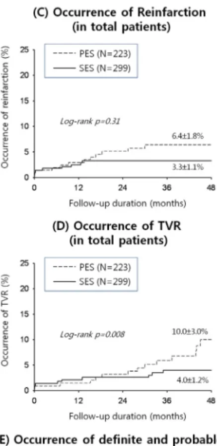
4. 전체 환자에서의 4년 동안의 임상 성적 (4-year clinical outcomes in total patients)
STEMI와 NSTEMI 모두를 포함한 전체 환자 522명 의 4년 추적 기간 동안의 death, cardiac death, reinfarction, 및 stent thrombosis의 발생은 양 군에서 차 이를 보이지 않았다 (그림 2). 하지만 4년 추적 기간 동 안 TVR의 시행 빈도는 PES 군에서 SES 군에 비하여 유의하게 많았으며 (PES=10.0±3.0% vs. SES=4.0±1.2%, p=0.008), MACE도 PES군에서 SES 군보다 유의하게 많이 발생하였다(PES=29.4±3.5% vs. SES=19.4±2.5%, p=0.003) (그림 2).
그림 2. 전체 환자에서의 4년 동안의 임상 성적 (4-year clinical outcomes in total patients). TVR; target vessel revascularization, MACE; major cardiac adverse events.
IV. 고찰 및 결론
급성심근경색증의 초기 치료에 사용된 1세대 약물 방출 스텐트인 SES와 PES의 4년 장기 임상 성적을 조 사한 본 연구를 통해 저자는 다음과 같은 결과를 얻을 수 있었다. (1) SES 및 PES 모두 안전하게 삽입술이 가 능하였고 시술 성공률이 높았다. (2) 전체 환자를 대상 으로 분석하였을 때 두 스텐트의 장기 사망률, 심장사 (cardiac death), reinfarction, 및 stent thrombosis의 발생은 차이가 없었으나 TVR 및 MACE의 발생은 PES 삽입 환자가 SES삽입 환자보다 유의하게 높았다. (3) STEMI 및 NSTEMI 환자를 각각 분석하였을 때 두 스텐트의 장기 사망률, 심장사 (cardiac death), reinfarction, stent thrombosis, 및 TVR의 발생은 양 스텐트 삽입군에서 차이가 없었으나 MACE의 발생은 PES 삽입 환자가 SES삽입 환자보다 유의하게 높았다.
참고문헌
[1] Moses JW, Leon MB, Popma JJ, Fitzgerald PJ, Holmes DR, O'Shaughnessy C, Caputo RP, Kereiakes DJ, Williams DO, Teirstein PS, Jaeger JL, Kuntz RE. Sirolimus-eluting stents versus standard stents in patients with stenosis in a native coronary artery. N Engl J Med.;Vol. 349, pp.1315-1323, 2003.
[2] Sabate M, Jimenez-Quevedo P, Angiolillo DJ, Gomez-Hospital JA, Alfonso F, Hernandez-Antolin R, Goicolea J, Banuelos C, Escaned J, Moreno R, Fernandez C, Fernandez-Aviles F, Macaya C. Randomized comparison of sirolimus-eluting stent versus standard stent for percutaneous coronary revascularization in diabetic patients: The diabetes and sirolimus-eluting stent (diabetes) trial. Circulation., Vol. 112, pp.2175-2183, 2005.
[3] Silber S, Colombo A, Banning AP, Hauptmann K, Drzewiecki J, Grube E, Dudek D, Baim DS. Final 5-year results of the taxus ii trial: A randomized study to assess the effectiveness of slow- and moderate-release polymer-based paclitaxel-eluting stents for de novo coronary artery lesions. Circulation, pp.120:1498-1504. 2009.
[4] Kastrati A, Dibra A, Eberle S, Mehilli J, Suarez de Lezo J, Goy JJ, Ulm K, Schomig A. Sirolimus-eluting stents vs paclitaxel-eluting stents in patients with coronary artery disease:
Meta-analysis of randomized trials. JAMA., Vol. 294, pp.819-825, 2003.
[5] Kastrati A, Dibra A, Spaulding C, Laarman GJ, Menichelli M, Valgimigli M, Di Lorenzo E, Kaiser C, Tierala I, Mehilli J,
Seyfarth M, Varenne O, Dirksen MT, Percoco G, Varricchio A, Pittl U, Syvanne M, Suttorp MJ, Violini R, Schomig A.
Meta-analysis of randomized trials on drug-eluting stents vs.
Bare-metal stents in patients with acute myocardial infarction.
Eur Heart J. Vol. 28, pp.2706-2713, 2007.
[6] De Luca G, Stone GW, Suryapranata H, Laarman GJ, Menichelli M, Kaiser C, Valgimigli M, Di Lorenzo E, Dirksen MT, Spaulding C, Pittl U, Violini R, Percoco G, Marino P.
Efficacy and safety of drug-eluting stents in st-segment elevation myocardial infarction: A meta-analysis of randomized trials. Int J Cardiol. Vol. 133, pp.213-222, 2009.
[7] Hao PP, Chen YG, Wang XL, Zhang Y. Efficacy and safety of drug-eluting stents in patients with acute st-segment-elevation myocardial infarction: A meta-analysis of randomized controlled trials. Tex Heart Inst J. Vol. 37, pp.516-524, 2010.
[8] Shafiq N, Malhotra S, Pandhi P, Behl A, Sharma YP. Cypher versus taxus: The stent war. Expert Opin Investig Drugs. Vol.
15, pp.1537-1544, 2006.
[9] Serruys PW, de Jaegere P, Kiemeneij F, Macaya C, Rutsch W, Heyndrickx G, Emanuelsson H, Marco J, Legrand V, Materne P, et al. A comparison of balloon-expandable-stent implantation with balloon angioplasty in patients with coronary artery disease.
Benestent study group. N Engl J Med. Vol. 331, pp.489-495, 1994.
[10] Cutlip DE, Windecker S, Mehran R, Boam A, Cohen DJ, van Es GA, Steg PG, Morel MA, Mauri L, Vranckx P, McFadden E, Lansky A, Hamon M, Krucoff MW, Serruys PW. Clinical end points in coronary stent trials: A case for standardized definitions. Circulation. Vol. 115, pp.2344-2351, 2007.