Safety and Efficacy of Overlapping Homogenous Drug-Eluting Stents in Patients with Acute Myocardial Infarction: Results from Korea Acute Myocardial Infarction Registry
The aim of this study was to compare safety and efficacy of 4 homogenous overlapping drug-eluting stents (DES) in acute myocardial infarction (AMI) patients. We selected 1,349 consecutive patients (62.1 ± 14.9 yr, 69.4% male) who received homogenous overlapping DESs in diffuse de novo coronary lesions from Korea Acute Myocardial Infarction Registry from April 2006 through September 2010. They were divided into 4 groups based on type of DES implanted – Paclitaxel (PES), Sirolimus (SES), Zotarolimus (ZES) and Everolimus (EES)-eluting stents. Primary endpoint was 12-month MACE. We also studied EES versus other DESs (PES + SES + ZES). Mean stent length was 26.2 ± 7.5 mm and mean stent diameter was 3.1 ± 0.4 mm. Average number of stents used per vessel was 2.2 ± 0.5.
Incidence of major adverse cardiac events (MACE) in PES, SES, ZES, and EES groups were 9.5%, 9.2%, 7.5%, and 3.8%, respectively (P = 0.013). In EES group, overall MACE and repeat revascularization were lowest, and no incidence of stent thrombosis was observed.
Non-fatal MI was highest in PES, almost similar in SES and EES with no incidence in ZES group (P = 0.044). Cox proportional hazard analysis revealed no differences in the incidence of primary endpoint (P = 0.409). This study shows no significant differences in 12-month MACE among 4 groups.
Key Words: Drug-Eluting Stents; Myocardial Infarction; Coronary Angioplasty Khurshid Ahmed1,2, Myung Ho Jeong1,
Rabin Chakraborty2, Young Joon Hong1, Doo Sun Sim1, Sumera Ahmed2, Seung Hwan Hwang1, Min Goo Lee1, Keun Ho Park1, Ju Han Kim1,
Youngkeun Ahn1, Myeong Chan Cho3, Chong Jin Kim4, Young Jo Kim5, Jong Chun Park1, Jung Chaee Kang1, and The Korea Acute Myocardial Infarction Registry Investigators
1The Heart Center of Chonnam National University Hospital, Chonnam National University Research Institute of Medical Sciences, Gwangju, Korea;
2Department of Cardiology, Apollo Gleneagles Hospital, Kolkata, India; 3Department of Cardiology, Chungbuk National University Hospital, Cheongju, Korea; 4Department of Cardiology, East West Neo Medical Center, Seoul, Korea; 5Department of Cardiology, Yeungnam University Hospital, Daegu, Korea
Received: 11 March 2012 Accepted: 5 September 2012 Address for Correspondence:
Myung Ho Jeong, MD
Principal Investigator of Korea Acute Myocardial Infarction Registry, Director of Heart Research Center Nominated by Korea Ministry of Health & Welfare, Director of Department of Education and Research, Chonnam National University Hospital, 671 Jaebong-ro, Dong-gu, Gwangju 501-757, Korea Tel: +82.62-220-6243, Fax: +82.62-228-7174 E-mail: [email protected]
This study was performed with the support of the Korean Society of Circulation in the memorandum of the 50th Anniversary of the Korean Society of Circulation and Korea Ministry of Health and Welfare (A084869), Republic of Korea.
http://dx.doi.org/10.3346/jkms.2012.27.11.1339 • J Korean Med Sci 2012; 27: 1339-1346 Cardiovascular Disorders
INTRODUCTION
Drug-eluting stents (DES) substantially reduce the risk of reste- nosis and target lesion revascularization (TLR) compared with bare metal stents (BMS), particularly in complex coronary dis- ease (1). Long lesions account for approximately 10% of con- temporary percutaneous coronary intervention (PCI) cases, and present specific challenges for stenting with DES. Data from randomized controlled trials and large observational registries highlight the increased risk of restenosis with longer lesions,
and demonstrate a significant reduction in restenosis and tar- get vessel revascularization (TVR) rates with DES use (2-5). As a result, use of DES in diffuse long lesions has become standard clinical practice, supported by international guidelines (6-9).
Multiple overlapping stents are often unavoidable to treat long diffuse lesions owing to excessive lesion length, edge dissection, or incomplete stent coverage. At the present time there is limited information about the safety and efficacy of overlapping DES in treating acute myocardial infarction (AMI) patients with severe diffuse coronary lesions. Experimental studies raised concerns
regarding the safety and efficacy of overlapping DES because of the increased density of polymer, drug, and stent material (10, 11). To further pursue this interesting subject we conducted this retrospective study to examine the clinical outcomes of over- lapping DESs and compared 4 most commonly used types of homogenous DES - Paclitaxel-eluting stents (PES), Sirolimus- eluting stents (SES), Zotarolimus-eluting stents (ZES) and Evero- limus-eluting stents (EES).
MATERIALS AND METHODS
This is a clinical analysis that was carried out in The Heart Cen- ter of Chonnam National University Hospital, Gwangju, Korea.
Ethics statement
The permission to carry out the study was obtained from the hospital authorities (Institutional Review Board number 05-49, I-2008-01-009) and written informed consent was taken from all patients.
Korea Acute Myocardial Infarction Registry
The Korea Acute Myocardial Infarction Registry (KAMIR) is a prospective, multicenter, observational registry designed to ex- amine current epidemiology in hospital management and out- come of patients with AMI in Korea in commemoration of the 50th anniversary of the Korean Circulation Society. The registry included 52 community and university hospitals for primary PCI with 1-yr clinical follow up. Data was collected at each site by a well-trained study coordinator based on standardized pro- tocol (12, 13).
Study population
Patients were enrolled in the registry after admission to partici- pating hospitals with a suspected diagnosis of AMI. Patients’
medical documents were used to note the demographic data, clinical characteristics and relevant laboratory results.
A total number of 14,329 patients with AMI (ST-segment ele- vation MI and non-ST segment elevation MI) undergoing PCI from April 2006 through September 2010 were assessed in this study. We selected 1,349 consecutive patients who received homogenous overlapping stents in diffuse de novo coronary le- sions and studied their 12-month clinical outcomes (follow-up rate 76.6%). Patients who received BMSs or 2 different types of overlapping DESs were excluded from this study. We divided these patients into 4 groups based on the type of homogenous DESs implanted –Paclitaxel-eluting stents, PES group (Taxus, Boston Scientific Corporation, Natick, MA, USA), n = 247; Siro- limus-eluting stents, SES (Cypher, Cordis Corporation, Miami Lakes, FL, USA), n = 280; Zotarolimus-eluting stents, ZES (En- deavor, Medtronic Vascular, Santa Rosa, CA, USA), n = 412 and Everolimus-eluting stents, EES (Xience V/Promus Element),
n = 410. The number of stents implanted, the type of DES used, and the degree of stent overlap were at the operator’s discre- tion.
Study definitions and clinical endpoints
A final diagnosis of AMI was made according to the European Society of Cardiology/American College of Cardiology diagnos- tic criteria of AMI (14). Hypertension was defined as systolic blood pressure > 140 mmHg and/or diastolic blood pressure
> 90 mmHg at rest, at repeated measurements or treatment with anti-hypertensive medications. Diabetes mellitus (DM) was defined as use of oral hypoglycaemic agent or insulin to lower blood glucose levels. Hyperlipidemia was defined as a total cho- lesterol level > 200 mg/dL or treatment with a lipid-lowering agent. CAD was defined as history of MI, revascularization pro- cedure or obstructive CAD.
Peripheral blood samples were obtained using direct veni- puncture. Blood samples were centrifuged and serum was re- moved and stored at a temperature of -70oC until the assay could be performed for proteins and sugar. Absolute creatine kinase- MB levels were determined by radioimmunoassay (Dade Beh- ring, Inc., Miami, FL, USA). Cardiac specific troponin I levels were measured by a paramagnetic particle, chemiluminescent immunoenzymatic assay (Beckham, Coulter, Inc. Fullerton, CA, USA). Twelve-hour fasting serum levels of total cholesterol, tri- glyceride, and low- and high-density lipoprotein cholesterol were measured by standard enzymatic methods. Blood samples for high-sensitivity C-reactive protein (hsCRP) were obtained on admission and analyzed turbidimetrically with sheep anti- bodies against human CRP; this has been validated against the Dade Behring method.
Two-dimensional echocardiography was performed in all patients and left ventricular ejection fraction (LVEF) was as- sessed using modified Simpson’s biplanar method. The mor- phology in coronary angiography was classified according to the criteria of American College of Cardiology/American Heart Association (ACC/AHA) (15). Degree of coronary flow was clas- sified by Thrombolysis In Myocardial Infarction (TIMI) score (16). Presence of left main coronary artery stenosis was defined as a luminal stenosis ≥ 50%. Multivessel disease was defined as the presence of a lesion with > 50% diameter stenosis in a non- infarct related coronary artery. Successful PCI was defined as TIMI flow 3 with residual stenosis ≤ 50% in the infarct related artery. In-hospital complications included any of atrio-ventric- ular block, bradycardia, ventricular tachycardia/ventricular fi- brillation, atrial fibrillation, cardiogenic shock, no re-flow, dis- section, acute renal failure, metabolic acidosis/lactic acidosis, cerebrovascular event, or infection/sepsis. All patients were ad- ministered loading doses of aspirin 325 mg and clopidogrel 300- 600 mg before PCI. Anticoagulation during PCI was performed according to the routine practices of each hospital. After the pro-
cedure, aspirin 100-200 mg (1 time per day) was prescribed in- definitely. Clopidogrel was prescribed continuously for 1 yr.
The primary endpoint was major adverse cardiac events (MACE) which included all-cause death, non-fatal myocardial infarction (MI), TLR and TVR during 12-month follow-up. Non- fatal MI was defined as the presence of clinical symptoms, elec- trocardiographic change, or abnormal imaging findings of MI combined with an increase in creatine kinase – MB fraction or troponin T/I more than the 99th percentile of the upper normal limit that was not related to an interventional procedure. TVR was defined as clinically driven repeat revascularization of a lesion in the same epicardial vessel treated in the index proce- dure at 12 month follow-up. TLR was defined as any revascu- larization of the target lesion due to restenosis or re-occlusion within 5 mm proximal/distal to the stent. All data were recorded on a standardized, electronic, web based registry at http://www.
kamir.or.kr.
Statistical analysis
SPSS 17.0 for Windows (SPSS, Inc., Chicago, IL, USA) was used for all analysis. Continuous variables were presented as mean ± SD; comparisons were conducted by one-way ANOVA. Discrete variables were presented as percentages and frequencies; com- parisons were conducted by chi-square statistics or Fischer’s exact test. A P value < 0.05 was considered statistically signifi- cant. Cox proportional hazard analysis was performed in a step-
wise manner to identify a model with independent predictive factors with determination of a hazard ratio and its 95% confi- dence interval (CI) for each variable in the model. Survival time of each patient to perform Cox analysis was calculated from date of admission/first presentation to the emergency room to the date of follow-up. A cut off P value < 0.20 were selected for en- try into the model. The results are presented as adjusted hazard ratios (HR) with 95% confidence intervals and P values.
RESULTS
A total number of 1,349 consecutive patients with AMI under- going PCI who received 2 or more homogenous overlapping DES in diffuse coronary lesions were included in the present study. These patients were divided into 4 groups based on the type of DESs implanted that is PES group, n = 247; SES group, n = 280; ZES group, n = 412 and EES group, n = 410. Mean age was 62.1 ± 14.9 yr and 69.4% were men. Mean stent length (in- cluding both stents) was 26.2 ± 7.5 mm and mean stent diame- ter was 3.1 ± 0.4 mm. Average number of stents used per vessel was 2.2 ± 0.5. Baseline clinical characteristics and concomitant medications of the 4 groups are presented in Table 1. Previous history of CAD was highest in PES group, followed by ZES, SES and lowest in EES group (P = 0.002). Clinical presentation with ST-segment elevation MI was found to be highest in ZES, fol- lowed by SES, EES and PES (P = 0.024). Use of beta blocker was
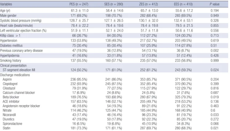
Table 1. Baseline clinical characteristics and concomitant medications
Variables PES (n = 247) SES (n = 280) ZES (n = 412) EES (n = 410) P value
Age (yr) 61.3 ± 11.0 58.4 ± 14.6 65.7 ± 13.0 55.6 ± 17.2 0.194
Male gender 171 (69.2%) 198 (70.7%) 282 (68.4%) 285 (69.5%) 0.949
Systolic blood pressure (mmHg) 128.7 ± 25.7 127.1 ± 26.5 130.1 ± 32.0 132.4 ± 53.1 0.326
Heart rate (beats/minute) 78.4 ± 22.2 78.4 ± 19.6 78.4 ± 19.8 79.5 ± 21.5 0.855
Left ventricular ejection fraction (%) 51.9 ± 11.1 52.1 ± 24.0 51.7 ± 11.8 50.6 ± 11.6 0.556
Killip class > 1 66 (26.7%) 84 (30.0%) 112 (27.2%) 124 (30.2%) 0.713
Hypertension 133 (53.8%) 138 (49.3%) 217 (52.7%) 203 (49.5%) 0.619
Diabetes mellitus 75 (30.4%) 85 (30.4%) 107 (25.9%) 114 (27.8%) 0.51
Previous coronary artery disease 47 (19.0%) 36 (12.8%) 54 (13.1%) 36 (8.7%) 0.002
Hyperlipidemia 41 (16.6%) 33 (11.8%) 57 (13.8%) 51 (12.4%) 0.426
Smoking history 137 (55.5%) 160 (57.1%) 235 (57.0%) 233 (56.8%) 0.999
Clinical presentation
ST-segment elevation MI 124 (50.2%) 171 (61.0%) 252 (61.2%) 243 (59.3%) 0.024
Discharge medications Aspirin
Clopidogrel Cilostazol
Calcium channel blocker Beta blocker
ACE inhibitor
Angiotensin receptor blocker Nitrate
Nicorandil Diuretics Spironolactone Statin
236 (95.5%) 232 (93.9%) 79 (31.9%) 17 (6.9%) 189 (76.5%) 157 (63.5%) 46 (18.6%) 114 (46.2%) 43 (17.4%) 47 (19.0%) 16 (6.5%) 181 (73.3%)
241 (86.0%) 245 (87.5%) 77 (27.5%) 24 (8.6%) 193 (68.9%) 146 (52.1%) 54 (19.3%) 125 (44.7%) 46 (16.4%) 50 (17.8%) 19 (6.8%) 171 (61.1%)
353 (85.7%) 352 (85.4%) 115 (27.9%) 24 (5.8%) 280 (67.9%) 205 (49.7%) 89 (21.6%) 185 (44.9%) 96 (23.3%) 92 (22.3%) 45 (10.9%) 287 (69.7%)
371 (90.5%) 370 (90.2%) 122 (29.7%) 31 (7.6%) 325 (79.3%) 218 (53.2%) 91 (22.2%) 168 (40.9%) 81 (19.7%) 85 (20.7%) 34 (8.3%) 280 (68.3%)
0.204 0.399 0.816 0.687 0.019 0.136 0.463 0.6 0.033 0.272 0.066 0.021 Data are expressed as mean ± SD or as number (percentage). ACE, Angiotensin-converting enzyme.
highest in EES (P = 0.019) and nicorandil in ZES (P = 0.033) group. Use of statin was found to be highest in PES group, fol- lowed by ZES, EES and SES groups (P = 0.021).
Laboratory findings of the 4 groups are presented in Table 2.
Triglyceride showed an increasing trend from PES to SES to ZES to EES (P = 0.024). Serum level of hsCRP was highest in ZES and lowest in SES group (P = 0.046). CK-MB and troponin-I were highest in ZES group and lowest in PES group (P = 0.002 and Table 2. Laboratory characteristics
Variables PES (n = 247) SES (n = 280) ZES (n = 412) EES (n = 410) P value
Glucose (mg/dL) 169.1 ± 82.1 171.8 ± 83.6 169.4 ± 75.7 171.5 ± 82.5 0.966
Creatinine (mg/dL) 1.2 ± 1.5 1.2 ± 1.4 1.3 ± 3.4 1.1 ± 1.2 0.705
Total cholesterol (mg/dL) 183.9 ± 43.5 188.5 ± 43.8 184.3 ± 43.9 187.2 ± 43.4 0.502
Triglyceride (mg/dL) 144.1 ± 143.9 125.4 ± 76.8 123.0 ± 79.4 122.4 ± 77.4 0.024
HDL-C (mg/dL) 43.2 ± 12.2 44.3 ± 12.2 43.4 ± 13.1 43.9 ± 14.6 0.766
LDL-C (mg/dL) 116.6 ± 34.7 120.2 ± 36.5 117.0 ± 35.8 118.3 ± 36.1 0.659
hsCRP (mg/dL) 8.1 ± 33.2 4.4 ± 11.8 11.3 ± 37.9 7.4 ± 24.8 0.046
NTpro BNP (pg/mL) 2,104.4 ± 4,804.3 2,642.1 ± 6,049.1 2,634.3 ± 5,323.1 2,654.2 ± 5,640.3 0.764
HbA1c (%) 6.7 ± 1.5 6.6 ± 1.6 6.7 ± 3.4 6.6 ± 1.4 0.936
CK-MB (U/L) 101.3 ± 147.0 130.8 ± 205.8 165.4 ± 257.4 138.1 ± 173.7 0.002
Troponin-I (ng/mL) 33.8 ± 58.5 55.7 ± 171.9 87.9 ± 430.1 38.6 ± 54.4 0.032
Data are expressed as mean ± SD. CK, Creatine kinase-MB; HbA1c, Hemoglobin A1c; HDL-C, High Density Lipoprotein Cholesterol; hsCRP, High-sensitivity C-Reactive Protein;
LDL-C, Low Density Lipoprotein Cholesterol; NTpro BNP, N-Terminal pro-Brain Natrieuretic Peptide.
Table 3. Coronary angiographic and procedural characteristics
Variables PES (n = 247) SES (n = 280) ZES (n = 412) EES (n = 410) P value
Intervened coronary artery Left main
Left anterior descending Left circumflex Right Coronary Multivessel involvement
2 (0.8%) 123 (49.8%)
23 (9.3%) 99 (40.1%) 142 (57.5%)
7 (2.5%) 167 (59.6%) 36 (12.9%) 70 (25.0%) 179 (63.9%)
5 (1.2%) 213 (51.7%) 45 (10.9%) 149 (36.2%) 226 (54.8%)
3 (0.7%) 234 (57.1%)
40 (9.8%) 133 (32.4%) 263 (64.1%)
0.188 0.056 0.518 0.001 0.019
ACC/AHA lesion Type C 140 (56.7%) 137 (48.9%) 178 (43.2%) 192 (46.8%) 0.06
Pre-TIMI flow grade 0 123 (49.8%) 109 (38.9%) 184 (44.7%) 182 (44.4%) 0.156
Post-TIMI flow grade 3 0 (0%) 0 (0%) 1 (0.2%) 1 (0.2%) 0.733
Total number of stents 2.2 ± 0.4 2.1 ± 0.5 2.2 ± 0.4 2.2 ± 0.5 0.713
Stent length (mm) 29.4 ± 8.8 27.1 ± 8.1 25.8 ± 7.2 23.9 ± 5.6 < 0.001
Stent diameter (mm) 3.1 ± 0.5 3.0 ± 0.4 3.1 ± 0.4 3.1 ± 0.4 0.004
Pre-PCI diameter stenosis (%) 83.1 ± 17.9 87.8 ± 13.1 85.6 ± 17.6 88.1 ± 15.3 0.015
Post-PCI diameter stenosis (%) 10.7 ± 7.8 13.1 ± 11.5 11.8 ± 11.0 12.4 ± 10.7 0.271
Glycoprotein IIb/IIIa inhibitor 32 (13.0%) 34 (12.1%) 45 (10.9%) 54 (13.2%) 0.728
Stent thrombosis 5 (2.0%) 4 (1.4%) 4 (0.9%) 0 (0%) 0.10
Thrombus aspiration 68 (27.5%) 73 (26.1%) 107 (25.9%) 116 (28.3%) 0.824
PCI success 241 (97.6%) 263 (93.9%) 402 (97.6%) 393 (95.8%) 0.453
In-hospital complications 27 (10.9%) 34 (12.1%) 45 (10.9%) 49 (11.9%) 0.952
Data are expressed as mean ± SD or as number (percentage). ACC/AHA, American College of Cardiology/American Heart Association; PCI, percutaneous coronary intervention;
TIMI, Thrombolysis In Myocardial Infarction.
Table 4. Twelve-month clinical outcomes
Outcomes PES (n = 210) SES (n = 229) ZES (n = 279) EES (n = 316) P value
MACE 20 (9.5%) 21 (9.2%) 21 (7.5%) 12 (3.8%) 0.013
All-cause death Cardiac death Non-cardiac death
2 (1.0%) 2 (1.0%) 0 (0%)
5 (2.2%) 0 (0%) 5 (2.2%)
8 (2.9%) 6 (2.2%) 2 (0.7%)
4 (1.3%) 2 (0.6%) 2 (0.6%)
0.508 0.149 0.062
Non-fatal MI 4 (1.9%) 1 (0.4%) 0 (0%) 2 (0.6%) 0.044
Repeat revascularization TVR
TLR
14 (6.7%) 3 (1.4%) 6 (2.9%)
15 (6.6%) 2 (0.9%) 2 (0.9%)
13 (4.6%) 1 (0.4%) 8 (2.9%)
6 (1.9%) 0 (0%) 1 (0.3%)
0.010 0.113 0.040
Stent thrombosis 5 (2.4%) 4 (1.7%) 4 (1.4%) 0 (0%) 0.10
Data are expressed as number (percentage). MACE, Major adverse cardiac events; MI, Myocardial Infarction; TVR, Target vessel revascularization; TLR, Target lesion revascular- ization.
P = 0.032, respectively).
Coronary angiographic and procedural characteristics of 4 groups are presented in Table 3. Multivessel involvement was highest in EES group, followed by SES, PES and ZES (P = 0.019).
Right coronary involvement was highest in PES, then ZES, EES and SES (P = 0.001). Percent diameter stenosis before PCI was highest in EES, followed by SES, ZES and PES (P = 0.015). Mean stent length decreased from PES to EES groups (P < 0.001). Mean stent diameter was found to be least in SES group and almost similar in other groups (P = 0.004). Other angiographic charac- teristics did not show statistically meaningful differences among the groups.
The rate of follow-up at 12 months was 76.6% (n = 1,034).
Twelve-month clinical outcomes are presented in Table 4. The incidence of MACE decreased from PES, SES, ZES to EES groups (9.5% vs 9.2% vs 7.5% vs 3.8%, P = 0.013). Non-fatal MI was high- est in PES group, almost similar in SES and EES groups with no incidence in ZES group (P = 0.044). The occurrence of 12-month cardiac death was highest in ZES, followed by PES and EES with no incidence in SES group (P = 0.149). Incidence of 12-month all-cause death and TVR were not statistically significant among the 4 groups. Repeat revascularization rate was found to be low- est in EES among 4 groups (P = 0.01). There was no significant difference in non-cardiac death and stent thrombosis among the 4 groups (P = 0.062 and P = 0.10 respectively). Two acute events of stent thrombosis (1 in PES and 1 in ZES groups), 5 sub- acute stent thrombosis (3 in PES, 1 in SES and 1 in ZES groups), 3 late stent thrombosis (2 in SES and 1 in ZES groups) and 2 very late stent thrombosis (1 in PES and 1 in SES groups) occurred during the hospital stay. The average rate of PCI success was 97.9% (P = 0.453).
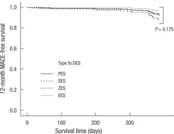
Cox proportional hazard analysis revealed no statistical dif-
ferences in the incidence of MACE when adjusted for age and gender (P = 0.826), and also when adjusted for multiple covari- ates (P = 0.175) (Fig. 1). Table 5 shows details of multivariate anal- ysis. ACC/AHA lesion type C (HR 0.431, CI 0.196 to 0.947, P = 0.036) was found to be the only independent factor of the pri- mary end point in this study. Covariates included in the model were age, gender, diameter stenosis before PCI, stent length and stent diameter, multivessel involvement, ACC/AHA lesion type C, pre-TIMI flow grade 0, clinical presentation ST-segment ele- vation MI, previous history of coronary artery disease (CAD), and statin use.
In this study, EES showed a lower incidence of MACE at 12- months compared to other 3 types of DESs in uni-variate analy- sis as shown in Table 4. Conversely, in multivariate analysis after adjustment for multiple confounders the same difference does not reach statistical significance (Fig. 1). This could be due to the limited power of multivariate analysis with 4 sub-groups.
The results on MACE are hypothesis-generating and warrant further long-term randomized evaluation.
DISCUSSION
The present study demonstrates no significant differences in safety and efficacy among 4 homogenous overlapping DESs in diffuse de novo coronary lesions in patients with AMI. Overlap- ping EES was found to have the lowest MACE rate and TLR-driv- en repeat revascularization rate with no reported events of stent thrombosis over the course of 12-month follow-up period in this cohort.
The development of DES, releasing anti-proliferative drugs into the vessel wall to inhibit neointimal hyperplasia, has revo- lutionized PCI dramatically reducing the incidence of ISR and TLR and permitting the treatment of more complex and exten- sive CAD, including diffuse or long lesions (17). The unique ad- Table 5. Cox regression analysis
Variable Hazard
Ratio
95% confidence interval P value
Lower Upper
Age 0.776 0.365 1.653 0.511
Gender 0.793 0.361 1.739 0.562
Multivessel involvement 1.321 0.625 2.791 0.466
ACC/AHA lesion type C 0.431 0.196 0.947 0.036
Previous history of CAD 1.151 0.473 2.799 0.756 stent length > 25 mm 1.111 0.509 2.423 0.791 stent diameter < 2.5 mm 1.278 0.365 4.474 0.701
Pre TIMI 0 2.036 0.877 4.726 0.098
Troponin I 1.089 0.492 2.411 0.833
Final diagnosis STEMI 0.475 0.212 1.068 0.072
Statin use 1.142 0.478 2.728 0.765
Pre-PCI diameter stenosis 2.205 0.827 5.875 0.114 ACC/AHA, American College of Cardiology/American Heart Association; CAD, Coro- nary artery disease; STEMI, ST-segment elevation myocardial infarction; PCI, Percu- taneous coronary intervention; TIMI, Thrombolysis In Myocardial Infarction.
12-month MACE-free survival
Survival time (days)
0 100 200 300
1.0
0.8
0.6
0.4
0.2
0.0
PES SES ZES EES Type fo DES
P = 0.175
Fig. 1. Cox model survival curves for 12-month major adverse cardiac events-free sur- vival among 4 overlapping homogenous drug-eluting stents. DES, drug-eluting stent;
PES, Paclitaxel-eluting stent; SES, Sirolimus-eluting stent; ZES, Zotarolimus-eluting stent; EES, Everolimus-eluting stent.
vantage of DES is that they allow higher local drug concentra- tions at lesion sites while avoiding systemic toxicity. DES showed signs of delayed arterial healing, more pronounced at the over- lapping sites with increased inflammation and fibrin deposition.
This leads to potential local toxicity caused by increased amounts of polymer and drug released at the overlapping stent site (10).
The importance of a significant delay in endothelialization at sites of overlapping DES is underscored by the recent report of McFadden et al. involving 4 cases of human coronary thrombo- sis occurring > 11 months after deployment of a single polymer- based PES or SES leading to more prolonged use of anti-platelet therapy (18). Lim et al. (19) studied porcine model and found that DESs inhibit neointimal hyperplasia, but inflammation and poor endothelization occur at the site of overlapping stents.
Moreover, Munoz et al. found no significant quantitative changes in intravascular ultrasound measurements within overlapped segment (20).
More than 60% of DESs are used in the treatment of complex coronary lesions and 10% of them are overlapping (21, 22). How- ever, there are sparse data available comparing 2 overlapping DESs, mainly PES and SES, with respect to important clinical outcomes such as death, MI, ST, TVR or TLR. This is the first clin- ical study comparing 4 types of overlapping homogenous DESs for diffuse long lesions in an established large interventional registry of AMI patients with prospective follow-up.
The impact of overlap of DES on clinical and angiographic outcomes is not well established. The Prospective, Randomized MultiCenter Comparison of the Cypher Sirolimus-Eluting and the Taxus Paclitaxel-Eluting Stent System (REALITY) Study (23), a 1,353–patient trial, demonstrated that binary restenosis rate, death, MI, TLR and TVR were similar between SES and PES sys- tems; whereas Sirolimus-eluting and Paclitaxel-eluting Stents for Coronary Revascularization (SIRTAX) Study (24), a 1,012-pa- tient trial, showed a favorable result in the SES arm in terms of TLR, late lumen loss, and binary restenosis.
In a 3-yr follow-up of SIRTAX trial, Raber et al. (22) demon- strated that patients with DES overlap are at greater risk of ex- periencing MACEs, particularly in needs of repeat revascular- ization and ischemic adverse events. Kastrati et al. (25) in a me- ta-analysis of 6 randomized trials compared clinical and angio- graphic outcomes of SES and PES. In this study subjects who received SES had a significantly lower risk of TVR and restenosis compared to PES. Shishehbor et al. (26) reported that overlap- ping SES are comparable with PES for early and late composite end points of death, ST, MI, and TLR; however those that received overlapping SES had a trend towards lower TLR. Degertekin et al. (27) demonstrated that overlapping SES implantation is safe and effective for very long coronary lesions. In their study, they used ≥ 2 overlapping stents at a minimum length of 41 mm to treat native coronary lesion. Their TLR, TVR, and MACE rates were compatible with our findings.
Kandzari et al. (28) in a prospective randomized trial studied comparison of the clinical efficacy, safety and angiographic out- comes among patients treated with ZES and SES, treatment with ZES was associated with increased neointimal hyperplasia re- sulting in greater angiographic late lumen loss. Patients treated with EES rather than PES experienced significantly improved event-free survival at a 2-yr follow-up in the SPIRIT III trial. (29) Serruys et al. (30) found ZES to be non-inferior to EES at 13- months follow-up. There is no data on safety of overlapping ZES and EES with regard to clinical outcomes. In our study cohort, EES appears to be relatively safe among overlapping DESs with lowest MACE and TLR-driven repeat revascularization rates, and no incidence of stent thrombosis. The higher rate of cardiac death in ZES group on univariate analysis could be attributable to high rates of several known predictors of mortality: higher prevalence of old age and ST-elevation myocardial infarction.
They also had higher serum levels of baseline hs-CRP, CK-MB and troponin I, which indicates that the area of infarction was broader in the ZES group.
The risk of compromising the flow in the side branches origi- nating from the main coronaries could have resulted in statisti- cally significant non-fatal myocardial infarction. The patients in our study group received dual anti-platelet therapy (87.9%) for 1 yr after DES implantation according to ACC/AHA/Society for Cardiovascular Angiography and Interventions guidelines for PCI (7) which may have remarkably reduced the rate of stent thrombosis. Lowest MACE rate in EES group could be attribut- able to longer duration of dual antiplatelet therapy, better stent technique with different delivery system and different polymer.
Moreover, the mean length of stents used was smallest in the EES group with highest percentage of pre-procedure diameter stenosis.
The present study is limited due to its retrospective and non- randomized nature, even though it was partially compensated for by adjustment in multivariable analysis. Our patients were analyzed in the short-term (12 months), and the follow-up rate was relatively low (76.6%). However, the number of follow-up patients at 12-months was substantial in this cohort of patients (n = 1,033). This study lacks systemic angiographic follow-up data and possible complications (restenosis or aneurysm) at the site of overlapping could not be ascertained due to registry lim- itation. There could be bias due to rapid evolution in DES strat- egies considering the duration of 4 yr for patient enrollment (SES might be used frequently in the early days of this study, whereas EES might be so in the late phase) and its pharmaco-therapeu- tic management in the recent years. The reason for using over- lapping DES is based on the operator’s discretion which is prone to bias. Nevertheless, inclusion of relatively large number of pa- tients and comparison of 4 most commonly used overlapping DES with appropriate method of statistical analysis is the strength of this study.
In conclusion this study demonstrates that any of the 4 types of overlapping homogenous DES (PES, SES, ZES, or EES) is ac- ceptable to treat AMI patients and there are no significant dif- ferences in their 12-month MACEs. Overlapping EES was found to have the lowest MACE rate and TLR-driven repeat revascu- larization rate with no reported events of stent thrombosis over the course of 12-month follow-up period. This is an important study comparing overlapping DESs where incidence of overall MACE, including TLR and TVR was quite low. We suggest fur- ther long-term randomized evaluation to establish the safety of our study findings.
Appendix: Korea Acute Myocardial Infarction Registry (KAMIR) Study Group of Korean Society of Cardiology Myung Ho Jeong, MD (Chonnam National University Hospital), Youngkeun Ahn, MD (Chonnam National University Hospital), Shung Chull Chae, MD (Kyungpook National University Hospi- tal), Jong Hyun Kim, MD (Busan Hanseo Hospital), Seung Ho Hur, MD (Keimyung University Dongsna Hospital), Young Jo Kim, MD (Yeungnam University Hospital), In Whan Seong, MD (Chungnam National University Hospital), Dong Hoon Choi, MD (Yonsei University Severance Hospital), Jei Keon Chae, MD (Chonnbuk University Hospital), Taek Jong Hong, MD (Pusan National University Hospital), Jae Young Rhew, MD (Chonju Presbyterian Hospital), Doo Il Kim, MD (Inje University Hospi- tal), In Ho Chae, MD (Seoul National University Hospital), Jung Han Yoon, MD (Wonju Christian Hospital), Bon Kwon Koo, MD (Seoul National University Hospital), Byung Ok Kim, MD (Inje University Hospital), Myoung Yong Lee, MD (Dankuk Universi- ty Hospital), Kee Sik Kim, MD (Daegu Catholic University Hos- pital), Jin Young Hwang, MD (Gyeongsang National , Myeong Chan Cho, MD (Chungbuk National University Hospital), Seok Kyu Oh, MD (Wonkwnag University Hospital), Nae Hee Lee, MD (Suncheonhyang University Hospital), Kyoung Tae Jeong, MD (Eulji University Hospital), Seung Jea Tahk, MD (Ahju Universi- ty Hospital), Jang Ho Bae, MD (Keonyang University Hospital), Seung Woon Rha, MD (Korea University Hospital), Keum Soo Park, MD (Inha University Hospital), Chong Jin Kim, MD (Kyung- hee University Hospital), Kyoo Rok Han, MD (Hanlym Univer- sity Hospital), Tae Hoon Ahn, MD (Gacheon University Gill Hos- pital), Moo Hyun Kim, MD (Dong University Hospital), Ki Bae Seung, MD (Catholic University Hospital), Wook Sung Chung, MD (Catholic University Hospital), Ju Young Yang, MD (Ilsan Hospital), Chong Yun Rhim, MD (Hanllym University Hospi- tal), Hyeon Cheol Gwon, MD (Samsung Medical Center), Seong Wook Park, MD (Asan Medical Center), Young Youp Koh, MD (Chosun University Hospital), Seung Jae Joo, MD (Cheju Uni- versity Hospital), Soo Joong Kim, MD (Kyung Hee University Hospital), Dong Kyu Jin, MD (Suncheonhyang University Hos- pital), Jin Man Cho, MD (Kyung Hee University Hospital), Byung Ok Kim, MD (Inje University Hospital), Sang-Wook Kim, MD
(Chungang University Hospital), Jeong Kyung Kim, MD (Dae- jon Sun Hospital), Tae Ik Kim, MD (Busan Maryknoll Hospital), Deug Young Nah, MD (Dongkuk University Hospital), Si Hoon Park, MD (Ihwa University Hospital), Sang Hyun Lee, MD (Mok- po Hankuk Hospital), Seung Uk Lee, MD (Kwangju Christian Hospital), Hang-Jae Chung, MD (Pohang Seonrin Hospital), Jang Hyun Cho, MD (Suncheon Carollo Hospital), Seung Won Jin, MD (Daejon Hankuk Hospital), Yang Soo Jang, MD (Yeon- sei University Severance Hospital), Jeong Gwan Cho, MD, and Seung Jung Park, MD (Asan Medical Center).
REFERENCES
1. Stettler C, Wandel S, Allemann S, Kastrati A, Morice MC, Schömig A, Pfisterer ME, Stone GW, Leon MB, de Lezo JS, et al. Outcomes associat- ed with drug-eluting and bare-metal stents: a collaborative network me- ta-analysis. Lancet 2007; 370: 937-48.
2. Kaltoft A, Jensen LO, Maeng M, Tilsted HH, Thayssen P, Bøttcher M, Lassen JF, Krusell LR, Rasmussen K, Hansen KN, et al. 2-year clinical outcomes after implantation of sirolimus-eluting, paclitaxel-eluting, and bare-metal coronary stents: results from the WDHR (Western Denmark Heart Registry). J Am Coll Cardiol 2009; 53: 658-64.
3. James SK, Stenestrand U, Lindback J, Carlsson J, Scherstén F, Nilsson T, Wallentin L, Lagerqvist B; SCAAR Study Group. Long-term safety and efficacy of drug-eluting versus bare-metal stents in Sweden. N Engl J Med 2009; 360: 1933-45.
4. Eisenberg MJ, Konnyu KJ. Review of randomized clinical trials of drug- eluting stents for the prevention of in-stent restenosis. Am J Cardiol 2006;
98: 375-82.
5. Roukoz H, Bavry AA, Sarkees ML, Mood GR, Kumbhani DJ, Rabbat MG, Bhatt DL. Comprehensive meta-analysis on drug-eluting stents versus bare-metal stents during extended follow-up. Am J Med 2009; 122: 581 e1-10.
6. Ramcharitar S, Hochadel M, Gaster AL, Onuma Y, Gitt A, Serruys PW.
An insight into the current use of drug eluting stents in acute and elective percutaneous coronary interventions in Europe. A report on the EuroPCI Survey. EuroIntervention 2008; 3: 429-41.
7. King SB 3rd, Smith SC Jr, Hirshfeld JW Jr, Jacobs AK, Morrison DA, Wil- liams DO; 2005 WRITING COMMITTEE MEMBERS, Feldman TE, Kern MJ, O’Neill WW, et al. 2007 Focused Update of the ACC/AHA/ SCAI 2005 Guideline Update for Percutaneous Coronary Intervention: a report of the American College of Cardiology/American Heart Association Task Force on Practice Guidelines: 2007 Writing Group to Review New Evi- dence and Update the ACC/AHA/SCAI 2005 Guideline Update for Per- cutaneous Coronary Intervention, Writing on Behalf of the 2005 Writing Committee. Circulation 2008; 117: 261-95.
8. Marroquin OC, Selzer F, Mulukutla SR, Williams DO, Vlachos HA, Wilen- sky RL, Tanguay JF, Holper EM, Abbott JD, Lee JS, et al. A comparison of bare-metal and drug-eluting stents for off-label indications. N Engl J Med 2008; 358: 342-52.
9. Messori A, Trippoli S. Drug-eluting stents for the treatment of coronary artery disease: NICE technology appraisal guidance. Heart 2009; 95: 847.
10. Finn AV, Kolodgie FD, Harnek J, Guerrero LJ, Acampado E, Tefera K, Skorija K, Weber DK, Gold HK, Virmani R. Differential response of de-
layed healing and persistent inflammation at sites of overlapping siroli- mus- or paclitaxel-eluting stents. Circulation 2005; 112: 270-8.
11. Wilson GJ, Polovick JE, Huibregtse BA, Poff BC. Overlapping paclitaxel- eluting stents: long-term effects in a porcine coronary artery model. Car- diovasc Res 2007; 76: 361-72.
12. Lee SR, Jeong MH, Ahn YK, Chae SC, Hur SH, Kim YJ, Seong IW, Chae JK, Hong TJ; Korea Acute Myocardial Infarction Registry Investigators.
Clinical safety of drug-eluting stents in the Korea acute myocardial infarc- tion registry. Circ J 2008; 72: 392-8.
13. Sim DS, Jeong MH, Kang JC. Current management of acute myocardial infarction: experience from the Korea Acute Myocardial Infarction Reg- istry. J Cardiol 2010; 56: 1-7.
14. Apple FS, Wu AH, Jaffe AS. European Society of Cardiology and Ameri- can College of Cardiology guidelines for redefinition of myocardial infarc- tion: how to use existing assays clinically and for clinical trials. Am Heart J 2002; 144: 981-6.
15. Kini AS. Coronary angiography, lesion classification and severity assess- ment. Cardiol Clin 2006; 24: 153-62.
16. Manginas A, Gatzov P, Chasikidis C, Voudris V, Pavlides G, Cokkinos DV.
Estimation of coronary flow reserve using the Thrombolysis In Myocardi- al Infarction (TIMI) frame count method. Am J Cardiol 1999; 83: 1562-5.
17. Kirtane AJ, Gupta A, Iyengar S, Moses JW, Leon MB, Applegate R, Bro- die B, Hannan E, Harjai K, Jensen LO, et al. Safety and efficacy of drug- eluting and bare metal stents: comprehensive meta-analysis of random- ized trials and observational studies. Circulation 2009; 119: 3198-206.
18. McFadden EP, Stabile E, Regar E, Cheneau E, Ong AT, Kinnaird T, Sud- dath WO, Weissman NJ, Torguson R, Kent KM, et al. Late thrombosis in drug-eluting coronary stents after discontinuation of antiplatelet thera- py. Lancet 2004; 364: 1519-21.
19. Lim SY, Jeong MH, Hong SJ, Lim DS, Moon JY, Hong YJ, Kim JH, Ahn Y, Kang JC. Inflammation and delayed endothelization with overlapping drug-eluting stents in a porcine model of in-stent restenosis. Circ J 2008;
72: 463-8.
20. Muñoz JS, Abizaid A, Mintz GS, Albertal M, Abizaid AS, Feres F, Cente- mero M, Staico R, Mattos LA, Pinto I, et al. Intravascular ultrasound study of effects of overlapping sirolimus-eluting stents. Am J Cardiol 2004; 93:
470-3.
21. Farb A, Boam AB. Stent thrombosis redux--the FDA perspective. N Engl J Med 2007; 356: 984-7.
22. Raber L, Juni P, Loffel L, Wandel S, Cook S, Wenaweser P, Togni M, Vogel R, Seiler C, Eberli F, et al. Impact of stent overlap on angiographic and long-term clinical outcome in patients undergoing drug-eluting stent im- plantation. J Am Coll Cardiol 2010; 55: 1178-88.
23. Morice MC, Colombo A, Meier B, Serruys P, Tamburino C, Guagliumi G, Sousa E, Stoll HP; REALITY Trial Investigators. Sirolimus- vs paclitaxel- eluting stents in de novo coronary artery lesions: the REALITY trial: a randomized controlled trial. JAMA 2006; 295: 895-904.
24. Windecker S, Remondino A, Eberli FR, Jüni P, Räber L, Wenaweser P, Togni M, Billinger M, Tüller D, Seiler C, et al. Sirolimus-eluting and pa- clitaxel-eluting stents for coronary revascularization. N Engl J Med 2005;
353: 653-62.
25. Kastrati A, Dibra A, Eberle S, Mehilli J, Suárez de Lezo J, Goy JJ, Ulm K, Schömig A. Sirolimus-eluting stents vs paclitaxel-eluting stents in pa- tients with coronary artery disease: meta-analysis of randomized trials.
JAMA 2005; 294: 819-25.
26. Shishehbor MH, Amini R, Raymond RE, Bavry AA, Brener SJ, Kapadia SR, Whitlow PL, Ellis SG, Bhatt DL. Safety and efficacy of overlapping sirolimus-eluting versus paclitaxel-eluting stents. Am Heart J 2008; 155:
1075-80.
27. Degertekin M, Arampatzis CA, Lemos PA, Saia F, Hoye A, Daemen J, Tanabe K, Lee CH, Hofma SJ, Sianos G, et al. Very long sirolimus-eluting stent implantation for de novo coronary lesions. Am J Cardiol 2004; 93:
826-9.
28. Kandzari DE, Leon MB, Popma JJ, Fitzgerald PJ, O’Shaughnessy C, Ball MW, Turco M, Applegate RJ, Gurbel PA, Midei MG, et al. Comparison of zotarolimus-eluting and sirolimus-eluting stents in patients with native coronary artery disease: a randomized controlled trial. J Am Coll Cardiol 2006; 48: 2440-7.
29. Stone GW, Midei M, Newman W, Sanz M, Hermiller JB, Williams J, Far- hat N, Caputo R, Xenopoulos N, Applegate R, et al. Comparison of evero- limus-eluting and paclitaxel-eluting stents: two-year clinical follow-up from the Clinical Evaluation of the Xience V Everolimus Eluting Coro- nary Stent System in the Treatment of Patients with de novo Native Cor- onary Artery Lesions (SPIRIT) III trial. Circulation 2009; 119: 680-6.
30. Serruys PW, Silber S, Garg S, van Geuns RJ, Richardt G, Buszman PE, Kelbaek H, van Boven AJ, Hofma SH, Linke A, et al. Comparison of zo- tarolimus-eluting and everolimus-eluting coronary stents. N Engl J Med 2010; 363: 136-46.