Paclitaxel-eluting Stent Implantation in Patients with Long Calcified Coronary Lesions
Seung-Ho Hur, MD, PhD, FACC; Yun-Kyeong Cho, MD, PhD; Chang-Wook, Nam, MD, PhD; Hyungseop Kim, MD, PhD; Seong-Wook Han, MD, PhD; Yoon-Nyun Kim, MD, PhD;
Hee-Joon Park, PhD; Jong-Seon Park, MD, PhD; Dong-Gu Shin, MD, PhD; Young-Jo Kim, MD, PhD; Bong-Sup Shim, MD, PhD; Tae-Hyun Yang, MD, PhD; Dae-Kyeong Kim, MD, PhD; Doo-Il Kim, MD, PhD; Dong-Soo Kim, MD, PhD; Kwon-Bae Kim, MD, PhD
Keimyung University Dongsan Medical Center, Cardiovascular Medicine (Hur, Cho, Nam, H. Kim, Han, Y.-N. Kim, K.-B. Kim); Keimyung University Dongsan Medical, Medical Informatics (H.-J. Park); Yeungnam University Hospital, Cardiovascular Medicine (J.-S. Park, Shin, Y.-J. Kim, Shim); Inje University Busan Paik Hospital, Cardiovascular Medicine (Yang, D.-K. Kim, D.-I. Kim, D.-S. Kim)
Jung-gu Daegu 700712 Republic of Korea [email protected]
Background: Although previously reported studies on coronary calcification mainly focused on its presence or absence in discrete focal target lesions, calcified coronary lesions (CCL) angiographically present as diffuse long lesions in some patients. The aim of our study was to evaluate the long-term efficacy of sirolimus-eluting stents (SES) and paclitaxel-eluting stents (PES) on long CCL.
Methods: A total of 122 patients with 134 lesions (77 patients with 88 lesions for SES and 45 patients with 46 lesions for PES) were enrolled from 3 centers. Long CCL was defined visually as a culprit lesion with type B or C that was mainly due to coronary calcification with >20 mm in total length by coronary angiography. Clinical follow-up was performed at 1 year and angiographic follow-up at 6 to 9 months after procedure. Major adverse coronary events (MACE) were defined as all-cause death, myocardial infarction (MI), and repeat target-lesion revascularization (TLR).
Results: There were no statistically significant differences in baseline, procedural, or angiographic character- istics and in 1-year rates of all-cause death, MI, and TLR between the 2 groups (all P = NS [not significant]).
Likewise, the cumulative incidence of MACE at 1 year was similar between the 2 groups (7.8% of patients in the SES group vs 4.4% of patients in the PES group, respectively, P = NS). In patients who underwent follow-up angiography, the angiographic binary restenosis rate was 6.2% in the SES group vs 12.1% in the PES group, respectively (P = NS).
Conclusion: In patients with long CCL, both SES and PES were comparably effective in either angiographic or clinical long-term outcomes.
Introduction
Several studies have demonstrated that calcified coronary lesion (CCL) is a predictor of restenosis or target-lesion revascularization (TLR) following bare-metal stent (BMS) implantation,
1,2most likely because the structure and rigidity of this lesion often adversely affects interventional device delivery or deployment and sometimes causes complications.
Drug-eluting stents (DES) have been used widely as a highly efficacious treatment for patients with coronary artery disease through neointimal hyperplasia inhibition
This work was supported by grant No. RTI04-01-01 from the Regional Technology Innovation Program of the Ministry Knowledge Economy (MKE).
and in-stent restenosis (ISR) reduction.
3,4Furthermore, several clinical trials have shown that DES are effective for patients with high-risk lesions or clinical conditions such as calcified lesions, left main coronary artery disease, multivessel disease, and diabetes.
5,6Calcified coronary lesions are now being routinely
treated with DES, namely with sirolimus-eluting stents
(SES) or paclitaxel-eluting stents (PES). Although previ-
ously reported data regarding CCL mainly addressed its
presence or absence in discrete focal target lesions, in
some patients, CCL appear angiographically as diffuse long
lesions. Technically, long as compared to focal CCL may
be associated with increased device delivery failure rates
and with higher incidences of stent underexpansion, which
in turn may translate into less favorable clinical outcomes
at long-term follow-up. The aim of the present study was
Clinical Investigations
continuedtherefore to evaluate the long-term efficacy of what is currently the most popular DES, namely SES and PES, on long CCL.
Methods Study Population
Patients who met the following criteria were identified from 3 hospitals (Keimyung University Dongsan Hospital, Yeung- nam University Hospital, and Inje University Busan Paik Hospital) between March 2003 and July 2006: (1) de novo coronary artery disease including long CCL; (2) predilation with conventional balloon before stenting; (3) successful DES implantation (SES or PES); and (4) available clinical follow-up data by telephone or office visits and/or high- quality quantitative coronary angiography at baseline and follow-up. Overall, a total of 122 patients (77 SES and 45 PES recipients) were enrolled in this study. Exclusion crite- ria included patients with left main coronary artery disease, cardiogenic shock, chronic renal failure requiring hemodial- ysis or peritoneal dialysis, target lesion containing focal calcification, performing rotational atherectomy or cutting balloon inflation before stenting, and aspirin or clopidogrel intolerance.
Interventional Procedure
Percutaneous coronary intervention (PCI) was performed by standard technique via femoral or radial approach. Vessel size was determined by quantitative coronary angiogra- phy for appropriate stent size selection. Predilation with a conventional balloon was successfully performed to facilitate stent passage across the lesion in all patients.
After predilation, patients received either single or multi- ple SESs (Cypher, Cordis, Johnson & Johnson, Warren, NJ) or PESs (Taxus, Boston Scientific, Natick, MA) with a stent/artery diameter ratio of 1.0 to 1.1. Additional high-pressure balloon inflation was allowed per physician discretion. All patients provided written informed consent prior to the procedure. Unfractionated intravenous heparin was given at doses of 70 to 100 IU/kg to achieve an activated clotting time of >250 seconds. Aspirin at 100 to 200 mg and a loading dose of 300 mg clopidogrel were administered prior to DES implantation and all patients were prescribed aspirin (100 mg/d) indefinitely and clopidogrel (75 mg/d) and/or cilostazol (200 mg/d) for at least 6 months.
Angiographic Analysis
All coronary angiograms were analyzed using standard def- initions and measurements. The external diameter of the contrast-filled catheter was used for calibration. Modified ACC/AHA criteria were used to classify coronary lesion morphology.
7Long CCL was defined visually as a culprit lesion with type B or C that was mainly due to coronary calcification (radio-dense configuration noted within the artery wall before contrast injection in the coronary artery)
with >20 mm in total length by coronary angiography. Ref- erence vessel diameters were measured at angiographically normal segments proximal and distal to the lesion. Mini- mum lumen diameter (MLD) was measured during diastole at the tightest lumen narrowing site pre-intervention and post-intervention, and at follow-up from multiple projec- tions. Angiographic percent diameter stenosis was defined as (1-MLD/reference vessel diameter) × 100. Acute gain was calculated as the pre-intervention MLD minus the post-intervention MLD. Late loss was calculated as the post- intervention MLD minus the MLD at follow-up. Loss index was calculated as late loss divided by acute gain. Residual diameter stenosis was defined as the ratio of lesion MLD and the averaged proximal and distal reference segment diameters. Angiographic binary restenosis was defined as diameter stenosis of ≥50% within the treated site at follow- up. Multivessel disease was defined as the presence of
≥50% stenosis of the luminal diameter in more than 2 major epicardial coronary arteries.
Clinical and Angiographic Follow-Up
Clinical follow-up was conducted by telephone or office interview at 30 days and 12 months after the intervention, and angiographic follow-up was performed at 6 to 9 months after procedure. Myocardial infarction (MI) was defined as a 2-time elevation of the upper limit of normal serum creatinine kinase (CK) levels and/or new pathological Q waves in 2 or more leads and with elevated (above normal) CK-MB levels. Target-lesion revascularizations were defined as any revascularization procedure performed on the treated segments and that were ischemia-driven.
Stent thrombosis was defined as angiographically proven thrombosis, unexplained death, or target-vessel MI. Stent thrombosis was also classified as acute if it occurred within 24 hours, subacute if it occurred between 1 day and 30 days, and late if it occurred between 30 days and 1 year after DES implantation. Major adverse cardiac events (MACE) were defined as the occurrence of death, MI, TLR, and stent thrombosis during the follow-up period. Angiographic follow-up was performed in 70.9% of SES patients and in 71.7% of PES patients.
Statistical Analysis
Statistical analyses were performed with the SPSS statistical
software, version 15.0 (SPSS, Inc., Chicago, IL). Quantitative
data are presented as mean ± SD and qualitative data are
presented as frequencies. For comparison of continuous
variables, a Student t test was used, and a χ
2test or Fisher
exact test was used to compare categorical variables. Event-
free survival curves were plotted using the Kaplan-Meier
method. A P value <0.05 was considered statistically
significant.
Male gender, % 62 49 0.19
ACS, % 69 73 0.68
Diabetes mellitus, % 34 31 0.84
Hypercholesterolemia, % 39 40 1.00
Hypertension, % 51 44 0.58
Current smoker, % 44 38 0.22
Prior MI, % 12 9 0.77
Prior CABG, % 0 4 0.13
Multivessel disease, % 60 76 0.11
LVEF, % 52.4± 10.9 54.7± 12.3 0.30
Abbreviations: ACS, acute coronary syndrome; CABG, coronary artery bypass graft; LVEF, left ventricular ejection fraction; MI, myocardial infarction.
Results
Patient Characteristics
Baseline characteristics of the SES and PES groups are shown in Table 1. Mean age, male gender proportion, clinical presentation, frequency of coronary risk factors, and history of myocardial infarction or coronary artery bypass surgery were similar between the 2 groups.
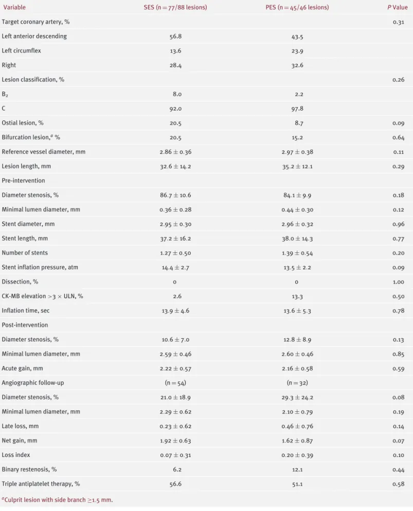
Procedural and Angiographic Results
Procedural characteristics and angiographic results are listed in Table 2. Reference vessel diameter, lesion length, stent diameter, stent length, and number of stents were not statistically different between the 2 groups. After the procedure, more than one half of the patients in both groups were treated with triple antiplatelet therapy. At follow-up, late loss and binary restenosis were not different between the 2 groups (0.23 ± 0.62 mm for SES vs 0.346 ± 0.76 mm for PES, 6.2% for SES vs 12.1% for PES, respectively; all
P= NS [not significant]).
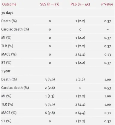
Clinical Outcomes
With respect to clinical outcomes (Table 3) for the SES and PES groups, no significant differences were noted in incidences of all-cause death (0% vs 2.2%, P = NS), MI (0%
vs 2.2%, P = NS), TLR (0% vs 2.2%, P = NS), and MACE (0% vs 4.4%, P = NS) at 30 days. At 1 year after SES or PES implantation, the incidences of all-cause death, MI, and TLR rates were not significantly different (3.9% vs 2.2%; 1.3%
vs 2.2%; 3.9% vs 4.4%, respectively, all P = NS), translating into similar cumulative MACE rates (7.8% vs 4.4%, P = NS)
Discussion
Although previous studies have reported late outcomes according to presence or absence of calcification in discrete target lesions, the present study evaluated the impact of SES and PES implantation on long-term angiographic and clinical outcomes in long CCL. Rates of angiographic binary restenosis and MACE were similar between the 2 groups, suggesting that both DES were comparably effective in reducing ISR and MACE in the complex lesion subset of long CCL.
Calcified coronary lesions have been associated with unfavorable early and late outcomes after PCI, such as dissection, less optimal stent expansion, and higher TLR or ISR rates.
1,8 – 10A study by Hoffmann et al
9showed that preatheroablation using rotational atherectomy, followed by adjunctive bare-metal stent implantation provided the largest final minimal lumen diameter compared to BMS implantation alone or rotational atherectomy plus balloon angioplasty, a feature that translated into the most favorable clinical outcomes at 9 months among the 3 modalities.
Drug-eluting stent use has been associated with dramatic reductions in neointimal proliferation and with improvement of clinical outcomes after stenting even in high-risk patients and complex lesion subsets.
5,6,11 – 13Recently, several studies have addressed the efficacy of SES in CCL treatment. Li et al
14reported that SES implantation in 189 patients with CCL conferred similar favorable results as compared with SES implantation in 264 patients with non- CCL. In-stent restenosis occurred in 3.8% of patients in CCL vs 4.2% of patients in non-CCL, and the TLR rate was 4.9%
in CCL vs 6.9% in non-CCL, respectively. A single center
study by Clavijo et al
15demonstrated efficacy of SES with
or without rotablator in 150 patients who had angiographic
evidence of heavy CCL. At 6 months after procedure, both
the SES alone and SES plus rotablator groups showed
low MACE rates (1.5% vs 1.3%) and no evidence of stent
thrombosis. In another study, ISR rate was significantly
lower and late loss was significantly smaller in the SES
group compared to the BMS group when predilatation was
successfully performed before stent implantation.
16These
studies suggested that CCL might not be associated with
unfavorable early or late outcomes after SES implantation,
which is in line with the results of the present study. In
contrast, Kawaguchi et al
17suggested that severe CCL may
be associated with a higher MACE rate at 1 year. In their
study in 195 lesions treated with SES, severe CCL was
associated with a higher TLR and angiographic restenosis
compared with non-CCL (9.2% vs 3.6%, P<0.05; 7.3% vs 2.8%,
Clinical Investigations
continuedTable 2. Procedural and Angiographic Characteristics
Variable SES (n= 77/88 lesions) PES (n= 45/46 lesions) P Value
Target coronary artery, % 0.31
Left anterior descending 56.8 43.5
Left circumflex 13.6 23.9
Right 28.4 32.6
Lesion classification, % 0.26
B2 8.0 2.2
C 92.0 97.8
Ostial lesion, % 20.5 8.7 0.09
Bifurcation lesion,a% 20.5 15.2 0.64
Reference vessel diameter, mm 2.86± 0.36 2.97± 0.38 0.11
Lesion length, mm 32.6± 14.2 35.2± 12.1 0.29
Pre-intervention
Diameter stenosis, % 86.7± 10.6 84.1± 9.9 0.18
Minimal lumen diameter, mm 0.36± 0.28 0.44± 0.30 0.12
Stent diameter, mm 2.95± 0.30 2.96± 0.32 0.96
Stent length, mm 37.2± 16.2 38.0± 14.3 0.77
Number of stents 1.27± 0.50 1.39± 0.54 0.20
Stent inflation pressure, atm 14.4± 2.7 13.5± 2.2 0.09
Dissection, % 0 0 1.00
CK-MB elevation >3× ULN, % 2.6 13.3 0.50
Inflation time, sec 13.9± 4.6 13.6± 5.3 0.78
Post-intervention
Diameter stenosis, % 10.6± 7.0 12.8± 8.9 0.13
Minimal lumen diameter, mm 2.59± 0.46 2.60± 0.46 0.85
Acute gain, mm 2.22± 0.57 2.16± 0.58 0.59
Angiographic follow-up (n= 54) (n= 32)
Diameter stenosis, % 21.0± 18.9 29.3± 24.2 0.08
Minimal lumen diameter, mm 2.29± 0.62 2.10± 0.79 0.19
Late loss, mm 0.23± 0.62 0.46± 0.76 0.14
Net gain, mm 1.92± 0.63 1.62± 0.87 0.07
Loss index 0.07± 0.31 0.20± 0.39 0.10
Binary restenosis, % 6.2 12.1 0.44
Triple antiplatelet therapy, % 56.6 51.1 0.58
aCulprit lesion with side branch≥1.5 mm.
Death (%) 0 1 (2.2) 0.37
Cardiac death (%) 0 0 –
MI (%) 0 1 (2.2) 0.37
TLR (%) 0 1 (2.2) 0.37
MACE (%) 0 2 (4.4) 0.13
ST (%) 0 1 (2.2) 0.37
1 year
Death (%) 3 (3.9) 1(2.2) 1.00
Cardiac death (%) 2 (2.6) 0 0.53
MI (%) 1 (1.3) 1 (2.2) 1.00
TLR (%) 3 (3.9) 2 (4.4) 1.00
MACE (%) 6 (7.8) 2 (4.4) 0.71
ST (%) 0 1 (2.2) 0.37
Abbreviations: MACE= major adverse cardiac event; MI = myocardial infarction; ST= stent thrombosis; TLR = target-lesion revascularization.
1.00
0.95
0.90
0.85
0.80
0.75
0.70
0 100 200 300 400
Days
SES PES Log rank, p = 0.48
Event-free survival
Figure 1. Kaplan-Meier estimates of event-free survival among patients who received SES and those who received PES.
P<0.05 respectively). This difference may be due to the
different baseline characteristics of the study populations.
Compared to SES, few data are available regarding efficacy of PES on CCL. A substudy of TAXUS-IV
18demonstrated that ISR rate at 9 months and ischemia-driven TLR rate at 1 year were similar between CCL and non- CCL in patients treated with PES, suggesting that PES
present study compared to that in the TAXUS-IV substudy (12.5% vs 7.5%, respectively). However, ischemia-driven TLR rates were similar between the 2 studies (5.7% vs 5.1%, respectively).
The impact of calcification within coronary lesions is still uncertain. Besides reducing vessel injury by a physical shield role, the relatively stable biomechanical properties of calcium may prevent activation of neointimal proliferation.
This theory was supported by an IVUS study of Shimada et al,
19showing that NIH was greater in non-CCL compared to CCL in 21 patients treated with BMS. On the other hand, higher ISR and TLR rates were observed in CCL following stenting.
17Severely calcified lesions pose a great technical challenge during the procedure and may induce stent underexpansion, which is a major predictor of ISR following DES or BMS implantation.
15,20Plaque calcification is mostly found in patients with higher risk factors such as diabetes, hypertension, high cholesterol, and old age.
21,22It may also be possible that fragments of small calcium plaque injure adjacent soft plaques or vessel lumens to cause plaque rupture or lumen damage. To clarify the clinical relevance of CCL during PCI amidst controversial theories, a larger study population with longer-term clinical follow-up is warranted.
Study Limitations
This retrospective study carries several potential limitations.
First, the duration of angiographic and clinical follow-up was limited to only 6 to 9 months and 1 year, respectively.
Further studies with longer-term follow-up are needed to assess the long-term effect of DES implantation in long CCL.
Second, because of the limited number of patients treated with rotational atherectomy or cutting balloon inflation before stenting, the present study included only patients who underwent successful plain old balloon angioplasty, which might be a source of selection bias. Third, cases of unsuccessful delivery of stent device due to long CCL have been excluded from this study. With the stent platform features of the current generation DES, however, the incidence of unsuccessful stent deployment would have been low. Finally, long CCL were not determined by intravascular ultrasound in the present study. Thus, the real length and degree of CCL may be underestimated.
Conclusions
Treatment of long CCL with either SES or PES was
associated with low cumulative incidences of MACE and
angiographic binary restenosis rates, which were not signif-
icantly different between the 2 groups. These data suggest
Clinical Investigations
continuedthat both types of DES were comparably effective for the treatment of long CCL.
Acknowledgments
The authors are grateful to Yong-Taik Son, BS and Roberto Patarca, MD, PhD for assistance in the preparation of this manuscript.
References
1. Fitzgerald PJ, Ports TA, Yock PG. Contribution of localized calcium deposits to dissection after angioplasty. An observational study using intravascular ultrasound. Circulation. 1992;86:64–70.
2. Hoffmann R, Mintz GS, Mehran R, et al. Intravascular ultrasound predictors of angiographic restenosis in lesions treated with Palmaz-Schatz stents. J Am Coll Cardiol. 1998;31:43–49.
3. Moses JW, Leon MB, Popma JJ, et al. Sirolimus-eluting stents versus standard stents in patients with stenosis in a native coronary artery. N Engl J Med. 2003;349:1315–1323.
4. Stone GW, Ellis SG, Cox DA, et al. A polymer-based, paclitaxel- eluting stent in patients with coronary artery disease. N Engl J Med. 2004;350:221–231.
5. Seung KB, Park DW, Kim YH, et al. Stents versus coronary-artery bypass grafting for left main coronary artery disease. N Engl J Med.
2008;358:1781–1792.
6. Stone GW, Ellis SG, Cannon L, et al. Comparison of a polymer- based paclitaxel-eluting stent with a bare metal stent in patients with complex coronary artery disease: a randomized controlled trial. JAMA. 2005;294:1215–1223.
7. Ellis SG, Vandormael MG, Cowley MJ, et al. Coronary morpho- logic and clinical determinants of procedural outcome with angio- plasty for multivessel coronary disease. Implications for patient selection. Multivessel Angioplasty Prognosis Study Group. Circu- lation. 1990;82:1193–1202.
8. Hoffmann R, Mintz GS, Popma JJ, et al. Treatment of calcified coro- nary lesions with Palmaz-Schatz stents. An intravascular ultrasound study. Eur Heart J. 1998;19:1224–1231.
9. Hoffmann R, Mintz GS, Kent KM, et al. Comparative early and nine-month results of rotational atherectomy, stents, and the com- bination of both for calcified lesions in large coronary arteries. Am J Cardiol. 1998;81:552–557.
10. Moussa I, Di Mario C, Moses J, et al. Coronary stenting after rotational atherectomy in calcified and complex lesions.
Angiographic and clinical follow-up results. Circulation. 1997;96:
128–136.
11. Kelbaek H, Klovgaard L, Helqvist S, et al. Long-term outcome in patients treated with sirolimus-eluting stents in complex coronary artery lesions: 3-year results of the SCANDSTENT (Stenting Coro- nary Arteries in Non-Stress/Benestent Disease) trial. J Am Coll Cardiol. 2008;51:2011–2016.
12. Yanagi D, Shirai K, Takamiya Y, et al. Results of provisional stent- ing with a sirolimus-eluting stent for bifurcation lesion: multicenter study in Japan. J Cardiol. 2008;51:89–94.
13. Kastrati A, Mehilli J, von Beckerath N, et al. Sirolimus-eluting stent or paclitaxel-eluting stent vs balloon angioplasty for prevention of recurrences in patients with coronary in-stent restenosis: a randomized controlled trial. JAMA. 2005;293:165–171.
14. Li JJ, Xu B, Yang YJ, et al. Effects of sirolimus-eluting stent on calcified coronary lesions. Chin Med J(Engl). 2008;121:6–11.
15. Clavijo LC, Steinberg DH, Torguson R, et al. Sirolimus-eluting stents and calcified coronary lesions: clinical outcomes of patients treated with and without rotational atherectomy. Catheter Cardio- vasc Interv. 2006;68:873–878.
16. Seo A, Fujii T, Inoue T, et al. Initial and long-term outcomes of sirolimus-eluting stents for calcified lesions compared with bare- metal stents. Int Heart J. 2007;48:137–147.
17. Kawaguchi R, Tsurugaya H, Hoshizaki H, Toyama T, Oshima S, Taniguchi K. Impact of lesion calcification on clinical and angio- graphic outcome after sirolimus-eluting stent implantation in real-world patients. Cardiovasc Revasc Med. 2008;9:2–8.
18. Moussa I, Ellis SG, Jones M, et al. Impact of coronary culprit lesion calcium in patients undergoing paclitaxel-eluting stent implantation (a TAXUS-IV sub study). Am J Cardiol. 2005;96:
1242–1247.
19. Shimada Y, Kataoka T, Courtney BK, et al. Influence of plaque calcium on neointimal hyperplasia following bare metal and drug- eluting stent implantation. Catheter Cardiovasc Interv. 2006;67:
866–869.
20. Virmani R, Farb A, Burke AP. Coronary angioplasty from the perspective of atherosclerotic plaque: morphologic predictors of immediate success and restenosis. Am Heart J. 1994;127:163–179.
21. Budoff MJ, Yu D, Nasir K, et al. Diabetes and progression of coro- nary calcium under the influence of statin therapy. Am Heart J.
2005;149:695–700.
22. Hata N, Kunimi T, Kishida H, Miyagawa H, Ikema Y, Hayakawa H.
Clinical significance of coronary artery calcification. Int Angiol.
1994;13:281–285.