Paclitaxel- Versus Sirolimus-Eluting Stents for Treatment of ST-Segment Elevation Myocardial Infarction
With Analyses for Diabetic and Nondiabetic Subpopulation
Youngjin Cho, MD,* Han-Mo Yang, MD,* Kyung-Woo Park, MD,*
Woo-Young Chung, MD,† Dong-Joo Choi, MD,† Won-Woo Seo, MD,*
Kyoung-Tae Jeong, MD,‡ Sung-Chul Chae, MD,§ Myoung-Yong Lee, MD,储 Seung-Ho Hur, MD,¶ Jei-Keon Chae, MD,# In-Whan Seong, MD,**
Jung-Han Yoon, MD,†† Suk-Kyu Oh, MD,‡‡ Doo-Il Kim, MD,§§ Keum-Soo Park, MD,储储 Seung-Woon Rha, MD,¶¶ Yang-Soo Jang, MD,## Jang-Ho Bae, MD,***
Taeg-Jong Hong, MD,††† Myeong-Chan Cho, MD,‡‡‡ Young-Jo Kim, MD,§§§
Myung-Ho Jeong, MD,储储储 Min-Jung Kim, MS,¶¶¶ Sue K. Park, MD,¶¶¶ In-Ho Chae, MD,†
Hyo-Soo Kim, MD,* for the KAMIR Investigators
Seoul, Seongnam, Daegu, Cheonan, Jeonju, Daejeon, Wonju, Iksan, Busan, Incheon, and Gwangju, Korea
Objectives The aim of this study was to determine which drug-eluting stent (DES) is preferable for the treatment of ST-segment elevation myocardial infarction (STEMI) and to elucidate the impact of diabetes mellitus on the outcome of each DES.
Background Recent studies have shown the benefit of DES in patients with STEMI. Diabetes melli- tus might differentially affect outcomes of each DES.
Methods We analyzed the large-scale, prospective, observational KAMIR (Korea Acute Myocardial Infarction Registry) study, which enrolled 4,416 STEMI patients (26% with diabetes) treated with paclitaxel-eluting stent (PES) or sirolimus-eluting stent (SES). Primary outcome was major adverse cardiac event (MACE), defined as a composite of mortality, nonfatal myocardial infarction, and target lesion revascularization (TLR).
Results In the overall population, the MACE rate at 1 year was significantly higher in the PES than the SES group (11.6% vs. 8.6%, p ⫽ 0.014), which was mainly due to increased TLR (3.7% vs. 1.8%, p ⬍ 0.001). In the diabetic subgroup, however, the MACE rate was not significantly different be- tween PES and SES (14.5% vs. 12.3%, p ⫽ 0.217), in contrast to the nondiabetic subgroup, where PES was inferior to SES as in the overall population. Matching by propensity-score did not signifi- cantly alter these results. For TLR, there was interaction between the type of stents and diabetes mellitus (unadjusted: p ⫽ 0.052; after propensity-score matching: p ⫽ 0.035).
Conclusions The PES was inferior to the SES in the overall population, with regard to the occur- rence of MACE and TLR. However, subgroup analysis for diabetic subjects showed no differences in clinical outcomes between PES and SES. These results suggest that diabetes differentially affects the outcome of first-generation DES. (J Am Coll Cardiol Intv 2010;3:498 –506) © 2010 by the American College of Cardiology Foundation
From the *Cardiovascular Center, Seoul National University Hospital, Seoul, Korea; †Division of Cardiology, Seoul National University Bundang Hospital, Seongnam, Korea; ‡EulJi General Hospital, Seoul, Korea; §Kyungpook National University Hospital, Daegu, Korea;储Dankook University Hospital, Cheonan, Korea; ¶Keimyung University Dongsan Hospital, Daegu, Korea; #Chonbuk National University Hospital, Jeonju, Korea; **Chungnam National University Hospital, Daejeon, Korea;
††Wonju Christian Hospital, Wonju, Korea; ‡‡Wonkwang University Hospital, Iksan, Korea; §§Busan Paik Hospital, Busan, Korea; 储储Inha University Hospital, Incheon, Korea; ¶¶Korea University Guro Hospital, Seoul, Korea; ##Yonsei University Severance Hospital, Seoul, Korea; ***Konyang University Hospital, Daejeon, Korea; †††Busan National University Hospital, Busan, Korea; ‡‡‡Choong-Buk National University, Cheong-Joo, Korea; §§§Young-Nam University Hospital, Daegu, Korea;
The presence of diabetes mellitus is 1 of the compelling indications to use drug-eluting stents (DES) over bare-metal stents during elective percutaneous coronary intervention (PCI) (1,2). Recently, data from randomized trials (3,4) and the large Massachusetts PCI registry (5) have shown the benefit and safety of DES even in patients with ST-segment elevation myocardial infarction (STEMI), which might lead to a significantly increased penetration of DES use in such patients. However, data compar- ing different DES in patients with STEMI are scarce, and furthermore, the impact of diabetes in the performance of each DES in these high-risk patients is mostly unknown.
Therefore, we sought to determine which DES is pref- erable for the treatment of STEMI in diabetic patients and to elucidate the impact of diabetes mellitus on outcome of each DES by analyzing the largest acute myocardial infarc- tion (AMI) database registry in Korea.
Methods
Study population and KAMIR (Korean Acute Myocardial Infarction Registry). In Korea, a nationwide effort was launched in 2005 to collect data from patients with AMI admitted to major cardiac centers capable of primary PCI, which was named the KAMIR. It is a large-scale, multi- center, Internet-based AMI registry, where 49 institutions are actively enrolling patients. It is the largest AMI registry in Korea and, to the best of our knowledge, one of the largest in the world. Since November 2005 to January 2008, 14,049 patients with AMI have been enrolled in the KAMIR. The general characteristics of KAMIR have been described elsewhere (6), and the study protocol was ap- proved by the ethics committee at each participating institution.
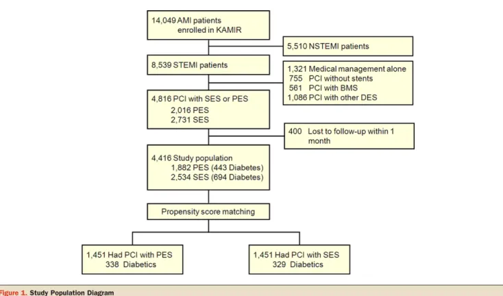
For the present study, patients who underwent PCI with paclitaxel-eluting stent (PES) or sirolimus-eluting stent (SES) for STEMI were selected, which comprised 4,816 of the 14,049 total registered patients. With exclusion of 400 patients lost to follow-up within 1 month, a total of 4,416 patients— of which 1,137 (26%) had diabetes mellitus—
were finally analyzed (Fig. 1). The proportions of patients receiving each DES at specified time periods were not significantly different (Online Fig. 1).
Study definitions. The presence of diabetes mellitus and type of diabetes treatment (insulin or oral antidiabetic agents) was documented through self-reporting by the patient. The level of glycosylated hemoglobin (HbA1c) during hospital stay and the information on the diabetic microvascular complications (nephropathy, retinopathy, or neuropathy) were additionally collected at the time of the
analysis, with retrospective review of medical records. Data on other cardiovascular risk factors such as hypertension, smoking, and prior ischemic heart disease were also re- ported by the patients themselves, except dyslipidemia, which was defined as a composite of self-reported history, prior statin usage, and fasting cholesterol ⱖ200 mg/dl.
Angiographic parameters such as Thrombolysis In Myocar- dial Infarction (TIMI) flow grade or American College of Cardiology/American Heart Association lesion type were assessed by the operator. Successful procedure was defined as ⬍20% residual stenosis after procedure in this study.
Major adverse cardiac event (MACE) was defined as a composite of mortality from any cause, nonfatal myocardial infarction, and target lesion revas-
cularization (TLR). Myocardial infarction was defined as the pres- ence of at least 2 of the following 3 conditions: 1) ischemic symp- toms; 2) elevation of cardiac markers at least twice the upper limit of normal; or 3) new ST- segment elevation. The TLR was defined as a repeated intervention (surgical or percutaneous) result- ing from restenosis or re-occlusion within the stent or in the adjacent 5-mm segments.
PCI and follow-up. Coronary in- terventions were performed ac- cording to current standard pro- cedural guidelines. The choice of PES or SES was left to the operator’s discretion. Aspirin and clopidogrel (loading dose, 300 mg, or 600 mg) were prescribed before or during the coronary in- tervention. After the procedure, clopidogrel was prescribed for at least 6 months, and aspirin was continued indefinitely. All pa- tients were scheduled to be fol-
lowed clinically at 1 month, 6 months, and 1 year after the index procedure. Routine angiographic follow-up for asymp- tomatic patients was not mandatory. Primary outcome in this study was cumulative MACE rate within 1 year after PCI.
Statistical analysis. Student t test and chi-square (or Fisher’s exact) test were used to compare means and proportion of baseline clinical and angiographic characteristics between 2 stent groups. After checking for the violation of propor-
储储储Chonnam National University Hospital, Gwangju, Korea; and the ¶¶¶Medical Research Collaborating Center, Department of Preventive Medicine, Seoul National University College of Medicine, Seoul, Korea. This work was supported by the Innovative Reserach Institute for Cell Therapy (IRICT) and the Clinical Research Center for Ischemic Heart Disease (0412-CR02-0704-0001). The first 3 authors contributed equally to this work.
Manuscript received February 1, 2010, accepted February 5, 2010.
Abbreviations and Acronyms
AMIⴝ acute myocardial infarction
CIⴝ confidence interval DESⴝ drug-eluting stent(s) HbA1cⴝ glycosylated hemoglobin HRⴝ hazard ratio MACEⴝ major adverse cardiac event
mTORⴝ mammalian target of rapamycin
PCIⴝ percutaneous coronary intervention PESⴝ paclitaxel-eluting stent(s)
SESⴝ sirolimus-eluting stent(s)
STEMIⴝ ST-segment elevation myocardial infarction
TIMIⴝ Thrombolysis In Myocardial Infarction TLRⴝ target lesion revascularization
J A C C : C A R D I O V A S C U L A R I N T E R V E N T I O N S , V O L . 3 , N O . 5 , 2 0 1 0 Cho et al.
M A Y 2 0 1 0 : 4 9 8 – 5 0 6 DES for Diabetic STEMI Patient
499
tional hazard assumption, the Cox proportional hazard model was used to estimate the hazard ratio (HR) and 95%
confidence interval (CI) for clinical outcomes between 2 stent groups in crude study population. The p value for interaction between the stent type and the presence of diabetes mellitus was calculated by testing significance with a likelihood ratio test in the Cox regression model, includ- ing an interaction term.
To address potential sources of bias and confounding in this observational study, rigorous adjustment was conducted by the use of propensity analysis (7,8). Propensity score for stent choice was computed by nonparsimonious logistic regression model (c-statistics ⫽ 0.640). Except outcome variables and prescribed medications after stenting, baseline clinical and angiographic features listed in Table 1 were incorporated for this model. Morbidity of diabetes mellitus was also excluded from this calculation, because it was an interested independent variable. We confirmed the model reliability with goodness-of-fit test (p ⫽ 0.101) and then performed 1:1 match iteration by propensity score from initial 8 to 1 digit. Baseline covariates were compared again in this matched population with paired t test or McNemar test (or marginal homeogeneity test). With the robust sandwich covariance matrix estimation, the HR (95% CIs) of clinical outcomes between PES and SES group were
estimated with the Cox proportional hazard model, adjusted for covariates as follows: lesion type B2/C, stent length, stent diameter, number of implanted stents that were well-known predictors for revascularization of DES (9), post-procedural TIMI flow grade 3, achievement of procedural success, and prescribed medication at the time of discharge. The HbA1c level, diabetic microvascular complication, and insulin require- ment were additionally adjusted for the diabetic subgroup.
Hazard ratio of recurrent myocardial infarction was estimated without adjustment for these covariates, because it interfered with the development of reliable regression model. All of these statistical analyses were performed with SAS version 9.1.3 (SAS Institute, Cary, North Carolina), and p value ⬍0.05 was considered as significant.
Results
Baseline characteristics of crude study population. Baseline clinical and angiographic characteristics of crude study population are presented in Table 1. The SES group was slightly younger, had a higher incidence of diabetes mellitus, and underwent stent implantation more frequently as pri- mary angioplasty. With respect to angiographic features, the left anterior descending artery was more often the infarct- related artery for the SES group, and B2/C lesions defined by
Figure 1.Study Population Diagram
AMI⫽ acute myocardial infarction; BMS ⫽ bare-metal stent(s); DES ⫽ drug-eluting stent(s); KAMIR ⫽ Korean Acute Myocardial Infarction Registry; NSTEMI ⫽ non–ST-segment elevation myocardial infarction; PCI⫽ percutaneous coronary intervention; PES ⫽ paclitaxel-eluting stent(s); SES ⫽ sirolimus-eluting stent(s);
STEMI⫽ ST-segment elevation myocardial infarction.
the American College of Cardiology was more frequent in the PES group. The SES group tended to use longer stents with smaller diameter, whereas the PES group tended to implant more stents during the index procedure. The use of glyco- protein IIb/IIIa inhibitor was higher in the PES group.
Clinical outcomes of crude study population up to 1-year follow-up. With median follow-up of 365 days in crude study population, a cumulative total of 396 events occurred.
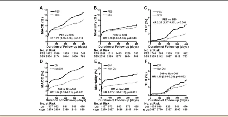
The estimated 1-year MACE rate was 9.5%. Unadjusted analysis revealed that the PES usage as well as diabetes mellitus was associated with increased MACE. Diabetes mellitus definitely augmented the risk (p ⬍ 0.001), with regard to mortality from any cause, but the type of DES was not a significant risk factor (p ⫽ 0.543). The PES usage was associated with higher TLR rate (p ⬍ 0.001), whereas diabetes mellitus increased the risk for TLR only with borderline statistical significance (p ⫽ 0.092) (Fig. 2). The interaction between diabetes mellitus and the type of DES was noteworthy only for TLR (p ⫽ 0.052).
Subgroup analysis in diabetic and nondiabetic population.
BASELINE CHARACTERISTICS.
Given the results described in the preceding text, we performed subgroup analysis for the diabetic and nondiabetic populations. Baseline charac- teristics of each population are presented in Online Table 1. In diabetic patients, diabetic microvascular disease was more frequent and the level of HbA1c tended to be higher in the SES group. In the nondiabetic population, which constituted 74% of the total study population, clinical and angiographic features followed the trend of the overall population.
CLINICAL OUTCOMES OF DIABETIC AND NONDIABETIC POPULATION.
In diabetic patients with STEMI, there was no significant difference in MACE (p ⫽ 0.217) or TLR (p ⫽ 0.448) between the 2 stent groups. However, in the nondiabetic population, PES use was associated with higher incidence of MACE (HR: 1.36; 95% CI: 1.06 to 1.74, p ⫽ 0.016), mainly due to increased TLR (HR: 3.21, 95% CI:
1.83 to 5.63, p ⬍ 0.001). The 2 groups did not differ from each other, with respect to the hard end points such as all-cause mortality, regardless of the presence of diabetes mellitus (for diabetic, p ⫽ 0.300; for nondiabetic, p ⫽ 0.820) (Fig. 3).
Analysis in patients matched for propensity score.
BASELINE CHARACTERISTICS.We performed propensity analysis as described in the Methods section, and 1,451 patients from each stent group were matched for propensity score. Of these, 667 (23%) were diabetic; other baseline characteristics are listed in Table 2. All baseline clinical and angiographic features became comparable after propensity score match- ing. Baseline characteristics of the diabetic and nondiabetic subpopulations are provided in Online Table 2.
CLINICAL OUTCOMES UP TO 1 YEAR.
With median follow-up of 368 days, a total of 212 MACE occurred in the matched population, and the estimated 1-year MACE rate was 9.4%
in the PES group and 6.5% in the SES group (p ⫽ 0.007). The
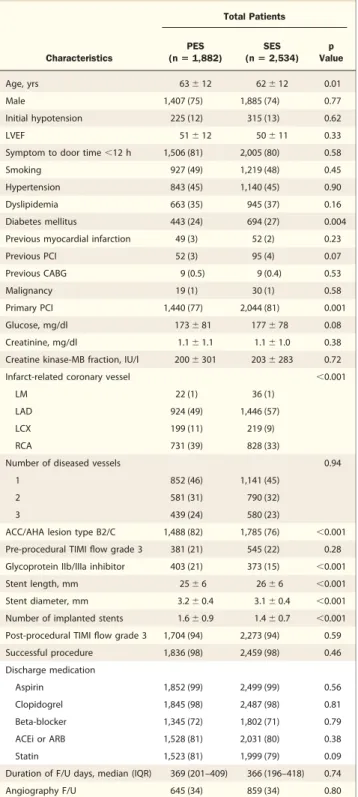
Table 1.Baseline Characteristics of Patients in Crude Population
Characteristics
Total Patients
PES (nⴝ 1,882)
SES (nⴝ 2,534)
p Value
Age, yrs 63⫾ 12 62⫾ 12 0.01
Male 1,407 (75) 1,885 (74) 0.77
Initial hypotension 225 (12) 315 (13) 0.62
LVEF 51⫾ 12 50⫾ 11 0.33
Symptom to door time⬍12 h 1,506 (81) 2,005 (80) 0.58
Smoking 927 (49) 1,219 (48) 0.45
Hypertension 843 (45) 1,140 (45) 0.90
Dyslipidemia 663 (35) 945 (37) 0.16
Diabetes mellitus 443 (24) 694 (27) 0.004
Previous myocardial infarction 49 (3) 52 (2) 0.23
Previous PCI 52 (3) 95 (4) 0.07
Previous CABG 9 (0.5) 9 (0.4) 0.53
Malignancy 19 (1) 30 (1) 0.58
Primary PCI 1,440 (77) 2,044 (81) 0.001
Glucose, mg/dl 173⫾ 81 177⫾ 78 0.08
Creatinine, mg/dl 1.1⫾ 1.1 1.1⫾ 1.0 0.38
Creatine kinase-MB fraction, IU/l 200⫾ 301 203⫾ 283 0.72
Infarct-related coronary vessel ⬍0.001
LM 22 (1) 36 (1)
LAD 924 (49) 1,446 (57)
LCX 199 (11) 219 (9)
RCA 731 (39) 828 (33)
Number of diseased vessels 0.94
1 852 (46) 1,141 (45)
2 581 (31) 790 (32)
3 439 (24) 580 (23)
ACC/AHA lesion type B2/C 1,488 (82) 1,785 (76) ⬍0.001 Pre-procedural TIMI flow grade 3 381 (21) 545 (22) 0.28 Glycoprotein IIb/IIIa inhibitor 403 (21) 373 (15) ⬍0.001
Stent length, mm 25⫾ 6 26⫾ 6 ⬍0.001
Stent diameter, mm 3.2⫾ 0.4 3.1⫾ 0.4 ⬍0.001
Number of implanted stents 1.6⫾ 0.9 1.4⫾ 0.7 ⬍0.001 Post-procedural TIMI flow grade 3 1,704 (94) 2,273 (94) 0.59
Successful procedure 1,836 (98) 2,459 (98) 0.46
Discharge medication
Aspirin 1,852 (99) 2,499 (99) 0.56
Clopidogrel 1,845 (98) 2,487 (98) 0.81
Beta-blocker 1,345 (72) 1,802 (71) 0.79
ACEi or ARB 1,528 (81) 2,031 (80) 0.38
Statin 1,523 (81) 1,999 (79) 0.09
Duration of F/U days, median (IQR) 369 (201–409) 366 (196–418) 0.74
Angiography F/U 645 (34) 859 (34) 0.80
Values given as percentage unless otherwise indicated. Data might not sum to total due to missed values.
ACC/AHA⫽ American College of Cardiology/American Heart Association; ACEi ⫽ angiotensin- converting enzyme inhibitor; ARB⫽ angiotensin receptor blocker; CABG ⫽ coronary artery bypass graft; F/U⫽ follow-up; IQR ⫽ interquartile range; LAD ⫽ left anterior descending; LCX ⫽ left circumflex; LM⫽ left main; LVEF ⫽ left ventricular ejection fraction; PCI ⫽ percutaneous coronary angioplasty; PES⫽ paclitaxel-eluting stent(s); RCA ⫽ right coronary artery; SES ⫽ sirolimus-eluting stent(s); TIMI⫽ Thrombolysis In Myocardial Infarction.
J A C C : C A R D I O V A S C U L A R I N T E R V E N T I O N S , V O L . 3 , N O . 5 , 2 0 1 0 Cho et al.
M A Y 2 0 1 0 : 4 9 8 – 5 0 6 DES for Diabetic STEMI Patient
501
Cox proportional hazard regression model for this matched population revealed results similar to the crude study popula- tion with more significant interaction p value between the
DES type and diabetes mellitus (Figs. 4 and 5). The estimated 1-year TLR rates for each group were as follows: 4.1% for PES and 4.4% for SES group in diabetic patients (p ⫽
Figure 2.Cumulative Incidences of MACE, All-Cause Mortality, and TLR in Crude Study Population
Cumulative incidences of major adverse cardiac events (MACE) (A, D), all-cause mortality (B, E), and target lesion revascularization (TLR) (C, F) in crude study population: comparison of PES versus SES or diabetes mellitus (DM) versus non-DM patients. The PES usage as well as DM was associated with increased MACE.
Diabetes mellitus augmented the risk for death, and PES usage increased the TLR rate. HR⫽ hazard ratio; other abbreviations as inFigure 1.
Figure 3.Clinical Outcomes of Diabetic and Nondiabetic Population
Note that the inferiority of PES to SES regarding MACE and TLR rates in the nondiabetic population disappears in the diabetic population. There is borderline significant interaction between DM and the type of DES for TLR (p⫽ 0.052). PY ⫽ patient-year; other abbreviations as inFigures 1and2.
0.767); and 4.0% for PES and 1.5% for SES group in nondiabetic patients (p ⫽ 0.003). And the incidence curve of TLR in the SES group of the nondiabetic population was located in the lowest area from index procedure to final
follow-up time (Fig. 6). The risk for the composite of all-cause mortality was not significantly different between 2 stent groups, with regard to the hard end point (p ⫽ 0.875 for diabetic, p ⫽ 0.259 for nondiabetic).
Discussion
The current study represents the largest multicenter com- parison of PES and SES implantation for patients with STEMI in Korea. The main finding of this study was that in the overall and nondiabetic population SES showed lower risk of MACE than PES up to 1 year, mainly due to a reduction in TLR. However, among diabetic patients there was no significant difference, suggesting that diabetes might differentially affect the outcomes of PES and SES in patients with STEMI. This was statistically tested by interaction p value, which revealed borderline significance.
Propensity analysis made this implication more convincing, with evident statistical significance.
The PES group showed, with respect to the hard end point represented by all-cause mortality in this study, slightly more events than the SES group, which was not significant. Regarding the nonfatal myocardial infarction, the incidence was too low to make any solid comparison between the 2 DES groups.
Comparison with previous studies. The estimated 1-year MACE rates for the overall population of this study were 11.6% in PES and 8.6% in SES group. These were comparable to the results from the AMI subgroup of the European RESEARCH/T-SEARCH (Rapamycin- Eluting Stent Evaluated at Rotterdam Cardiology Hospital/
Taxus-Stent Evaluated At Rotterdam Cardiology Hospital) registry, which reported 1-year MACE as 15.4% in PES and 9.7% in SES group (10). After matching for propensity score the estimated 1-year MACE rates became 9.4% in PES and 6.5% in SES group, which were also similar to the results of randomized trials conducted for AMI patients such as the PASSION (Paclitaxel-Eluting Stent Versus Conventional Stent in ST-Segment Elevation Myocardial Infarction) (8.8% in PES group), TYPHOON (Trial to Assess the Use of the Cypher Stent in Acute Myocardial Infarction Treated with Angioplasty) (7.3% in SES group) or SESAMI (Sirolimus-Eluting Stent Versus Bare-Metal Stent in Acute Myocardial Infarction) trials (6.8% in SES group). Comparing the baseline characteristics of the cur- rent study with these randomized trials also suggested that the study populations were not different for the most part (3,4,11).
There had been many studies about relative efficacy of PES versus SES (12,13), and recent collaborative network meta-analysis comprising 40 randomized trials concluded that the patients receiving SES had a lower risk for repeat
Table 2.Baseline Characteristics of Patients Matched for Propensity Score
Characteristics
Total Patients
PES (nⴝ 1,451)
SES
(nⴝ 1,451) p Value
Age, yrs 62⫾ 12 63⫾ 12 0.781
Male 1,094 (75) 1,095 (76) 1.000
Initial hypotension 162 (11) 163 (11) 1.000
LVEF 50⫾ 12 50⫾ 11 0.702
Symptom to door time⬍12 h 1,173 (81) 1,157 (80) 0.478
Smoking 728 (50) 735 (71) 0.824
Hypertension 634 (44) 643 (44) 0.761
Dyslipidemia 523 (36) 538 (37) 0.591
Diabetes mellitus 338 (23) 329 (23) 0.725
Previous myocardial infarction 35 (2) 33 (2) 0.901
Previous PCI 40 (3) 54 (4) 0.175
Previous CABG 8 (0.6) 6 (0.4) 0.791
Malignancy 15 (1) 15 (1) 1.000
Primary PCI 1,101 (76) 1,107 (76) 0.819
Glucose, mg/dl 170⫾ 74 168⫾ 68 0.451
Creatinine, mg/dl 1.1⫾ 0.9 1.1⫾ 1.2 0.608
Creatine kinase-MB fraction, IU/l 205⫾ 323 199⫾ 305 0.589
Infarct-related coronary vessel 0.489
LM 15 (1) 10 (1)
LAD 760 (52) 753 (52)
LCX 150 (10) 145 (10)
RCA 526 (36) 543 (37)
Number of diseased vessels 0.797
1 693 (48) 689 (48)
2 446 (31) 443 (31)
3 312 (22) 319 (22)
ACC/AHA lesion type B2/C 1,172 (81) 1,164 (80) 0.739 Pre-procedural TIMI flow grade 3 309 (22) 339 (24) 0.215 Glycoprotein IIb/IIIa inhibitor 283 (20) 268 (19) 0.476
Stent length, mm 25⫾ 6 26⫾ 6 0.485
Stent diameter, mm 3.2⫾ 0.4 3.2⫾ 0.3 0.955
Number of implanted stents 1.5⫾ 0.8 1.5⫾ 0.8 0.895 Post-procedural TIMI flow grade 3 1,367 (95) 1,367 (95) 0.797
Successful procedure 1,426 (99) 1,414 (98) 0.203
Discharge medication
Aspirin 1,433 (99) 1,435 (99) 0.864
Clopidogrel 1,429 (99) 1,428 (98) 1.000
Beta-blocker 1,059 (73) 1,051 (72) 0.775
ACEi or ARB 1,201 (83) 1,197 (83) 0.886
Statin 1,195 (82) 1,188 (82) 0.776
Duration of F/U days, median (IQR) 372 (224–412) 368 (207–413) 0.880
Angiography F/U 532 (37) 545 (38) 0.646
Values given as percentage unless otherwise indicated.
Abbreviations as inTable 1.
J A C C : C A R D I O V A S C U L A R I N T E R V E N T I O N S , V O L . 3 , N O . 5 , 2 0 1 0 Cho et al.
M A Y 2 0 1 0 : 4 9 8 – 5 0 6 DES for Diabetic STEMI Patient
503
revascularization compared with patients receiving PES (14). Only a few studies directly compared SES and PES previously, with regard to the patients with AMI, mostly being limited in size (10,15). Our group previously reported that SES, compared to PES, was associated with remark- ably lower 1-year MACE rate in the AMI subgroup of the Korean VERITAS (VERIfy Thrombosis risk ASsessment) registry (16), and the current study again confirmed the superior efficacy of SES over PES in STEMI patients.
Meanwhile, superiority of SES was not apparent in patients with diabetes mellitus. Although Dibra et al. (17) reported that SES had lower late loss than PES in diabetic patients, several registry data showed conflicting results
(15,18,19). The most recently reported meta-analysis of the diabetic patients concluded that there were no significant differences in clinical outcomes between SES and PES (20).
We also addressed comparability between 2 DES in diabetic STEMI patients in this study, which implied extrapolation of the results from the overall diabetic population to the diabetic STEMI population.
Insights into the possible mechanisms. Each drug eluted by DES has its specific site of action to suppress proliferation and migration of vascular smooth muscle cells, thus reduc- ing neointimal hyperplasia. It is microtubule for paclitaxel and mammalian target of rapamycin (mTOR) for sirolimus (21,22).
Figure 4.Cumulative Incidences of MACE, All-Cause Mortality, and TLR in Patients Matched for Propensity Score
Cumulative incidences of MACE (A), all-cause mortality (B), and TLR (C) in patients matched for propensity score: comparison between PES and SES. The PES usage was associated with increased MACE and TLR, as in the crude study population. Abbreviations as inFigure 2.
Figure 5.Clinical Outcomes of Patients Matched for Propensity Score
The analysis for the propensity-score matched population shows results similar to those of the crude study population. Note that the interaction between the DES type and DM for target lesion revascularization is significant. Abbreviations as inFigures 1and3.
There are 2 possible explanations for the different re- sponse to diabetes mellitus according to the stent type. One is that phosphatidylinositol-3-kinase/AKT/mTOR signal axis activated via insulin receptor substrate 1 is already weakened by the characteristic insulin resistance of type II diabetes (23), and this attenuates the effect of sirolimus, which blocks mTOR. The other is that mTOR blockade by sirolimus induces AKT activation paradoxically and en- hances migration of vascular smooth muscle cells via signal pathways bypassing mTOR, like FOXO1 or p27. Patterson et al. (24) reported that this paradoxical AKT activation becomes even more striking under insulin resistance, which emphasizes the attenuated efficacy of sirolimus in diabetic patients.
Theoretically, reduced efficacy of sirolimus in the diabetic condition could be a class effect of “-limus” drugs. In fact, there was a positive interaction between diabetes and stent type for MACE in a recent randomized trial comparing everolimus-eluting stent versus PES (25), supporting the aforementioned explanation.
Clinical implications. There might be differences in the relative efficacy between PES and SES according to the specific subgroups. And for the patients who have 2 of the major high-risk factors, diabetes and STEMI, it had not been fully evaluated due to the difficulty in enrollment of sufficient patients to test the difference in clinical outcomes between 2 stents. Therefore, the current study analyzing the large AMI registry, KAMIR, has important implications as the first study that compared PES with SES for the treatment of diabetic STEMI patients in the real-world
setting with adequate power. In addition, it adds evidence to previous observations on the possible interaction between diabetes and the type of DES.
Furthermore, it is noteworthy that theoretically the im- pact of diabetes on TLR of SES could be extrapolated to newer “-limus”-coated stents: zotarolimus-eluting and everolimus-eluting stents. Thus, the current study empha- sizes dedicated analysis in a diabetic subgroup when verify- ing the efficacy of newer “-limus”-eluting stents, because it might differ from the results of the overall population.
Study limitations. This is a nonrandomized registry-based study. Although we conducted propensity analysis to over- come this limitation, there still remains the possibility of bias from unmeasured variables.
Another limitation is the lack of long-term follow-up data. Our recent data revealed that the late “catch-up”
phenomenon was more prominent in SES than in PES (26). Moreover, the SIRTAX (Sirolimus-Eluting Stent Compared With Paclitaxel-Eluting Stent for Coronary Revascularization) trial at 5 years showed no difference in clinical outcomes between SES and PES, in contrast to the superior efficacy of SES at 1-year follow-up (27). Thus, further observation is needed to draw more definite conclu- sions on the long-term clinical outcomes. Also the KAMIR lacks quantitative coronary angiography data. Instead, stent diameter and length, which reflect vessel diameter and lesion length, were adjusted.
Incomplete data on stent thrombosis is another limita- tion. At the beginning of the KAMIR investigation, occur- rence of stent thrombosis was not measured. With increas- ing concerns about DES thrombosis (28,29), the KAMIR database was updated to include this item in November 2006. Available data from 1,812 STEMI patients indicated that PES did not differ from SES with regard to the risk for stent thrombosis within 1 year (1.7% vs. 1.4%, p ⫽ 0.650).
Among 1,506 patients whose data on antiplatelet drug prescription during follow-up was fully available, 103 pa- tients ceased clopidogrel before 6 months from index procedure (6.7% in PES vs. 6.9% in the SES group; p ⫽ 0.641), and instead, cilostazol or ticlopidine was prescribed for 36 patients.
Although angiographic follow-up was not mandatory, more than 30% of the study population underwent follow-up angiography. We cannot absolutely rule out the possibility of bias caused by “oculo-stenotic reflex,” which could have influenced unfavorably on the outcomes of the PES group, due to more late-luminal loss.
Finally, because almost all patients enrolled in the KAMIR were Korean, it might be difficult to generalize the results of the present study to other ethnic groups. However, because data from oriental patients were relatively insufficient to date, our data are at least a good supplementation.
Figure 6.Cumulative TLR Rates of Propensity Score Matched Population up to 1-Year Follow-Up
The incidence curve of TLR in the nondiabetic SES group was located in the lowest area from index procedure to final follow-up time. Abbreviations as inFigures 1and2.
J A C C : C A R D I O V A S C U L A R I N T E R V E N T I O N S , V O L . 3 , N O . 5 , 2 0 1 0 Cho et al.
M A Y 2 0 1 0 : 4 9 8 – 5 0 6 DES for Diabetic STEMI Patient
505
Conclusions
In the overall and nondiabetic Korean STEMI population, PES was inferior to SES in terms of MACE, mostly due to a higher rate of TLR up to 1 year after PCI. However, in a diabetic Korean STEMI population, PES was as good as SES with regard to mortality, recurrent myocardial infarc- tion, TLR, and a composite of these. Our data suggest that diabetes mellitus might differentially affect the risk of TLR according to the type of DES.
Reprint requests and correspondence: Dr. Hyo-Soo Kim, Car- diovascular Center, Seoul National University Hospital, 28 Yongon-dong Chongno-gu, Seoul 110-744, Korea. E-mail:
[email protected]. OR Dr. In-Ho Chae, Cardiovascular Center, Seoul National University Bundang Hospital, Seongnam, Gyeonggi-do 464-707, Korea. E-mail: [email protected].
REFERENCES
1. Sabate M, Jimenez-Quevedo P, Angiolillo DJ, et al. Randomized comparison of sirolimus-eluting stent versus standard stent for percu- taneous coronary revascularization in diabetic patients: the diabetes and sirolimus-eluting stent (DIABETES) trial. Circulation 2005;112:
2175– 83.
2. Hermiller JB, Raizner A, Cannon L, et al. Outcomes with the polymer-based paclitaxel-eluting TAXUS stent in patients with diabe- tes mellitus: the TAXUS-IV trial. J Am Coll Cardiol 2005;45:1172–9.
3. Spaulding C, Henry P, Teiger E, et al. Sirolimus-eluting versus uncoated stents in acute myocardial infarction. N Engl J Med 2006;
355:1093–104.
4. Laarman GJ, Suttorp MJ, Dirksen MT, et al. Paclitaxel-eluting versus uncoated stents in primary percutaneous coronary intervention. N Engl J Med 2006;355:1105–13.
5. Mauri L, Silbaugh TS, Garg P, et al. Drug-eluting or bare-metal stents for acute myocardial infarction. N Engl J Med 2008;359:1330 – 42.
6. Lee KH, Jeong MH, Ahn YK, et al. Gender differences of success rate of percutaneous coronary intervention and short term cardiac events in Korea Acute Myocardial Infarction Registry. Int J Cardiol 2008;130:
227–34.
7. Rosenbaum P, Rubin D. Constructing a control group using multivar- iate matched sampling methods that incorporate the propensity score.
Am Stat 1985;39:33– 8.
8. D’Agostino RB Jr. Propensity score methods for bias reduction in the comparison of a treatment to a non-randomized control group. Stat Med 1998;17:2265– 81.
9. Kastrati A, Dibra A, Mehilli J, et al. Predictive factors of restenosis after coronary implantation of sirolimus- or paclitaxel-eluting stents.
Circulation 2006;113:2293–300.
10. Daemen J, Tanimoto S, Garcia-Garcia HM, et al. Comparison of three-year clinical outcome of sirolimus- and paclitaxel-eluting stents versus bare metal stents in patients with ST-segment elevation myo- cardial infarction (from the RESEARCH and T-SEARCH Regis- tries). Am J Cardiol 2007;99:1027–32.
11. Menichelli M, Parma A, Pucci E, et al. Randomized trial of Sirolimus- Eluting Stent Versus Bare-Metal Stent in Acute Myocardial Infarction (SESAMI). J Am Coll Cardiol 2007;49:1924 –30.
12. Windecker S, Remondino A, Eberli FR, et al. Sirolimus-eluting and paclitaxel-eluting stents for coronary revascularization. N Engl J Med 2005;353:653– 62.
13. Kim YH, Park SW, Lee SW, et al. Sirolimus-eluting stent versus paclitaxel-eluting stent for patients with long coronary artery disease.
Circulation 2006;114:2148 –53.
14. Stettler C, Wandel S, Allemann S, et al. Outcomes associated with drug-eluting and bare-metal stents: a collaborative network meta- analysis. Lancet 2007;370:937– 48.
15. Simonton CA, Brodie B, Cheek B, et al. Comparative clinical outcomes of paclitaxel- and sirolimus-eluting stents: results from a large prospective multicenter registry—STENT Group. J Am Coll Cardiol 2007;50:1214 –22.
16. Suh JW, Park JS, Cho HJ, et al. Sirolimus-eluting stent showed better one-year outcomes than paclitaxel-eluting stent in a real life setting of coronary intervention in Koreans. Int J Cardiol 2007;117:31– 6.
17. Dibra A, Kastrati A, Mehilli J, et al. Paclitaxel-eluting or sirolimus- eluting stents to prevent restenosis in diabetic patients. N Engl J Med 2005;353:663–70.
18. Daemen J, Garcia-Garcia HM, Kukreja N, et al. The long-term value of sirolimus- and paclitaxel-eluting stents over bare metal stents in patients with diabetes mellitus. Eur Heart J 2007;28:26 –32.
19. Buch AN, Javaid A, Steinberg DH, et al. Outcomes after sirolimus- and paclitaxel-eluting stent implantation in patients with insulin- treated diabetes mellitus. Am J Cardiol 2008;101:1253– 8.
20. Mahmud E, Bromberg-Marin G, Palakodeti V, Ang L, Creanga D, Demaria AN. Clinical efficacy of drug-eluting stents in diabetic patients: a meta-analysis. J Am Coll Cardiol 2008;51:2385–95.
21. Marx SO, Jayaraman T, Go LO, Marks AR. Rapamycin-FKBP inhibits cell cycle regulators of proliferation in vascular smooth muscle cells. Circ Res 1995;76:412–7.
22. Axel DI, Kunert W, Goggelmann C, et al. Paclitaxel inhibits arterial smooth muscle cell proliferation and migration in vitro and in vivo using local drug delivery. Circulation 1997;96:636 – 45.
23. Krook A, Kawano Y, Song XM, et al. Improved glucose tolerance restores insulin-stimulated Akt kinase activity and glucose transport in skeletal muscle from diabetic Goto-Kakizaki rats. Diabetes 1997;46:
2110 – 4.
24. Patterson C, Mapera S, Li HH, et al. Comparative effects of paclitaxel and rapamycin on smooth muscle migration and survival: role of AKT-dependent signaling. Arterioscler Thromb Vasc Biol 2006;26:
1473– 80.
25. Stone GW, Midei M, Newman W, et al. Comparison of an everolimus-eluting stent and a paclitaxel-eluting stent in patients with coronary artery disease: a randomized trial. JAMA 2008;299:1903–13.
26. Park KW, Kim CH, Lee HY, et al. Does “late catch-up” exist in drug-eluting stents: insights from a serial quantitative coronary angiog- raphy analysis of sirolimus versus paclitaxel-eluting stents. Am Heart J 2010;159:446 –53.e3.
27. Räber L, Togni M, Wandel S, et al. SIRTAX-LATE: Five-Year Clinical and Angiographic Follow-up from a Prospective Randomized Trial of Sirolimus-Eluting and Paclitaxel-Eluting Stents. Paper pre- sented at: Transcatheter Cardiovascular Therapeutics meeting, Sep- tember 21–26, 2009: San Francisco, CA.
28. Virmani R, Guagliumi G, Farb A, et al. Localized hypersensitivity and late coronary thrombosis secondary to a sirolimus-eluting stent: should we be cautious? Circulation 2004;109:701–5.
29. Pfisterer M, Brunner-La Rocca HB, Buser PT, et al. Late clinical events after clopidogrel discontinuation may limit the benefit of drug-eluting stents: an observational study of drug-eluting stents versus bare-metal stents. J Am Coll Cardiol 2006;48:2584 –91.
Key Words: diabetes mellitus 䡲 myocardial infarction 䡲 stents.
APPENDIX
For supplementary figures and tables, please see the online version of this article.