© 2014 The Korean Ophthalmological Society
This is an Open Access article distributed under the terms of the Creative Commons Attribution Non-Commercial License (http://creativecommons.org/licenses /by-nc/3.0/) which permits unrestricted non-commercial use, distribution, and reproduction in any medium, provided the original work is properly cited.
Original Article
Intravitreal Anti-vascular Endothelial Growth Factor for Typical Exudative Age-related Macular Degeneration in Eyes with Good
Baseline Visual Acuity
Young Suk Chang1, Jung Il Han2, Su Jin Yoo2, Young Ju Lew2, Jae Hui Kim2
1Department of Ophthalmology, Konyang University College of Medicine, Daejeon, Korea
2Department of Ophthalmology, Kim’s Eye Hospital, Konyang University College of Medicine, Seoul, Korea
Purpose: To investigate 12-month treatment outcomes of anti-vascular endothelial growth factor therapy in eyes with typical exudative age-related macular degeneration with good baseline visual acuity.
Methods: This retrospective observational case series included 18 eyes (18 patients) with typical exudative age-related macular degeneration with a baseline best-corrected visual acuity of 20 / 25 or better. Patients were treated with anti-vascular endothelial growth factor monotherapy during the 12-month follow-up period.
Baseline visual acuity and central foveal thickness were compared to the values at 12 months.
Results: Patients received an average of 4.4 ± 1.3 intravitreal anti-vascular endothelial growth factor injections.
The mean logarithm of minimum angle of resolution visual acuity was 0.08 ± 0.04, 0.08 ± 0.07, 0.12 ± 0.09, and 0.16 ± 0.11 at baseline, three months, six months, and 12 months, respectively. Visual acuity at 12 months was significantly worse than the baseline value at diagnosis (p = 0.017), and the mean central foveal thickness at the defined time points was 270.2 ± 55.6, 204.4 ± 25.4, 230.1 ± 56.3, and 216.8 ± 48.7 µm, respectively. The central foveal thickness at 12 months was significantly less than the baseline value at diagnosis (p = 0.042).
Conclusions: Deterioration in visual acuity was noted in eyes with typical exudative age-related macular degen- eration with good baseline visual acuity, suggesting the need for close patient monitoring and prompt treat- ment even in patients with good baseline visual acuity.
Key Words: Anti-vascular endothelial growth factor, Bevacizumab, Good visual acuity, Macular degeneration, Ranibizumab
Intravitreal anti-vascular endothelial growth factor (VEGF) therapy has been extremely effective in maintain- ing or improving visual acuity in patients with exudative age-related macular degeneration (AMD) [1-3]. This thera-
py has also been shown to be effective in patients with good baseline visual acuity [4-7]. However, in most previ- ous studies, treatment outcome in eyes with good baseline visual acuity were analyzed together, regardless of the ex- udative AMD subtype [4-6,8].
Treatment outcome may differ among exudative AMD subtypes [9,10]. One study examined 12-month visual out- comes in eyes with polypoidal choroidal vasculopathy (PCV) and good baseline visual acuity [7], but outcomes in eyes with typical exudative AMD and good baseline visual
Received: March 24, 2014 Accepted: April 28, 2014
Corresponding Author: Jae Hui Kim, MD. Department of Ophthalmolo- gy, Kim’s Eye Hospital, #136 Yeongsin-ro, Yeongdeungpo-gu, Seoul 150- 034, Korea. Tel: 82-2-2639-7664, Fax: 82-2-2639-7824, E-mail: kimoph@
gmail.com
acuity remain to be elucidated.
The purpose of the present study was to evaluate the 12-month treatment outcome of intravitreal anti-VEGF therapy in eyes with typical exudative AMD and good baseline visual acuity (20 / 25 or better).
Materials and Methods
This retrospective, observational case series was per- formed at a single center and adhered to the tenets of the Declaration of Helsinki. The study was approved by our institutional review board. The medical records of patients with newly diagnosed exudative AMD between September 2009 and December 2012 were reviewed.
To be included in the study, subjects were required to have undergone a comprehensive initial ophthalmologic examination, including best-corrected visual acuity (BCVA) measurement, 90-diopter lens slit-lamp biomi- croscopy, fundus photography, fluorescein angiography, spectral domain optical coherence tomography (OCT;
Spectral OCT/SLO, OTI Ophthalmic Technologies Inc., Miami, FL, USA), and indocyanine green angiography (ICGA; HRA-2 confocal laser-scanning system, Heidel- berg Engineering, Dossenheim, Germany). Eyes with branching vascular networks and/or terminating polypoi- dal lesions on ICGA were considered to have PCV and were excluded from analyses. All remaining eyes were di- agnosed with typical exudative AMD. Patients diagnosed with typical exudative AMD and treated with intravitreal anti-VEGF for up to 12 months following diagnosis were included. Only eyes with baseline BCVA of 20 / 25 or bet- ter were included.
Exclusion criteria included less than 12 months of fol- low-up, severe media opacity, previous history of intraocu- lar surgery (except cataract surgery), macroaneurysm, pro- liferative diabetic retinopathy, central retinal vascular occlusion, or any other retinal disorder that could influence macular microstructure and/or function. Eyes that had un- dergone other treatment for exudative AMD (e.g., photo- dynamic therapy) were also excluded so that only treat- ment-naïve eyes were included in our analyses.
All included eyes were treated with intravitreal ranibi- zumab for three consecutive months as an initial treat- ment. Afterward, patients were scheduled to visit the hos- pital once every one to three months, at clinician discretion.
An OCT examination was also performed every one to three months, at the treating physician’s discretion. Fluo- rescein angiography and ICGA were also performed after initial treatment, again at the treating physician’s discre- tion. Re-treatment with intravitreal anti-VEGF (either ran- ibizumab or bevacizumab) was performed when intrareti- nal fluid, subretinal fluid, or retinal/subretinal hemorrhage developed accompanied by a central foveal thickness (CFT) exceeding approximately 300 µm. Re-treatment was also performed when visual acuity deteriorated, even if the CFT was less than 300 µm.
CFT was measured on OCT images and was defined as the distance between the internal limiting membrane and Bruch’s membrane at the fovea; it was manually measured using the built-in calipers in the OCT software program.
The BCVA was converted to logarithm of minimal angle of resolution (logMAR) scale for analyses. Baseline BCVA and CFT were compared with those measured three, six, and 12 months after treatment.
Data are presented as mean ± standard deviation, where applicable. Statistical analyses were performed using a commercially available software package (SPSS ver. 12.0;
SPSS Inc., Chicago, IL, USA). Differences in values at var- ious time points were analyzed using a repeated-measures analysis of variance with a Bonferroni correction. A p-value <0.05 was considered statistically significant.
Results
A total of 26 eyes (26 patients) were newly diagnosed with typical exudative AMD with an initial visual acuity of 20 / 25 or better. Eight of 26 eyes (30.7%) were exclud- ed: five for a follow-up duration of less than 12 months and three for inadequate OCT images. Ultimately, 18 eyes from 18 patients (69.2%) were included in analyses (Table 1).
Thirteen patients (72.2%) were men, and five (27.8%) were women. The mean patient age was 66.1 ± 7.3 years (range, 51 to 75 years). On fluorescein angiography, 11 (61.1%) and 7 (38.9%) eyes showed classic and occult-type choroidal neovascularization, respectively. The mean size of lesions on fluorescein angiography was measured as 1.0 ± 0.5 optic disc areas. Retinal cysts, subretinal fluid, and retinal pig- ment epithelial detachment were noted in 9 (50.0%), 16 (88.9%), and 14 (77.8%) eyes, respectively. The mean base- line BCVA was 0.08 ± 0.04 (Snellen equivalent, 20 / 24;
range, 20 / 25 to 20 / 20), and the mean baseline CFT was 270.2 ± 55.5 µm (range, 180 to 376 µm). Fluorescein angi- ography revealed subfoveal lesions in eight eyes (44.4%) and juxtafoveal lesions in the remaining ten eyes (55.6%).
Patients were treated with an average of 4.4 ± 1.3 intrav- itreal anti-VEGF injections (range; 3 to 7 injections, 3.9 ± 1.1 ranibizumab injections, 0.4 ± 0.9 bevacizumab injec-
tions) during the 12-month follow-up period. Fifteen eyes (83.3%) were treated with ranibizumab only, and the re- maining three eyes (16.7%) were treated with both ranibi- zumab and bevacizumab. After the initial three monthly ranibizumab injections, patients were examined an average of 4.5 ± 0.9 times (range, 3 to 7) at our hospital during the 12-month follow-up period. The mean interval between each follow-up visit was 2.0 months.
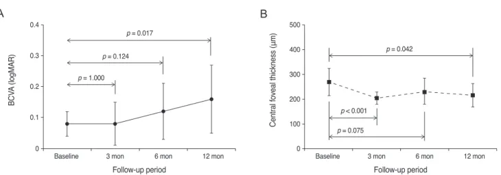
Fig. 1 shows changes in BCVA and CFT, according to the follow-up period. The BCVA was 0.08 ± 0.04, 0.08 ± 0.07, 0.12 ± 0.09, and 0.16 ± 0.11 at baseline, three months, six months, and 12 months, respectively (Fig. 1A) and sig- nificantly differed among the four time points (p = 0.009).
The BCVA at diagnosis was not different from that mea- sured at three or six months (p = 1.000 and p = 0.124, re- spectively). However, the BCVA at 12 months was signifi- cantly worse than that measured at baseline (p = 0.017).
Deterioration in BCVA of 0.1 to 0.2 logMAR BCVA was noted in seven eyes (38.9%) and a ≥0.2 logMAR BCVA decrease was found in two eyes (11.1%) (Fig. 2). The re- maining nine eyes (50.0%) had stable BCVA (Fig. 3).
The mean CFT at baseline, three months, six months, and 12 months was 270.2 ± 55.6, 204.4 ± 25.4, 230.1 ± 56.3, and 216.8 ± 48.7 µm, respectively (Fig. 1B). The CFT sig- nificantly differed among the four time points (p = 0.001) examined. Baseline CFT was significantly different from the CFT at 3 and 12 months (p < 0.001 and p = 0.042, re- spectively) but not at 6 months (p = 0.075).
Table 1. Baseline characteristics of eyes diagnosed with typi- cal exudative age-related macular degeneration and good ini- tial visual acuity (n = 18)
Variable Value
Age (yr) 66.1 ± 7.3 (54 to 80) Sex
Male 13 (72.2) Female 5 (27.8)
logMAR BCVA 0.08 ± 0.04 (20 / 25 to 20 / 20) Central foveal thickness (µm) 270.2 ± 55.6 (180 to 376) Location of lesion
Subfoveal 8 (44.4) Juxtafoveal 10 (55.6) Type of lesion
Classic 11 (61.1) Occult 7 (38.9) Size of lesion (disc areas) 1.0 ± 0.5
Data presented as mean ± standard deviation (range) or number (%).
logMAR = logarithm of minimum angle of resolution; BCVA = best-corrected visual acuity.
Fig. 1. Changes in mean logarithm of minimum angle of resolution (logMAR) best-corrected visual acuity (BCVA, A) and central foveal thickness (B) in eyes diagnosed with typical exudative age-related macular degeneration with good baseline visual acuity. Statistical anal- yses were performed using repeated measures analysis of variances with Bonferroni’s correction.
Baseline
BCVA (logMAR)
3 mon p = 0.017
p = 0.042
p < 0.001 p = 0.075 p = 0.124
p = 1.000
6 mon 12 mon
0 0.1 0.2 0.3 0.4
Baseline
Central foveal thickness (μm)
Follow-up period Follow-up period
3 mon 6 mon 12 mon
0 200 100 300 400
A B 500
Baseline
BCVA (logMAR)
3 mon p = 0.017
p = 0.042
p < 0.001 p = 0.075 p = 0.124
p = 1.000
6 mon 12 mon
0 0.1 0.2 0.3 0.4
Baseline
Central foveal thickness (μm)
Follow-up period Follow-up period
3 mon 6 mon 12 mon
0 200 100 300 400 500
Discussion
In the present study, we observed a relatively unfavor- able outcome with intravitreal anti-VEGF therapy in eyes with typical exudative AMD with good baseline visual acuity. Twelve months into the follow-up, a significant de- terioration in BCVA was noted, even though CFT had sig- nificantly decreased. Deterioration in visual acuity was noted in nine of 18 (50.0%) eyes.
The good initial visual acuity observed in our patients may be partially associated with the fact that the lesion sizes in the present study were relatively smaller than those in previous clinical trials [1,11]. In addition, retinal cysts were noted less frequently in our patients (50.0%) compared to those in a previous study (90.0%) [11]. It is no- table that visual acuity remained stable during the first three months when ranibizumab injections were adminis- tered. Deterioration in visual acuity was only noted after this period, which may have been due to lesion progres- sion. Lesion size generally increases in untreated exuda- tive-AMD [12]. Although multiple anti-VEGF injections
have been shown to prevent lesion progression [1,13,14], the efficacy of less frequent injections has not yet been stud- ied. Because follow-up fluorescein angiography and ICGA were not routinely performed, we do not know for certain whether lesion progression occurred in our patient cohort.
Further studies that include angiographic examination during the follow-up period are needed to verify whether lesion progression plays a role in vision loss.
Exudative AMD may have been undertreated because of treatment delays or an insufficient number of anti-VEGF injections. Because our study was retrospective, a strict uniform follow-up visit schedule was not employed. Thus, the monthly follow-up examination after initial treatment used in previous well-controlled clinical trials, all of which included OCT imaging [11,15], was not used in our study.
Patients were followed up once every one to three months, and OCT examination was not routinely performed at each follow-up visit. It is possible that mild exudative recur- rence occurred but was not detected due to omission of OCT. Therefore, prompt treatment may not have been per- formed in some cases. There is a close positive association Fig. 2. Fluorescein angiography (A) and optical coherence tomography (B,C,D) findings in an eye with typical exudative age-related macular degeneration. The best-corrected visual acuity at the time of diagnosis was 20 / 25 (A,B). The eye received six ranibizumab in- jections during the 12-month follow-up period, but the subretinal lesion enlarged, as seen on optical coherence tomography at six (C) and 12 (D) months. A decrease in visual acuity to 20 / 50 was observed at 12 months.
A B
C D
between frequency of anti-VEGF injections and visual im- provement in exudative AMD [16]. Eyes receiving injec- tions at least every two months regained more vision than eyes receiving injections less frequently [16]. In previous clinical trials that showed favorable visual outcome of an- ti-VEGF therapy in exudative AMD, a mean of 5.6 [11], 6.9 [15], and 7.7 injections [15] were administered during the 12-month follow-up period. However, in the present study, the mean number of anti-VEGF injection was only 4.4 during the same time period, with a mean interval between injections of 2.7 months. Thus, it is possible that our pa- tients may have been undertreated. However, we postulate that the difference in the number of injections may also be influenced by differences in disease characteristics. For in- stance, the baseline lesion size in our patients was marked- ly smaller than that of previous clinical trials [1,11]. In ad- dition, exudation recurrence was not noted in five of 18 eyes (27.8%) after the initial three monthly injections, whereas the proportion was approximately 18% in a previ- ous clinical trial [11]. These results suggest that the exuda- tive AMD patients included in the present study may have
had a relatively favorable course.
Mori et al. [7] investigated the 12-month treatment out- come of eyes with PCV and a good baseline visual acuity.
A favorable visual outcome was achieved with an average of 4.7 injections over 12 months, which is a comparable treatment frequency to that in our study. This outcome discrepancy may originate from the different natures of typical exudative AMD and PCV. In addition, their study was a prospective study with more frequent follow-ups, suggesting that prompt detection of exudation recurrence and subsequent prompt treatment may have been adminis- tered. Our study results suggest the need for intensive pa- tient monitoring and treatment, including monthly fol- low-up examinations, for patients with typical exudative AMD and good baseline visual acuity, as in previous clini- cal trials.
In addition to its retrospective nature and small sample size, our study had some other limitations. Patient fol- low-up and re-treatment were conducted at the discretion of clinicians, rather than according to defined criteria.
Moreover, a strict monthly follow-up schedule accompa- Fig. 3. Fluorescein angiography (A) and optical coherence tomography (B,C,D) findings of an eye diagnosed with typical exudative age-related macular degeneration. The best-corrected visual acuity at the time of diagnosis was 20 / 25 (A,B). After three consecutive ra- nibizumab injections, exudation recurrence was not noted during the 12-month follow-up period, as verified by optical coherence tomog- raphy at six (C) and 12 (D) months. The best-corrected visual acuity at 12 months was maintained at 20 / 25.
A B
C D
nied by routine OCT examination, which allows prompt detection of exudation recurrence, was not required. Last- ly, two different anti-VEGF agents (ranibizumab and bev- acizumab) were used to treat patients. However, knowing that the two agents have comparable efficacies in treating exudative AMD [17], the influence of using different agents is likely minimal.
In conclusion, a significant deterioration in visual acuity was noted 12 months after anti-VEGF therapy initiation in eyes diagnosed with typical exudative AMD and good baseline visual acuity. We postulate that this unfavorable outcome is partially attributable to less frequent follow-up and treatment. Frequent follow-ups accompanied by OCT examination, which is generally recommended for exuda- tive AMD treatment, may be required even in cases with typical exudative AMD showing good baseline visual acu- ity. Further prospective studies with a larger study popula- tion and more frequent follow-ups are required to demon- strate more accurate treatment outcomes for this condition.
Conflict of Interest
No potential conflict of interest relevant to this article was reported.
Acknowledgements
This study is supported by Kim’s Eye Hospital Research Center.
References
1. Rosenfeld PJ, Brown DM, Heier JS, et al. Ranibizumab for neovascular age-related macular degeneration. N Engl J Med 2006;355:1419-31.
2. Lee SY, Kim JG, Joe SG, et al. The therapeutic effects of bevacizumab in patients with polypoidal choroidal vascu- lopathy. Korean J Ophthalmol 2008;22:92-9.
3. Cho HJ, Baek JS, Lee DW, et al. Short-term effectiveness of intravitreal bevacizumab vs. ranibizumab injections for patients with polypoidal choroidal vasculopathy. Korean J Ophthalmol 2012;26:157-62.
4. Mones J, Biarnes M, Trindade F, Casaroli-Marano R. FU-
SION regimen: ranibizumab in treatment-naïve patients with exudative age-related macular degeneration and rela- tively good baseline visual acuity. Graefes Arch Clin Exp Ophthalmol 2012;250:1737-44.
5. Takahashi M, Sato T, Kishi S. Intravitreal bevacizumab for age-related macular degeneration with good visual acuity.
Jpn J Ophthalmol 2010;54:565-70.
6. Axer-Siegel R, Bor E, Bourla DH, et al. Intravitreal bevaci- zumab treatment for exudative age-related macular degen- eration with good visual acuity. Retina 2012;32:1811-20.
7. Mori R, Yuzawa M, Akaza E, Haruyama M. Treatment re- sults at 1 year of ranibizumab therapy for polypoidal cho- roidal vasculopathy in eyes with good visual acuity. Jpn J Ophthalmol 2013;57:365-71.
8. Shona O, Gupta B, Vemala R, Sivaprasad S. Visual acuity outcomes in ranibizumab-treated neovascular age-related macular degeneration; stratified by baseline vision. Clin Experiment Ophthalmol 2011;39:5-8.
9. Matsumiya W, Honda S, Kusuhara S, et al. Effectiveness of intravitreal ranibizumab in exudative age-related macular degeneration (AMD): comparison between typical neovas- cular AMD and polypoidal choroidal vasculopathy over a 1 year follow-up. BMC Ophthalmol 2013;13:10.
10. Yamashiro K, Tomita K, Tsujikawa A, et al. Factors associ- ated with the response of age-related macular degeneration to intravitreal ranibizumab treatment. Am J Ophthalmol 2012;154:125-36.
11. Fung AE, Lalwani GA, Rosenfeld PJ, et al. An optical co- herence tomography-guided, variable dosing regimen with intravitreal ranibizumab (Lucentis) for neovascular age-re- lated macular degeneration. Am J Ophthalmol 2007;143:
566-83.
12. Liu TY, Shah AR, Del Priore LV. Progression of lesion size in untreated eyes with exudative age-related macular de- generation: a meta-analysis using Lineweaver-Burk plots.
JAMA Ophthalmol 2013;131:335-40.
13. Arias L, Ruiz-Moreno JM, Gomez-Ulla F, et al. A 1-year retrospective review of ranibizumab for naive nonsubfove- al choroidal neovascularization secondary to age-related macular degeneration. Retina 2009;29:1444-9.
14. You JY, Chung H, Kim HC. Evaluation of changes in cho- roidal neovascularization secondary to age-related macular degeneration after anti-VEGF therapy using spectral do- main optical coherence tomography. Curr Eye Res 2012;37:
438-45.
15. CATT Research Group, Martin DF, Maguire MG, et al.
Ranibizumab and bevacizumab for neovascular age-related macular degeneration. N Engl J Med 2011;364:1897-908.
16. Dadgostar H, Ventura AA, Chung JY, et al. Evaluation of injection frequency and visual acuity outcomes for ranibi- zumab monotherapy in exudative age-related macular de- generation. Ophthalmology 2009;116:1740-7.
17. Comparison of Age-related Macular Degeneration Treat- ments Trials (CATT) Research Group, Martin DF, Maguire MG, et al. Ranibizumab and bevacizumab for treatment of neovascular age-related macular degeneration: two-year re- sults. Ophthalmology 2012;119:1388-98.