© 2015 The Korean Ophthalmological Society
This is an Open Access article distributed under the terms of the Creative Commons Attribution Non-Commercial License (http://creativecommons.org/licenses /by-nc/3.0/) which permits unrestricted non-commercial use, distribution, and reproduction in any medium, provided the original work is properly cited.
Original Article
Clinical Outcomes of Eyes with Submacular Hemorrhage Secondary to Age-related Macular Degeneration Treated with Anti-vascular
Endothelial Growth Factor
Kun Hae Kim1, Jae Hui Kim1, Young Suk Chang2, Tae Gon Lee1, Jong Woo Kim1, Young Ju Lew1
1Department of Ophthalmology, Kim’s Eye Hospital, Konyang University College of Medicine, Seoul, Korea
2Department of Ophthalmology, Konyang University College of Medicine, Daejeon, Korea
Purpose: To evaluate the long-term outcomes of intravitreal anti-vascular endothelial growth factor (VEGF) monotherapy for patients diagnosed with submacular hemorrhage secondary to exudative age-related macu- lar degeneration.
Methods: This retrospective, observational study included 49 patients (49 eyes) who initially presented with sub- macular hemorrhage associated with exudative age-related macular degeneration and who were followed-up for at least 24 months. Only eyes that were treated with intravitreal anti-VEGF monotherapy were included in the study. Best-corrected visual acuity (BCVA) measurements obtained at diagnosis, six months, and the final visit were compared. The associations of BCVA at the final visit with baseline BCVA, BCVA at six months, symptom duration, hemorrhage extent, and central foveal thickness were also analyzed.
Results: Over the course of follow-up (mean, 32.1 ± 8.5 months), an average of 5.1 ± 2.2 anti-VEGF injections were administered. Recurrent hemorrhage was noted in 13 eyes (26.5%). The mean logarithm of the minimal angle of resolution BCVA at diagnosis, six months, and the final visit were 1.40 ± 0.52, 0.87 ± 0.64, and 1.03 ± 0.83, respectively. Both baseline BCVA (p = 0.012) and BCVA at six months (p < 0.001) were significantly as- sociated with BCVA at the final visit.
Conclusions: Improved visual acuity was maintained for more than two years with intravitreal anti-VEGF mono- therapy. BCVA at six months is a useful clinical index to predict long-term visual prognosis.
Key Words: Anti-vascular endothelial growth factor, Exudative age-related macular degeneration, Long-term outcome, Polypoidal choroidal vasculopathy, Submacular hemorrhage
Intravitreal anti-vascular endothelial growth factor (VEGF) is an effective treatment for exudative age-related macular degeneration (AMD) [1-6]. Recent studies have
shown that the effect of intravitreal anti-VEGF is also demonstrated in eyes with submacular hemorrhage [7-12].
However, the previous studies that investigated anti-VEGF monotherapy efficacy in eyes with submacular hemor- rhage only followed their patients for 12 months, despite the fact that outcome changes have been reported 2 to 3 years after therapy for submacular hemorrhages that occur secondarily to exudative AMD [13-17]. These prior studies mostly date to the era before anti-VEGF therapy. Little in-
Received: December 5, 2014 Accepted: April 14, 2015
Corresponding Author: Jae Hui Kim, MD. Department of Ophthalmol- ogy, Kim’s Eye Hospital, Konyang University College of Medicine, #136 Yeongsin-ro, Yeongdeungpo-gu, Seoul 07301, Korea. Tel: 82-2-2639- 7664, Fax: 82-2-2639-7824, E-mail: [email protected]
formation is available regarding long-term outcomes after treatment with anti-VEGF therapy [17].
Recently, we reported on the efficacy of anti-VEGF monotherapy in 91 eyes with submacular hemorrhage after an initial presentation of exudative AMD [11]. In the pres- ent study, we report the extended long-term outcomes as- sociated with treatment administered as part of the previ- ous study. We also discuss the recurrence of hemorrhage and factors predictive of long-term visual prognosis.
Materials and Methods
This retrospective, observational case series was per- formed at a single center according to the tenets of the Declaration of Helsinki. The study was approved by the institutional review board of Kim’s Eye Hospital.
The present study included patients from the same co- hort and used similar inclusion and exclusion criteria as were used in our previous study [11]. A computerized search for patients who were newly diagnosed with exuda- tive AMD from September 2009 to November 2012 at our institution was conducted. Fovea-involving subretinal hemorrhages extending over at least 50% of the lesion area or at least three disc areas were included. Additionally, only patients who exhibited initial visual acuity of 20 / 30 or worse and who were treated with intravitreal ranibi- zumab were included. Only newly diagnosed, treat- ment-naïve eyes were included. Patients who had complet- ed two years or more of follow-up were included in the result analysis.
All subjects underwent a comprehensive ophthalmologic examination, including best-corrected visual acuity (BCVA) measurement, 90-diopter lens slit-lamp biomi- croscopy, fundus photography, fluorescein angiography, and spectral domain optical coherence tomography (either Spectral OCT/SLO, OTI Ophthalmic Technologies, Ontar- io, Canada; or Spectralis, Heidelberg Engineering, Heidel- berg, Germany). Indocyanine green angiography was per- formed using a confocal laser-scanning system (HRA-2, Heidelberg Engineering) at the discretion of each physi- cian. The exclusion criteria included less than six months of follow-up, duration of symptoms longer than six months, severe media opacity, evidence of end-stage AMD such as central geographic atrophy or disciform scarring, evidence of a macroaneurysm, proliferative diabetic reti-
nopathy, central retinal vascular occlusion, or history of intraocular surgery other than cataract surgery. Eyes that had undergone pneumatic displacement or photodynamic therapy, as well as eyes that had received intravitreal injec- tions of tissue plasminogen activator or photodynamic therapy during the follow-up period, were also excluded.
The number of eyes that underwent vitrectomy or cataract surgery during the follow-up period due to the develop- ment of severe vitreous hemorrhage was recorded; howev- er, all data pertaining to these eyes were excluded from the analyses. If a submacular hemorrhage developed in both eyes, the eye that was affected first was included; thus, only one eye was included for each patient.
Visual acuities were converted to logarithm of minimal angle of resolution (logMAR). As recommended by Holla- day [18], “counting fingers” and “hand-motion” were con- verted to logMAR equivalents 2 and 3, respectively. The extent of hemorrhage was estimated using disc area as the basic unit. Central foveal thickness was defined as the dis- tance between the internal limiting membrane and Bruch’s membrane at the fovea and was manually measured using the calipers provided by an optical coherence tomography software program. We were concerned about the accuracy of hemorrhage-extent measurements exceeding 20 disc ar- eas and central foveal thickness values exceeding 1,500 µm and therefore set these as threshold values. Lesions exceed- ing these measurements were recorded as 20 discs or 1,500 µm thickness, respectively. All extent-of-hemorrhage and central foveal thickness measurements were estimated by a single examiner (JHK).
The indocyanine green angiography results were ana- lyzed by two independent examiners (JHK and YSC). Cas- es of exudative AMD were classified as typical exudative AMD or polypoidal choroidal vasculopathy (PCV) based on the indocyanine green findings. Cases exhibiting branching vascular networks and/or terminating polypoi- dal lesions were diagnosed as PCV. In some cases, the presence of late geographic hyperfluorescence on indocy- anine green angiography [19] and/or the double-layer sign on optical coherence tomography [20,21] were observed in association with a branching vascular network on indocy- anine green angiography. These cases were classified as PCV even if no definite polypoidal lesions were identified.
All other cases were classified as typical exudative AMD.
For cases in which a definite initial diagnosis was not pos- sible using indocyanine green angiography, indocyanine
green angiography images collected within six months af- ter diagnosis were reviewed. Any disagreements were set- tled by discussion between the examiners.
Patients were initially treated with either ranibizumab (Lucentis; Genentech, San Francisco, CA, USA) or bevaci- zumab (Avastin, Genentech). As an initial treatment, 1 to 3 monthly intravitreal anti-VEGF injections were adminis- tered. Following initial treatment, patients were scheduled to visit the hospital once every 1 to 4 months based on sta- tus. Optical coherence tomography examinations were performed once every 1 to 6 months at the discretion of the clinician. Retreatment with intravitreal anti-VEGF usually occurred when intraretinal/subretinal fluid was present af- ter the initial injections or when intraretinal/subretinal flu- id or retinal/subretinal hemorrhage recurred and was ac- companied by an increase in macular thickness. Some PCV cases were treated with photodynamic therapy at the discretion of the treating physician.
Similar to our prior study, we also analyzed values of central foveal thickness, extent of hemorrhage, and BCVA up to 12 months of follow-up. The BCVA in the period be- tween 12 months of follow-up and the final visit was newly measured in the present study. The analyses are described in the following sections.
Changes in BCVA and factors associated with BCVA at the final visit
Baseline BCVA, BCVA at six months, BCVA at 12 months, and BCVA at the final visit were compared. Eyes with BCVA of 20 / 40 or better, between 20 / 400 and 20 / 40, and 20 / 400 or worse were classified into the fair vi- sion group, moderate vision group, and poor vision group, respectively. The distributions of eyes into the three groups at six months and at the final visit were compared. The as- sociations of BCVA at the final visit with baseline BCVA, BCVA at six months, symptom duration, hemorrhage ex- tent, and central foveal thickness were analyzed.
Comparison between eyes with typical exudative AMD and eyes with PCV
Values for symptom duration, central foveal thickness, hemorrhage extent, and number of anti-VEGF injections were compared between the typical exudative AMD group and the PCV group. Additionally, baseline BCVA, BCVA
at six months, BCVA at 12 months, and BCVA at the final visit were compared within each group.
Hemorrhage recurrence
The number of eyes that experienced a recurrence of fo- vea-involving submacular hemorrhage of at least one disc area during the follow-up period was noted. Baseline BCVA and BCVA at the final visit were compared between eyes that did and did not experience recurrent hemorrhage.
Baseline characteristics, including age, diagnosis, central foveal thickness, and hemorrhage extent, were compared between the groups.
Other analyses
The proportion of cases for each diagnosis (typical exu- dative AMD vs. PCV vs. unclassified), baseline BCVA, duration of symptoms, hemorrhage extent, and central fo- veal thickness were compared between the included eyes and the excluded eyes. The association between the num- ber of anti-VEGF injections received throughout the entire follow-up period in patient with BCVA at the final visit and overall change in BCVA during the follow-up period was also analyzed.
Statistics
Data are presented as mean ± standard deviation when applicable. Statistical analyses were performed with a com- mercially available software package (SPSS ver. 12.0; SPSS Inc., Chicago, IL, USA). Differences among various time points were analyzed using a repeated-measures analysis of variance, and individual comparisons were performed using Bonferroni’s method. Differences between groups were an- alyzed using an independent-samples t-test or a chi-square test. Multivariable analysis was performed using a multiple stepwise linear regression model. Pearson correlation anal- ysis was conducted to verify any associations between variables. A p-value <0.05 was considered significant.
Results
Among the 159 patients that initially presented with sub- macular hemorrhage, 91 satisfied all eligibility criteria and
were followed-up for six months or longer. The six-month clinical outcomes of these patients were presented in a pre- vious study [11]. Among the 91 patients, 55 completed two or more years of follow-up. Two of these 55 patients under- went cataract surgery, two underwent vitrectomy, and two underwent photodynamic therapy between the first six months post-therapy and the final visit. Finally, 49 of the 91 enrolled patients (53.8%) were included in the analyses for this manuscript (Table 1). Thirty of the patients (61.2%) were men and 19 (38.8%) were women. The mean age was 68.6 ± 8.6 years (range, 51 to 86 years), and the mean symptom duration was 15.5 ± 17.1 days (range, 1 to 90 days). The mean BCVA was 1.40 ± 0.52 (Snellen equiva- lent, 20 / 502; range, counting fingers to 20 / 40). The mean hemorrhage extent was 7.7 ± 6.1 disc areas (range, 3 to 20), and the mean central foveal thickness was 601.4 ± 228.9 µm (range, 331 to 1,300 µm). The mean follow-up period was 32.1 ± 8.5 months from diagnosis. During this period, patients were treated with 5.1 ± 2.2 (range, 1 to 11) intravitreal anti-VEGF injections. A mean of 3.7 ± 1.2 in- jections were administered during the first 12 months, and a mean of 1.4 ± 1.4 injections were administered during the period extending from the 12-month follow-up to the final visit. Thirty-five eyes were treated with ranibizumab only, another 14 eyes were treated with both ranibizumab and bevacizumab, and the single remaining eye was treat- ed with bevacizumab only. All the included eyes received at least one intravitreal anti-VEGF injection during the first 12 months. Thirty-one eyes (63.3%) received addition- al treatment at some point between the 12-month follow-up visit and the final follow-up visit.
For the 42 excluded eyes, measurement values were:
baseline BCVA = 1.37 ± 0.60 (Snellen equivalent, 20 / 468;
range, hand motion to 20 / 30), symptom duration = 41.8 ± 51.9 days, hemorrhage extent = 7.8 ± 5.2 disc areas, and central foveal thickness = 620.3 ± 274.1 µm. The group of excluded eyes had a significantly longer mean symptom duration than the included eyes (p = 0.003). None of the differences in baseline BCVA, hemorrhage extent, and central foveal thickness between the two groups were sig- nificant (p = 0.744, p = 0.827, and p = 0.909, respectively).
Changes in BCVA
Fig. 1 shows a representative case of long-term change in the macular microstructure of an eye with submacular
hemorrhage. The mean BCVA values at baseline, six months post-diagnosis, 12 months post-diagnosis, and at the final visit were 1.40 ± 0.52 (Snellen equivalent, 20 / 502), 0.87 ± 0.64 (Snellen equivalent, 20 / 148), 0.88 ± 0.68 (Snellen equivalent, 20 / 151), and 1.03 ± 0.83 (Snellen equivalent, 20 / 214), respectively (Fig. 2A). BCVA values differed significantly among the four time points (p <
0.001). The mean BCVA at the final visit showed signifi- cant improvement compared to the baseline value (p = 0.012), whereas the differences between the BCVA values at six months and at 12 months were not significantly dif- ferent from BCVA at the final visit (p = 0.156 and p = 0.113, respectively). Compared to baseline values, a BCVA im- provement of three lines or more was noted in 28 eyes (57.1%) at the final visit. A deterioration of three or more lines was noted in nine eyes (18.4%). The remaining 12 eyes (24.5%) exhibited stable BCVA throughout the fol- low-up period.
Compared to the six-month values, a BCVA improve- ment of three or more lines was noted in seven eyes (14.3%) at the final visit. A deterioration of three or more lines was noted in 15 eyes (30.6%). The remaining 27 eyes (55.1%) exhibited stable BCVA during the follow-up peri-
Table 1. Baseline characteristics of patients with exudative AMD with submacular hemorrhage as an initial presentation (n = 49)
Characteristics Value
Age (yr) 68.6 ± 8.6 (51 to 86)
Sex
Male 30 (61.2)
Female 19 (38.8)
Diagnosis
Typical exudative AMD 15 (30.6) Polypoidal choroidal
vasculopathy 31 (63.3)
Definitive diagnosis was not
possible 3 (6.1)
BCVA (logMAR) 1.40 ± 0.52 (CF to 20 / 40) Duration of symptoms (day) 15.5 ± 17.1 (1 to 90) Extent of hemorrhage (disc area) 7.7 ± 6.1 (3 to 20) Central foveal thickness (µm) 601.4 ± 228.9 (331 to 1,300) Values are presented as mean ± standard deviation (range) or number (%).
AMD = age-related macular degeneration; BCVA = best-cor- rected visual acuity; logMAR = logarithm of minimal angle of resolution; CF = finger counting.
od. The number of anti-VEGF injections was not associat- ed with BCVA at the final visit (p = 0.470) or the degree of change in BCVA during the follow-up period (p = 0.151).
At six months, the numbers of eyes included in the fair vision group (BCVA 20 / 40 or better), moderate vision group (BCVA from 20 / 400 to 20 / 40), and poor vision group (20 / 400 or worse) were 15 (30.6%), 20 (40.8%), and 14 (28.6%), respectively. The mean number of eyes in these groups at the final visit were 15 (30.6%), 16 (32.7%), and 18 (36.7%), respectively. The distribution of eyes among the three groups was not different between the six-month and final visits (p = 0.766). When classified based on BCVA at six months, six eyes (40.0%) in the fair vision group re- quired additional treatment at some point between 12 months and the final follow-up. The number of eyes that received additional treatment was 15 (75.0%) in the moder- ate vision group and 10 (71.4%) in the poor vision group.
The proportion of eyes that required additional treatment during the aforementioned period was not significantly different among the three groups (p = 0.065). In the fair vision group, only one eye (6.7%) experienced three lines of deterioration in BCVA between the six-month visit and
the final follow-up. In the remaining 14 eyes (93.3%), BCVA remained stable throughout the entire follow-up pe- riod.
Comparison among eyes with typical exudative AMD and eyes with PCV
Indocyanine green angiography results obtained at the time of diagnosis or within six months after diagnosis were available for all included eyes. Among them, 15 (30.6%) and 31 eyes (63.3%) were ultimately diagnosed
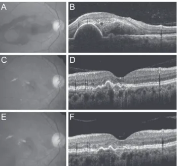
Fig. 1. Fundus photography and optical coherence tomography findings of an eye with submacular hemorrhage secondary to polypoidal choroidal vasculopathy. At the time of diagnosis, visual acuity was measured as 20 / 100 (A,B). At 6 months, the hemorrhage had resolved completely, and visual acuity had im- proved to 20 / 25 (C,D). The eye was treated with 5 ranibizumab injections during the 28-month follow-up period. At 28 months, visual acuity was maintained at 20 / 25 (E,F).
A B
C
E
D
F
Fig. 2. Changes in the mean logarithm of minimal angle of res- olution (logMAR) best-corrected visual acuity (BCVA) among eyes that received anti-vascular endothelial growth factor mono- therapy for submacular hemorrhage secondary to exudative age-related macular degeneration, according to the follow-up pe- riod. (A) In all 39 eyes, BCVA at the final visit was significantly better than baseline BCVA (p = 0.012). The difference between BCVA at the final visit and BCVA at six months or 12 months was not significant (p = 0.156 and 0.113, respectively). (B) Chang- es in values when the patients were divided into two groups ac- cording to diagnosis. Solid line (closed circles) indicates eyes di- agnosed with typical exudative age-related macular degeneration (n = 15); dashed line (closed squares) indicates eyes diagnosed with polypoidal choroidal vasculopathy (n = 31).
0.0 Baseline 6 mon 12 mon Final
Follow-up period p = 0.012
p = 0.156 p = 0.113
BCVA (logMAR)
2.5 2.0 1.5 1.0 0.5
0.0 Baseline 6 mon 12 mon Final
Follow-up period
BCVA (logMAR)
2.5 2.0 1.5 1.0 0.5
A
B
with typical exudative AMD and PCV, respectively. A definite diagnosis was not possible in the remaining three cases (6.1%) because a thick subretinal hemorrhage pre- cluded obtainment of reliable indocyanine green angiogra- phy images of the lesion.
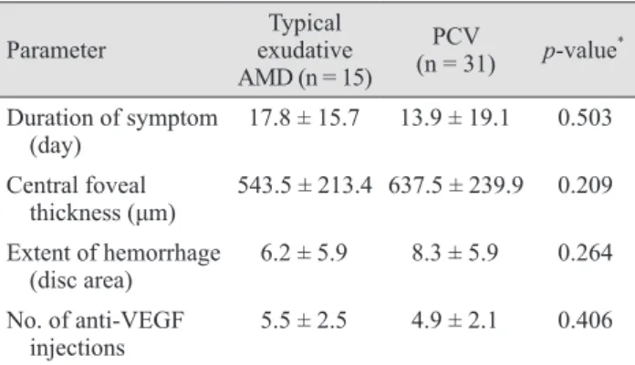
In the typical exudative AMD group, symptom duration, central foveal thickness, hemorrhage extent, and number of anti-VEGF injections were 17.8 ± 15.7 days, 543.5 ± 213.4 μm, 6.2 ± 5.9 disc areas, and 5.5 ± 2.5, respectively.
In the PCV group, the values were 13.9 ± 19.1 days, 637.5 ± 239.9 μm, 8.3 ± 5.9 disc areas, and 4.9 ± 2.1, respectively.
There were no significant differences between the two groups for these four parameters (p = 0.503, p = 0.209, p = 0.264, and p = 0.406, respectively) (Table 2). In the typical exudative AMD group, the mean baseline BCVA, at six months, at 12 months, and at the final visit were 1.49 ± 0.49, 0.94 ± 0.65, 0.97 ± 0.77, and 1.13 ± 0.87, respectively (Fig.
2B). In the PCV group, the values were 1.36 ± 0.54, 0.80 ± 0.64, 0.77 ± 0.64, and 0.87 ± 0.86, respectively. In unclassi- fied eyes, the values were 1.43 ± 0.56, 0.19 ± 0.67, 1.20 ± 0.62, and 1.28 ± 0.53, respectively.
Compared with the baseline value, BCVA at six months was significantly improved for both the typical exudative AMD (p = 0.031) and PCV (p = 0.001) groups. For the PCV group, the changes in BCVA at 12 months (p < 0.001) and at final follow-up (p = 0.007) compared to baseline were also statistically significant, but these same compari- sons were not significant in the AMD group (12 months, p
= 0.177; final follow-up, p = 0.145).
Factors associated with BCVA at the final visit
Both baseline BCVA and BCVA at six months were sig- nificantly associated with BCVA at the final visit (p = 0.012 and p < 0.001, respectively) (Table 3). No other factors, in- cluding symptom duration, hemorrhage extent, and central foveal thickness, were associated with BCVA at the final visit (p = 0.841, p = 0.083, and p = 0.121, respectively) (Table 2). In multivariate analysis, BCVA at six months was found to be the factor most strongly associated with BCVA at the final visit (p < 0.001) (Table 2).
Hemorrhage recurrence
Fifteen recurrences of fovea-involving submacular hem- orrhage occurred in 13 eyes (26.5%) during the follow-up
period (Fig. 3). Two of these eyes experienced multiple re- currences. On average, hemorrhages reoccurred by 15.0 ± 11.1 months (range, 4 to 42) after diagnosis. Four (30.8%) and six eyes (46.2%) were diagnosed with typical exuda- tive AMD and PCV, respectively. The remaining three eyes (23.1%) were unclassified. The results of comparisons between eyes with and without hemorrhage recurrence are
Table 2. Comparisons between parameters of typical exudative AMD and PCV
Parameter Typical
exudative AMD (n = 15)
(n = 31)PCV p-value* Duration of symptom
(day) 17.8 ± 15.7 13.9 ± 19.1 0.503
Central foveal
thickness (μm) 543.5 ± 213.4 637.5 ± 239.9 0.209 Extent of hemorrhage
(disc area) 6.2 ± 5.9 8.3 ± 5.9 0.264 No. of anti-VEGF
injections 5.5 ± 2.5 4.9 ± 2.1 0.406 AMD = age-related macular degeneration; PCV = polypoidal choroidal vasculopathy; VEGF = vascular endothelial growth factor.
*Statistics were analyzed using independent samples t-test.
Table 3. The associations of BCVA at the final visit with base- line BCVA, BCVA at six months, symptom duration, hemor- rhage extent, and central foveal thickness
Characteristics p-value*
Baseline BCVA 0.012
BCVA at six months 0.001†
Duration of symptoms 0.841
Extent of hemorrhage 0.083
Central foveal thickness 0.121
BCVA = best-corrected visual acuity.
*Statistics were analyzed using Pearson correlation analysis; †Sta- tistically significant when tested using stepwise multiple linear regression.
Fig. 3. A timetable showing the timing of recurrences of fo- vea-involving submacular hemorrhage according to the follow-up period. Inverted triangles indicate the first recurrence. Asterisks indicate the second recurrence. Fifteen recurrences were noted in 13 eyes. Two eyes experienced two episodes of recurrence.
Follow-up period (mon)
0 10
*
20 30*
40 50summarized in Table 4. There was no difference in base- line BCVA (p = 0.304), BCVA at the final visit (p = 0.709), diagnosis (p = 0.573), symptom duration (p = 0.361), cen- tral foveal thickness (p = 0.970), or hemorrhage extent (p
= 0.509) between eyes with or without hemorrhage recur- rence.
Discussion
In this study, visual acuity in the majority of eyes that initially presented with submacular hemorrhage had im- proved or remained relatively stable between six months after diagnosis and the final visit (mean, 32.1 months). As a result, BCVA values at the final visit were still significant- ly improved compared to baseline. This result suggests that, although the development of submacular hemorrhage induces retinal damage, any remaining retinal function can be maintained long-term through continuous an- ti-VEGF therapy.
Notably, the number of injections received between 12 months post-diagnosis and the final visit was markedly lower than the number of injections received during the first 12 months after diagnosis. Only 63.3% of patients re- ceived additional injections during this period. The mean
number of injections after 12 months was far less than that administered during the 12- to 24-month period in prior controlled clinical trials [3,4], despite our use of a longer time interval between the 12-month follow-up point and the final visit (20.1 months). The lower injection frequency after 12 months may reflect several factors. To facilitate the early detection and prompt treatment of recurrent exu- dation in patients with exudative AMD, it is generally rec- ommended to perform monthly follow-ups, including monthly OCT examinations [3,22]. In the present study, OCT examination was not routinely performed during ev- ery visit, and the follow-up period varied from 1 to 4 months at the discretion of the treating physician. Thus, recurrent exudate may not have been detected promptly.
Moreover, the re-accumulation of small amounts of intra- retinal/subretinal fluid may have been missed. Some pa- tients, particularly those with very low visual acuity, re- fused additional treatment without a guarantee that their vision would subsequently improve. Similarly, treating physicians decided not to administer additional treatments in some cases when no definite benefit was anticipated.
Regardless of these possible explanations, however, the findings in patients with relatively good visual acuity (20 / 40 or better) at six months after diagnosis are noteworthy.
In this group, almost all the eyes maintained relatively sta- ble BCVA, although only 40% of them required additional treatment. This particular finding requires further expla- nation. We postulate that relatively large, active vascular lesions, such as major polyps, may contribute to frequent re-accumulation of intraretinal/subretinal fluid that may have ruptured. Although other vascular abnormalities per- sisted, the activity of the entire exudative AMD lesion may have decreased after the rupture of a major vascular lesion and thus reduced the frequency of recurrent exudation.
In a study by Hwang et al. [17] that evaluated the inci- dence of recurrent submacular hemorrhage in exudative AMD, the recurrence rate was 51.1% during the follow-up period (mean, 36.8 months). That rate is much higher than the rate reported in the present study (26.5%). A direct comparison between this study and that of Hwang et al.
[17] may not be appropriate because they evaluated various treatment modalities, including photodynamic therapy, in- travitreal anti-VEGF, pneumatic displacement, and vitrec- tomy, whereas all the patients in the present study were treated with only intravitreal anti-VEGF monotherapy. In Hwang et al. [17], the use of intravitreal anti-VEGF was Table 4. Comparisons of parameters between eyes with and
without recurring fovea-involving submacular hemorrhage
Parameter Eyes with
recurrence (n = 13)
Eyes without recurrence
(n = 36) p-value Baseline BCVA 1.53 ± 0.57 1.36 ± 0.49 0.304* BCVA at the final visit 1.10 ± 0.89 1.00 ± 0.82 0.709*
Diagnosis† 0.573‡
Typical exudative
AMD 4 (40.0) 11 (30.6)
PCV 6 (60.0) 25 (69.4)
Duration of symptoms 19.2 ± 15.8 14.1 ± 17.6 0.361* Central foveal
thickness 599.3 ± 264.3 602.1 ± 219.0 0.970* Extent of hemorrhage 6.8 ± 5.6 8.1 ± 6.3 0.509* Values are presented as mean ± standard deviation or number (%).
BCVA = best-corrected visual acuity; AMD = age-related macu- lar degeneration; PCV = polypoidal choroidal vasculopathy.
*Statistics were analyzed using independent samples t-test;
†Analyses were performed for 46 eyes in which an accurate indocyanine-green angiography-based classification was possible;
‡Statistics were analyzed using chi-square test.
associated with a reduced risk of recurrent hemorrhage.
The relatively low recurrence rate found in the present study may also suggest a potential role of anti-VEGF monotherapy in preventing recurrent hemorrhage. Most hemorrhage recurrences developed within two years of di- agnosis. However, some recurrences occurred after more than three years, suggesting that a lack of recurrence over this relatively long-term period may not guarantee com- plete stabilization of the lesion.
We also found a strong association between BCVA at six months and BCVA at the final visit. BCVA at six months was more closely associated with BCVA at the final visit than with baseline BCVA. A subretinal hemorrhage typi- cally resolves or at least decreases markedly in size during the first six months [7,8,11]. Because a hemorrhage can block the visual stimulus, BCVA at six months rather than baseline BCVA may more accurately reflect underlying retinal function. In approximately two-thirds of eyes, visu- al acuity at six months was relatively unchanged from baseline or even improved over the course of a mean 32.1 months of follow-up. As a result, BCVA at the final visit had not significantly decreased compared to the values at six months post-diagnosis. This suggests that the six- month visual acuity measurement can be used as a refer- ence value in any discussion with patients regarding long- term visual prognosis.
We also found that typical exudative AMD and PCV ex- hibited different visual outcomes. More specifically, a fa- vorable long-term visual outcome was achieved only in the PCV group. PCV is a distinct entity from typical exudative AMD [23], and the incidence of PCV is generally higher in Asian populations than in European populations [24,25].
Additionally, hemorrhage is a frequent finding in PCV cases [26,27]. The majority of our patients were diagnosed with PCV. We believe that this high frequency is a result of the two aforementioned characteristics. While an- ti-VEGF monotherapy has been considered the most effec- tive first-line therapy for typical exudative AMD [1,6], its efficacy in PCV is still a matter of controversy [28]. Al- though anti-VEGF therapy has been found to be effective in previous studies [29,30], its efficacy is generally inferior to that of photodynamic therapy for polyp regression [31].
For this reason, some experts recommend photodynamic therapy rather than anti-VEGF monotherapy as first-line therapy in PCV [28]. Despite this controversy, the treat- ment outcomes of anti-VEGF monotherapy in our patients
with PCV were encouraging. As discussed above, it is pos- sible that this favorable outcome may be partially in re- sponse to the rupture of a major polyp after a hemorrhage.
In contrast, the outcomes of typical exudative AMD were less favorable. Although marked improvement in BCVA was noted during the first six months, deterioration of vi- sual acuity typically occurred thereafter. As a result, BCVA values at 12 months and later were not different from those at baseline. We think that the symptom dura- tion may have had an influence. Although statistical signif- icance was not reached, symptom duration in the typical exudative AMD group was relatively longer than that of the PCV group, suggesting more accumulated damage to the retina in the AMD group. Another reason may be a possible difference in the degree of retinal degeneration between typical exudative AMD and PCV patients. It is well known that age-related degenerative changes in the retina, including drusen and pseudodrusen, are more markedly present in typical exudative AMD cases than in PCV cases [32,33]. Additionally, the choroid of eyes with typical exudative AMD is generally thinner than that of eyes with PCV [34]. We think that retinas with pre-exist- ing degeneration may be more vulnerable to damage re- sulting from the development of subretinal hemorrhage.
Furthermore, a thin choroid may not provide sufficient perfusion to the retina. Although testing these hypotheses is beyond the scope of this study, the less favorable out- comes associated with typical exudative AMD suggest the need for other treatments (e.g., pneumatic displacement).
Further studies are needed to establish the appropriate treatment method to improve long-term outcomes of sub- macular hemorrhages that occur secondary to typical exu- dative AMD.
This study has several limitations. First, it was a retro- spective study and included only a small number of pa- tients who exhibited submacular hemorrhage as an initial presentation. Second, there was no common treatment protocol or follow-up/optical coherence tomography exam- ination schedule. Thus, differences in treatment decisions among clinicians may have influenced the study results.
Because optical coherence tomography, fluorescein angi- ography, and indocyanine green angiography were not routinely performed during the follow-up period, the long- term anatomical outcomes were not analyzed.
In conclusion, relatively stable long-term visual outcome can be achieved for the majority of eyes exhibiting subma-
cular hemorrhage as a secondary symptom to exudative AMD when treated with intravitreal anti-VEGF monother- apy, despite recurrence of exudation and/or submacular hemorrhage. BCVA at six months was found to be a useful clinical index predictive of long-term visual prognosis.
Conflict of Interest
No potential conflict of interest relevant to this article was reported.
Acknowledgements
This study is supported by Kim’s Eye Hospital Research Center.
References
1. Rosenfeld PJ, Brown DM, Heier JS, et al. Ranibizumab for neovascular age-related macular degeneration. N Engl J Med 2006;355:1419-31.
2. Avery RL, Pieramici DJ, Rabena MD, et al. Intravitreal bevacizumab (Avastin) for neovascular age-related macular degeneration. Ophthalmology 2006;113:363-72.e5.
3. Fung AE, Lalwani GA, Rosenfeld PJ, et al. An optical co- herence tomography-guided, variable dosing regimen with intravitreal ranibizumab (Lucentis) for neovascular age-re- lated macular degeneration. Am J Ophthalmol 2007;143:
566-83.
4. Comparison of Age-related Macular Degeneration Treat- ments Trials (CATT) Research Group, Martin DF, Maguire MG, et al. Ranibizumab and bevacizumab for treatment of neovascular age-related macular degeneration: two-year re- sults. Ophthalmology 2012;119:1388-98.
5. Kwon YH, Lee DK, Kim HE, Kwon OW. Predictive find- ings of visual outcome in spectral domain optical coher- ence tomography after ranibizumab treatment in age-relat- ed macular degeneration. Korean J Ophthalmol 2014;28:
386-92.
6. Brown DM, Kaiser PK, Michels M, et al. Ranibizumab versus verteporfin for neovascular age-related macular de- generation. N Engl J Med 2006;355:1432-44.
7. Cho HJ, Koh KM, Kim HS, et al. Anti-vascular endothelial
growth factor monotherapy in the treatment of submacular hemorrhage secondary to polypoidal choroidal vasculopa- thy. Am J Ophthalmol 2013;156:524-31.e1.
8. Iacono P, Parodi MB, Introini U, et al. Intravitreal ranibi- zumab for choroidal neovascularization with large subma- cular hemorrhage in age-related macular degeneration.
Retina 2014;34:281-7.
9. Shienbaum G, Garcia Filho CA, Flynn HW Jr, et al. Man- agement of submacular hemorrhage secondary to neovas- cular age-related macular degeneration with anti-vascular endothelial growth factor monotherapy. Am J Ophthalmol 2013;155:1009-13.
10. Stifter E, Michels S, Prager F, et al. Intravitreal bevacizum- ab therapy for neovascular age-related macular degenera- tion with large submacular hemorrhage. Am J Ophthalmol 2007;144:886-92.
11. Kim JH, Chang YS, Kim JW, et al. Intravitreal anti-vascu- lar endothelial growth factor for submacular hemorrhage from choroidal neovascularization. Ophthalmology 2014;
121:926-35.
12. McKibbin M, Papastefanou V, Matthews B, et al. Ranibi- zumab monotherapy for sub-foveal haemorrhage second- ary to choroidal neovascularisation in age-related macular degeneration. Eye (Lond) 2010;24:994-8.
13. Avery RL, Fekrat S, Hawkins BS, Bressler NM. Natural history of subfoveal subretinal hemorrhage in age-related macular degeneration. Retina 1996;16:183-9.
14. Bressler NM, Bressler SB, Childs AL, et al. Surgery for hemorrhagic choroidal neovascular lesions of age-related macular degeneration: ophthalmic findings: SST report no.
13. Ophthalmology 2004;111:1993-2006.
15. Scupola A, Coscas G, Soubrane G, Balestrazzi E. Natural history of macular subretinal hemorrhage in age-related macular degeneration. Ophthalmologica 1999;213:97-102.
16. Bennett SR, Folk JC, Blodi CF, Klugman M. Factors prog- nostic of visual outcome in patients with subretinal hemor- rhage. Am J Ophthalmol 1990;109:33-7.
17. Hwang JU, Yang SJ, Yoon YH, et al. Recurrent submacular hemorrhage in patients with neovascular age-related macu- lar degeneration. Retina 2012;32:652-7.
18. Holladay JT. Visual acuity measurements. J Cataract Re- fract Surg 2004;30:287-90.
19. Kang SW, Chung SE, Shin WJ, Lee JH. Polypoidal choroi- dal vasculopathy and late geographic hyperfluorescence on indocyanine green angiography. Br J Ophthalmol 2009;
93:759-64.
20. Kim JH, Kang SW, Kim TH, et al. Structure of polypoidal choroidal vasculopathy studied by colocalization between tomographic and angiographic lesions. Am J Ophthalmol 2013;156:974-80.e2.
21. Sato T, Kishi S, Watanabe G, et al. Tomographic features of branching vascular networks in polypoidal choroidal vasculopathy. Retina 2007;27:589-94.
22. CATT Research Group, Martin DF, Maguire MG, et al.
Ranibizumab and bevacizumab for neovascular age-related macular degeneration. N Engl J Med 2011;364:1897-908.
23. Yannuzzi LA, Sorenson J, Spaide RF, Lipson B. Idiopathic polypoidal choroidal vasculopathy (IPCV). Retina 1990;10:1-8.
24. Ciardella AP, Donsoff IM, Huang SJ, et al. Polypoidal cho- roidal vasculopathy. Surv Ophthalmol 2004;49:25-37.
25. Coscas G, Yamashiro K, Coscas F, et al. Comparison of ex- udative age-related macular degeneration subtypes in Japa- nese and French Patients: multicenter diagnosis with multi- modal imaging. Am J Ophthalmol 2014;158:309-18.e2.
26. Sho K, Takahashi K, Yamada H, et al. Polypoidal choroidal vasculopathy: incidence, demographic features, and clini- cal characteristics. Arch Ophthalmol 2003;121:1392-6.
27. Byeon SH, Lee SC, Oh HS, et al. Incidence and clinical patterns of polypoidal choroidal vasculopathy in Korean patients. Jpn J Ophthalmol 2008;52:57-62.
28. Koh AH; Expert PCV Panel, Chen LJ, et al. Polypoidal choroidal vasculopathy: evidence-based guidelines for clin-
ical diagnosis and treatment. Retina 2013;33:686-716.
29. Oishi A, Kojima H, Mandai M, et al. Comparison of the ef- fect of ranibizumab and verteporfin for polypoidal choroi- dal vasculopathy: 12-month LAPTOP study results. Am J Ophthalmol 2013;156:644-51.
30. Lee SY, Kim JG, Joe SG, et al. The therapeutic effects of bevacizumab in patients with polypoidal choroidal vascu- lopathy. Korean J Ophthalmol 2008;22:92-9.
31. Koh A, Lee WK, Chen LJ, et al. EVEREST study: efficacy and safety of verteporfin photodynamic therapy in combi- nation with ranibizumab or alone versus ranibizumab monotherapy in patients with symptomatic macular polyp- oidal choroidal vasculopathy. Retina 2012;32:1453-64.
32. Yoneyama S, Sakurada Y, Mabuchi F, et al. Genetic and clinical factors associated with reticular pseudodrusen in exudative age-related macular degeneration. Graefes Arch Clin Exp Ophthalmol 2014;252:1435-41.
33. Fujimura S, Ueta T, Takahashi H, et al. Characteristics of fundus autofluorescence and drusen in the fellow eyes of Japanese patients with exudative age-related macular de- generation. Graefes Arch Clin Exp Ophthalmol 2013;251:1-9.
34. Kim SW, Oh J, Kwon SS, et al. Comparison of choroidal thickness among patients with healthy eyes, early age-re- lated maculopathy, neovascular age-related macular degen- eration, central serous chorioretinopathy, and polypoidal choroidal vasculopathy. Retina 2011;31:1904-11.