ISSN 0378-6471 (Print)⋅ISSN 2092-9374 (Online)
http://dx.doi.org/10.3341/jkos.2016.57.7.1093
Original Article
세 가지 유형의 삼출성 나이관련황반변성에서 빛간섭단층촬영의 특징 비교
Comparison of Optical Coherence Tomography Characteristics among Three Subtypes of Exudative Age-related Macular Degeneration
안소은⋅안동섭⋅양 헌⋅윤희성
Soh-Eun Ahn, MD, Dong Seob Ahn, MD, Heon Yang, MD, Hee Seong Yoon, MD, PhD
성모안과병원
Sungmo Eye Hospital, Busan, Korea
Purpose: To compare the characteristics of optical coherence tomography in eyes with treatment-naïve typical neovascular age-related macular degeneration (typical nAMD), polypoidal choroidal vasculopathy (PCV), and retinal angiomatous pro- liferation (RAP).
Methods: One hundred fifty-three eyes newly diagnosed with exudative AMD were retrospectively collected. All study eyes were classified into three subtypes: typical nAMD, PCV, and RAP. Subfoveal choroidal thickness (SFCT) was measured using en- hanced depth imaging optical coherence tomography (EDI-OCT). Central macular thickness (CMT) and other OCT features in- cluding intraretinal cystoid fluid and subretinal fluid were also evaluated in all eyes. SFCT, CMT and other OCT features were compared among the three subtypes of exudative AMD.
Results: Seventy-four eyes with typical nAMD, 55 eyes with PCV, and 24 eyes with RAP were included. SFCT was significantly thickest in PCV and thinnest in RAP (p < 0.001). RAP showed the thickest CMT and the highest frequency of intraretinal cystoid fluid (p = 0.004, p < 0.001, respectively).
Conclusions: In patients with exudative AMD, different characteristics of OCT were observed according to the three subtypes.
Identification of OCT characteristics could help differentiate the subtypes of exudative AMD.
J Korean Ophthalmol Soc 2016;57(7):1093-1101
Keywords: Age-related macular degeneration, Optical coherence tomography, Polypoidal choroidal vasculopathy, Retinal an- giomatous proliferation
■Received: 2016. 3. 24. ■ Revised: 2016. 4. 14.
■Accepted: 2016. 5. 17.
■Address reprint requests to Hee Seong Yoon, MD, PhD Sungmo Eye Hospital, #409 Haeun-daero, Haeundae-gu, Busan 48064, Korea
Tel: 82-51-743-0775, Fax: 82-51-743-0776 E-mail: [email protected]
ⓒ2016 The Korean Ophthalmological Society
This is an Open Access article distributed under the terms of the Creative Commons Attribution Non-Commercial License (http://creativecommons.org/licenses/by-nc/3.0/) which permits unrestricted non-commercial use, distribution, and reproduction in any medium, provided the original work is properly cited.
나이관련황반변성은 고령층에서 심각한 시력손실 및 실 명을 야기하는 주된 원인 질환으로 알려져 있다.1 혈관내피 세포성장인자(vascular endothelial growth factor, VEGF)를
포함한 혈관신생인자들이 분비되어 맥락막신생혈관이 발생 한 경우를 삼출성 나이관련황반변성이라 하며 맥락막신생 혈관으로부터 부종과 출혈이 발생함으로써 급격하고 심각 한 시력저하가 야기된다.2 결절맥락막혈관병증(polypoidal choroidal vasculopathy, PCV)은 삼출성 나이관련황반변성 의 하위 유형으로 인도시아닌그린혈관조영상에서 맥락막 혈관의 결절성 확장과 분지혈관망의 존재를 특징으로 한 다.3,4 또 다른 하위 유형인 망막혈관종성증식(retinal angiom- atous proliferation, RAP)은 망막에서 유래한 것으로 생각되 는 신생혈관을 특징으로 한다.5,6 PCV와 RAP는 전형적인
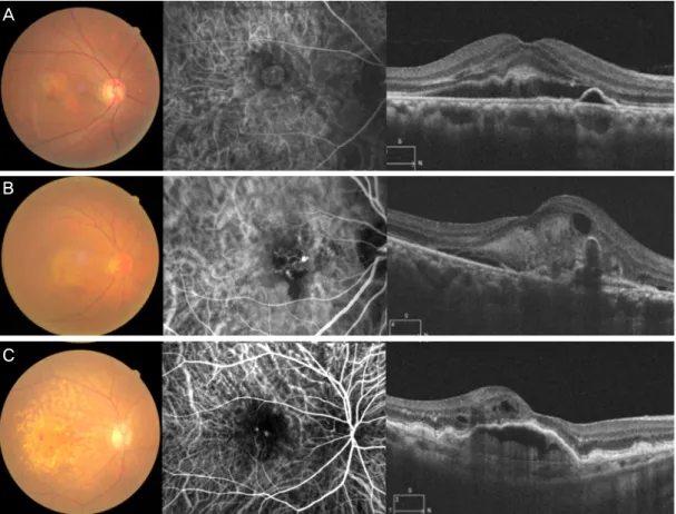
Figure 1. Representative images of three subtypes of exudative age-related macular degeneration (AMD). Fundus photograph, in-
docyanine green angiography and optical coherence tomography images of typical neovascular AMD (A), polypoidal choroidal vas- culopathy (B), and retinal angiomatous proliferation (C).삼출성 나이관련황반변성(typical neovascular age-related macular degeneration, typical nAMD)과 비교하여 서로 다 른 임상경과와 치료반응을 나타낸다고 알려져 있으며,7-9 진 단 당시 유형을 구분하는 것이 연구 목적뿐만 아니라 임상 에서의 치료 방향 설정이나 환자 설명에 있어서도 중요하게 되었다.
현재까지 형광안저혈관조영술(fluorescein angiography)과 인도시아닌그린혈관조영술(indocyanine green angiography) 이 삼출성 나이관련황반변성을 확진하는 데 있어서 핵심적 인 역할을 수행하고 있으며, 빛간섭단층촬영(optical coher- ence tomography, OCT) 역시 진단 및 치료 반응을 평가함 에 있어서 중요하게 자리매김하고 있다. 비침습적인 진단 장비라는 측면에서 혈관조영술에 비해 OCT의 활용도는 매 우 높으며, 기술적인 발전이 더해지면서 망막의 미세구조 를 더욱 자세하게 관찰할 수 있게 되어 삼출성 나이관련황 반변성의 진단 및 치료에 있어 차지하는 역할이 점차 커지 고 있다.10,11
OCT의 발전으로 깊이증강모드(enhanced depth imaging [EDI] mode)가 개발되면서 맥락막층을 더욱 정교하게 평 가할 수 있게 되었고, 이를 통해 측정된 중심하 맥락막두께
가 맥락막 및 맥락막모세혈관의 순환상태를 반영하는 하나 의 지표로 사용되고 있다.12,13 또한 해상도가 높아지면서 황 반부 미세구조의 관찰이 더욱 용이해졌고 삼출성 나이관련 황반변성의 병태생리를 이해하는 데 있어서 큰 역할을 하 고 있다.14
나이관련황반변성과 관련하여 맥락막두께를 비롯한 빛 간섭단층촬영의 특징적 소견을 연구한 논문은 다양하게 존
재하지만,15-19 typical nAMD, PCV, RAP와 같이 유형별로
분류하고 각각의 특징을 비교 분석한 논문은 찾아볼 수 없 었다. 빛간섭단층촬영을 통한 삼출성 나이관련황반변성 유 형별 비교는 각각의 병태생리를 이해하고 감별진단하는 데 있어서 큰 도움이 될 것이라 생각된다. 이에 저자들은 삼출 성 나이관련황반변성 환자에서 typical nAMD, PCV, 그리 고 RAP의 세 가지 유형에 따른 빛간섭단층촬영의 특징을 비교하고자 하였다.
대상과 방법
대상 선정 및 자료 획득
2013년 1월부터 2015년 8월까지 본원을 내원한 삼출성
A
B
C
Figure 2. Measurement of subfoveal choroidal thickness. The
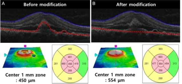
image was obtained using enhanced-depth imaging optical co- herence tomography (OCT). Choroidal thickness (double-head arrow) was defined as the vertical distance drawn from the outer border of the retinal pigment epithelium to the inner bor- der of the sclera using Cirrus High-Definition-OCT software.Figure 3. Central macular thickness modification using a built-in segmentation-modifying tool of optical coherence tomography.
Segmentation lines for retinal pigment epithelium (red dotted line) are determined automatically for each of the volume scans, and the central macular thickness is provided (A). Using the modifying tool, the cursor line on retinal pigment epithelium is moved to the level of Bruch’s membrane. W ith this movement, the modified central macular thickness is determined (B).
나이관련황반변성 환자 중에서 치료 기왕력이 없는 153명 153안을 대상으로 하였다. 모든 환자는 내원 당시 최대교 정시력, 안압측정, 굴절검사, 안저검사, 형광안저혈관조영, 인도시아닌그린혈관조영 및 빛간섭단층촬영을 시행하였다.
최대교정시력은 Snellen 시력표를 사용하여 측정한 뒤 log- MAR로 변환하였다.
삼출성 나이관련황반변성은 황반부의 맥락막신생혈관, 망막하액, 망막색소상피박리, 망막하 삼출물과 출혈을 시사 하는 과형광 및 후기 누출을 보이는 경우로 정의하였으며,20 안저소견, 형광안저혈관조영 소견, 인도시아닌그린혈관조 영 결과를 종합적으로 판단하여 3개의 군으로 나누었다. 분 지하는 혈관망과 그 말단부의 결절 병변을 보이는 특징적 소견이 있는 경우 결절맥락막혈관병증(polypoidal choroidal vasculopathy, PCV)으로 진단하였고 특징적인 망막-망막 혹 은 망막-맥락막 문합이 발견되는 경우 망막혈관종성증식 (RAP)으로 진단하였으며 이를 제외한 경우를 전형적삼출성 황반변성(typical neovascular AMD, typical nAMD)으로 분 류하였다(Fig. 1).
Lens Opacities Classification System III 분류에서 NO3, C3, P3 이상의 백내장을 동반한 경우와 고도근시(6디옵터 이상) 환자, 시력에 영향을 줄 수 있는 다른 질환이나 병태 를 동반한 경우 본 연구에서 제외하였다. 이전에 유리체망 막 수술을 시행 받았거나, 나이관련황반변성으로 진단 받 고 안구내주사 또는 레이저치료를 시행 받은 경우 역시 연 구에서 제외하였다.
빛간섭단층촬영은 spectral-domain optical coherence to-
mography (SD-OCT; CirrusTM HD-OCT 4000, Carl Zeiss Medi- tec, Dublin, CA, USA)를 사용하였고, 황반부 큐브 스캔(mac- ular cube scan mode) 및 고해상도 라스터 스캔(high-defi- nition raster scan mode) 방법으로 촬영하였으며, 7 이상의 신호 강도(signal strength)를 보이는 영상만을 분석하였다.
중심하 맥락막두께의 측정
중심하 맥락막두께는 EDI 기법으로 촬영된 고해상도 라 스터 스캔 영상에서 Cirrus 소프트웨어에서 제공하는 직선 측정도구(linear measurement tool)를 이용하여, 망막색소상 피(retinal pigment epithelium, RPE)의 외측경계에서부터 공 막의 내측경계까지 수직선을 그어 측정하였다(Fig. 2).12,16,21 두 명의 저자(ASE, ADS)가 독립적으로 측정하여 도출한
A B
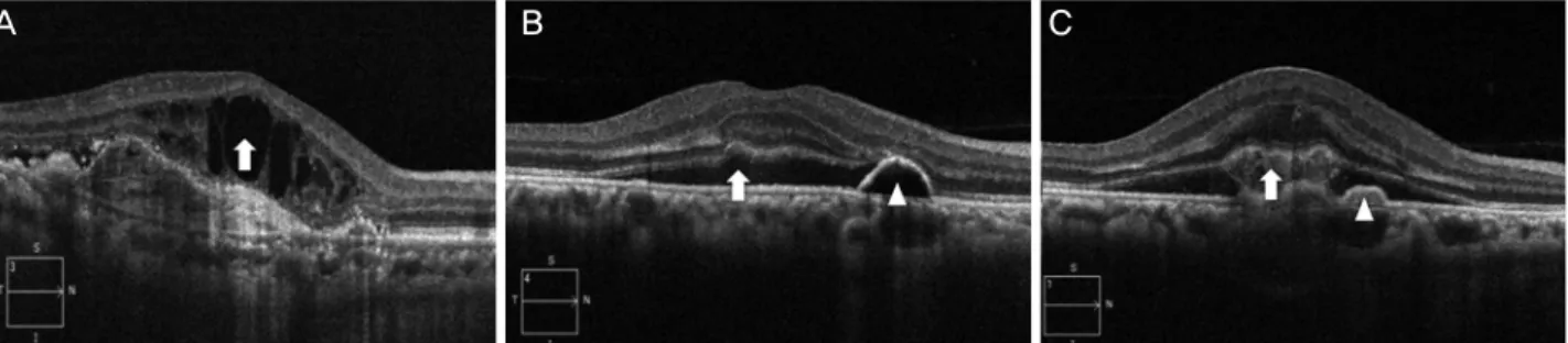
Figure 4. Morphologic features of optical coherence tomography related to age-related macular degeneration. Intraretinal cystoid
fluid is defined as round, hyporeflective cystic spaces within the neurosensory retina (A, arrow). Subretinal fluid is identified as non- reflective space between the posterior boundary of the neurosensory retina and the retinal pigment epithelium (B, arrow). Subretinal hyperreflective material appears as poorly defined, medium- to hyperreflective mass between neurosensory retina and retinal pig- ment epithelium (C, arrow). Pigment epithelial detachment is defined as a focal elevation of the reflective RPE band over an opti- cally clear or moderately reflective space (B, C; arrowhead).Table 1. Demographic data of the patients
Characteristics Data
Number of eyes (n) 153
Age (years) 71.9 ± 8.6
Sex (M:F) 96:57
Mean BCVA (log MAR) 0.662 ± 0.458
Mean SFCT (μm) 218.71 ± 64.47
Mean central macular thickness (μm) 465.09 ± 161.17 OCT characteristics (eyes, n [%] )
Intraretinal cytroid fluid 64 (41.8)
Subretinal fluid 120 (78.4)
Subretinal hyperreflective material 62 (40.5) Pigment epithelial detachment 142 (92.8) Values are presented as mean ± SD or n (%)
BCVA = best corrected visual acuity; log MAR=logarithm of the minimum angle of resolution; SFCT = subfoveal choroidal thick- ness; OCT = optical coherence tomography.
맥락막 두께의 평균값을 분석에 사용하였다. 중심하 맥락 막두께의 측정자 간 일치도를 분석한 결과, 급내상관계수 (intra-class correlation coefficient) 0.984 (95% confidence in- terval [CI]: 0.978-0.988, p<0.001)로 높은 신뢰도를 보였다.
수정된 중심망막두께
중심망막두께는 cube scan data로부터 계산된 Early Treat- ment Diabetic Retinopathy Study 영역 중에서 중심 1 mm 영 역의 평균망막두께로 정의하였다. 중심망막두께의 경우 삼출 성 황반변성 질병의 특성상 SD-OCT 촬영에서 흔하게 발생 하는 망막내경계 및 망막외경계 설정오류를 built-in segmen- tation-modifying tool을 이용하여 보정한 후 새롭게 도출된 중심 1 mm 영역의 평균망막두께를 사용하였다(Fig. 3).
기타 빛간섭단층촬영의 특징적 소견
OCT에서 보이는 특징적 소견을 망막내낭포액(intrareti- nal cystoid fluid, IRCF), 망막하액(subretinal fluid), 망막하 고반사물질(subretinal hyperreflective material), 망막색소상 피박리(pigment epithelial detachment, PED)로 분류하고 존 재유무를 확인하였다(Fig. 4).22
통계 분석
Typical nAMD, PCV, RAP 세 가지 유형에 따른 중심하 맥락막두께, 중심망막두께 및 기타 빛간섭단층촬영의 특징 적 소견들의 발생 빈도를 비교 분석하였다. 통계분석은 SPSS version 18.0 (SPSS Inc., Chicago, IL, USA)을 사용 하였다. 세 가지 유형 간의 비교분석은 연속형 변수의 경우 one way analysis of variance (ANOVA) test, 범주형 변수 의 경우 카이제곱 검정을 이용하였으며, p-value가 0.05보 다 작은 경우를 통계적으로 유의한 것으로 간주하였다. 유 의한 차이가 있는 경우 각각의 집단을 본페로니 검정
(bonferroni test)을 통해 확인하였다.
결 과
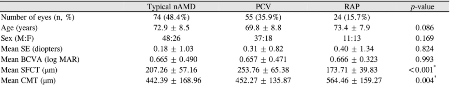
전체 대상군은 총 153명, 153안으로 남자 96안, 여자 57 안이었으며 평균 나이는 71.9 ± 8.6세였다(Table 1). 이 중 에서 전형적삼출성나이관련황반변성(typical nAMD군) 74 안(48.4%), 결절맥락막혈관병증(PCV군) 55안(35.9%), 망 막혈관종성증식(RAP군) 24안(15.7%)이었다. 유형별 연령, 성별, 진단 당시 굴절력 및 최대교정시력은 Table 2에 제시 하였으며 통계학적 유의한 차이는 보이지 않았다.
진단 당시 중심하 맥락막두께는 typical nAMD군이 207.26 ± 57.163 μm, PCV군이 253.76 ± 65.38 μm, RAP군 이 173.71 ± 39.83 μm로 통계적으로 유의한 차이를 보였다 (p<0.001). PCV군이 가장 두꺼운 맥락막두께를 보였고, typical nAMD군이 뒤를 이었으며 RAP군이 가장 얇은 맥 락막두께를 보였다(Table 2, 3). 각각의 그룹에서 중심하 맥
A B C
Table 2. Comparison of baseline characteristics among three subtypes of exudative age-related macular degeneration
Typical nAMD PCV RAP p-value
Number of eyes (n, %) 74 (48.4%) 55 (35.9%) 24 (15.7%)
Age (years) 72.9 ± 8.5 69.8 ± 8.8 73.4 ± 7.9 0.086
Sex (M:F) 48:26 37:18 11:13 0.169
Mean SE (diopters) 0.18 ± 1.03 0.31 ± 0.82 0.40 ± 1.34 0.824
Mean BCVA (log MAR) 0.665 ± 0.490 0.657 ± 0.471 0.666 ± 0.323 0.993
Mean SFCT (μm) 207.26 ± 57.16 253.76 ± 65.38 173.71 ± 39.83 <0.001*
Mean CMT (μm) 442.39 ± 168.96 452.27 ± 135.87 564.46 ± 159.27 0.004*
Values are presented as mean ± SD unless otherwise indicated. One way ANOVA for continuous variables; χ2 test for categorical variables.
nAMD = neovascular age-related macular degeneration; PCV = polypoidal choroidal vasculopathy; RAP = retinal angiomatous proliferation;
SE = spherical equivalent; BCVA = best corrected visual acuity; log MAR = logarithm of the minimum angle of resolution; SFCT = sub- foveal choroidal thickness; CMT = central macular thickness.
*p-value<0.05 was set as statistical significance.
Table 3. Post-hoc analysis p-value among three subtypes
Typical nAMD vs. PCV p-value PCV vs. RAP p-value RAP vs. typical nAMD p-value
Subfoveal choroidal thickness <0.001* <0.001* 0.010*
Central macular thickness 1.000 0.012* 0.003*
Bonferroni’s method for post-hoc analysis.
nAMD = neovascular age-related macular degeneration; PCV = polypoidal choroidal vasculopathy; RAP = retinal angiomatous proliferation.
*p-value<0.05 was set as statistical significance.
Figure 5. Comparative incidence of morphological features on
optical coherence tomography in each subtypes of exudative age-related macular degeneration. Accounting for a Bonferroni correction, p-values less than 0.017 were considered to in- dicate statistical significance. nAMD = neovascular age-re- lated macular degeneration; PCV = polypoidal choroidal vas- culopathy; RAP = retinal angiomatous proliferation; IRCF = intraretinal cytroid fluid; SRF = subretinal fluid; HRM = subretinal hyperreflective material; PED = pigment epithelial detachment. *The frequency of morphological characteristics which have p-values less than 0.017 when compared with oth- er subtype using Chi-square test.락막두께와 환자 연령 사이에 의미 있는 연관관계를 보이지 는 않았다(Pearson’s correlation coefficient, r=0.043, -0.017 and 0.103, p=0.823, 0.900 and 0.631, respectively). 맥락막 두께와 굴절력 또한 유의한 상관관계를 보이지 않았다 (Pearson’s correlation coefficient, r=0.174, 0.257 and 0.231, p=0.318, 0.158 and 0.277, respectively).
수정된 중심망막두께는 RAP군이 564.46 ± 159.27 μm로 typical nAMD군 442.39 ± 168.96 μm, PCV군 452.27 ± 135.87 μm에 비해 유의하게 두꺼운 중심망막두께를 보였 다(one way ANOVA test with Bonferroni’s correction, p=0.003, 0.012, respectively) (Table 2, 3).
기타 OCT에서 보이는 특징적 소견은 유형별로 서로 다 른 빈도를 보였다. 망막내낭포액의 경우 RAP군에서 typical nAMD군과 PCV군에 비해 빈번하게 관찰되었으며(typical nAMD vs. PCV, p-value=0.469; PCV vs. RAP, p-value<0.001;
RAP vs. typical nAMD, p-value<0.001), 망막하액은 상대 적으로 RAP군에서 낮은 빈도로 관찰되었다(typical nAMD vs. PCV, p-value=0.110; PCV vs. RAP, p-value=0.001; RAP vs. typical nAMD, p-value=0.021). 망막색소상피박리는 typical nAMD군에서 PCV군에 비해 낮은 빈도로 관찰되었 고(typical nAMD vs. PCV, p-value=0.005; PCV vs. RAP, p-value=0.304; RAP vs. typical nAMD, p-value=0.285), 망 막하 고반사물질은 세 군에서 비슷한 빈도로 관찰되었다 (typical nAMD vs. PCV, p-value=0.763; PCV vs. RAP, p-value
=0.990; RAP vs. typical nAMD, p-value=0.829) (Fig. 5).
고 찰
본 연구는 삼출성 나이관련황반변성을 전통적인 방법인 형광안저혈관조영술, 인도시아닌그린혈관조영술 결과에 따
라 typical nAMD, PCV, RAP 세 가지 유형으로 나누고, 빛 간섭단층촬영을 통해 측정한 중심하 맥락막두께, 중심망막 두께 및 기타 특징적 소견들을 유형별로 비교분석해 본 결 과, 유의한 차이를 보인다는 것을 확인하였다.
Verteporfin을 이용한 광역학요법(photodynamic therapy) 이 나이관련황반변성의 표준치료법이었던 시기에는 형광 안저혈관조영술을 통해 맥락막신생혈관의 유형을 잠복형 (occult)과 전형적(classic) 형태로 구분하는 것이 치료반응 을 예측하는 측면에서 중요하게 여겨졌다. 그렇지만 현재 는 항혈관내피성장인자(VEGF)의 유리체강내주사가 표준 치료방법으로서 널리 이용되고 있으며, 맥락막신생혈관의 형태에 관계없이 전반적으로 좋은 치료 효과를 나타냄이 알려져 있다.23-25 이로 인해 실제 임상에서 삼출성 나이관 련황반변성 환자를 진단하고 치료할 때 맥락막신생혈관의 형태에 따른 전통적인 분류는 그 유용성이 낮아지고 있으 며, 그보다는 예후나 치료경과에 있어서 차이를 보이는 PCV와 RAP를 typical nAMD와 구분하는 것이 임상적으로 더욱 의미 있을 것으로 생각된다.
결절맥락막혈관병증(PCV)는 일반적으로 전형적인 맥락 막신생혈관에 비해 양호한 자연경과를 보이지만, 반복적인 출혈이나 삼출 변화로 만성, 재발성 경과를 거치게 되어 결 국 시력저하를 보이게 되는 경우도 많다.7,8 PCV의 병인은 아직 완전히 밝혀져 있지 않으나, 망막-맥락막 기능부전 및 이로 인한 혈관이상으로 시력 저하를 일으키는 황반부 질환 이라는 점과 유전학적으로 보체인자와 관련된 유사한 발생 배경을 지닌다는 점에서 AMD의 한 유형으로 보여진다.26
망막혈관종성증식(RAP)은 다른 종류의 삼출성 나이관련 황반변성에 비해 예후가 나쁘며 양안성으로 발생하는 경우 가 많은 것으로 알려졌다.9 전형적인 삼출황반변성과 비교 하였을 때, 망막혈관종성증식으로 진단된 환자는 비교적 나이가 많고 맥락막이 더 얇으며, 드루젠이나 거짓드루젠 과 같은 망막의 연령관련 변성 소견이 더 높은 빈도로 나타 나는 것으로 알려져 있다.27,28
본 연구에서는 안저소견, 형광안저혈관조영술 및 인도시 아닌그린혈관조영술의 결과를 토대로 typical nAMD, PCV, RAP의 세 가지 유형으로 삼출성 나이관련황반변성을 분류 하였다. typical nAMD, PCV, RAP 순으로 48.4%, 35.9%, 15.7%의 분포율을 보였으며 국내 환자를 대상으로 한 이제 까지의 연구결과와 비교하였을 때,29 PCV의 경우 비슷한 빈도를 보였으나 RAP의 경우 본 연구에서 다소 높은 빈도 를 나타냈다.
이제까지 여러 연구를 통해 맥락막의 순환과 나이관련황 반변성과의 연관성이 언급되어 왔고, 맥락막의 순환 부전 이 삼출성 나이관련황반변성의 병태생리에 있어서 중요한
원인 중의 하나로 보고되었다.30,31 본 연구에서는 PCV와 RAP 환자에서 typical nAMD와 비교하여 맥락막 두께에 있어서 유의한 차이를 보였다. PCV군이 가장 두꺼운 맥락 막두께를 보였고 typical nAMD, RAP 순으로 얇은 맥락막 두께를 보였다. Kim et al27은 RAP와 typical nAMD를 비교 한 연구에서 RAP가 139.0 ± 65.5 μm로 184.9 ± 68.5 μm의 typical nAMD에 비해 더 얇은 맥락막두께를 보인다고 보 고하였으며, 본 연구에서도 유사한 결과를 보였다. Kim et al27은 RAP에서 얇은 맥락막두께와 더불어 더 넓은 범위의 드루젠 분포를 보이며 이는 RAP 발생기전에 있어서 맥락 막 순환부전이 더 크게 작용함을 시사한다고 하였다. PCV 의 경우, Chung et al16의 연구에서 정상안 및 AMD군에 비 해 PCV가 438.3 ± 87.8 μm로 보다 두꺼운 맥락막두께를 가짐이 보고되었고, Kim et al17의 연구에서도 PCV가 319.9
± 68.7 μm로 AMD군에 비해 두꺼운 맥락막두께를 보였다.
Rishi el al32 또한 PCV가 285.9 μm로 정상안과 wet AMD 안에 비해 두꺼운 맥락막두께를 가진다고 보고하였다. 본 연구에서도 PCV군이 253.76 ± 65.38 μm로 통계적으로 유 의하게 typical nAMD와 RAP에 비해 두꺼운 맥락막 두께 를 보였다. 이는 PCV의 발생기전에 있어서 맥락막 혈관의 위축보다는 확장, 그리고 이로 인한 혈류 순환의 장애가 관 여할 것임을 시사한다. 세 가지 유형의 AMD 모두 맥락막 순환 부전이 발생한다는 점에서 같은 맥락의 질환으로 볼 수 있으나, 그 원인에 있어서 typical nAMD나 RAP의 경우 얇아진 맥락막두께로 미루어 짐작하건대 맥락막의 전반적 인 위축으로 인한 혈류량의 저하가 신생혈관의 발생과 연 관되어 있을 것으로 보인다. 반면 PCV의 경우 두꺼운 맥락 막두께로 판단하건대 혈관벽의 확장 및 얇아짐으로 인해 혈류 속도의 저하가 초래되고 이로 인해 맥락막 순환 부전 을 일으키는 것으로 생각된다.
정상안을 대상으로 한 연구에서는 연령이 증가함에 따라 맥락막 두께가 감소함이 알려져 있으나,33 본 연구에서는 세 가지 유형의 삼출성 나이관련황반변성 모두 연령과의 상관성은 보이지 않았으며 굴절력과도 유의한 상관관계가 도출되지 않았다. 정상안과는 달리 삼출성 나이관련황반변 성의 경우 연령이나 굴절력에 비해 질병의 활성도(activity) 또는 단계(stage)와 같은 변수들이 맥락막두께에 더 큰 영 향을 미쳤을 것으로 판단된다. 또한 본 연구에 포함된 대상 들의 연령 및 굴절률의 범위가 상대적으로 좁기 때문에 연 관성이 masking되었을 가능성도 배제할 수 없다.
본 연구에서는 typical nAMD, PCV군에 비해 RAP군에 서 유의하게 두꺼운 중심망막두께를 보였다. RAP군에서 다른 군에 비해 망막하액은 낮은 빈도로 관찰되고 망막내 낭포액이 높은 빈도로 관찰된 것으로 미루어보아 RAP군에
서 중심망막두께가 두꺼운 것은 망막하액보다는 망막내낭 포액에 의한 것으로 보인다.
망막내낭포액은 빛간섭단층촬영에서 관찰되는 저반사도 의 낭포성 공간으로 정의하며 망막내층에 축적된 누출액으 로 인한 부종과 연관된다.22 기존의 연구결과를 살펴보면, IRCF는 삼출성 나이관련황반변성에서 초기 시력과 치료 후 최대교정시력의 예측인자로 보고되고 있으며, classic choroidal neovascularization (CNV), RAP 또는 후기 단계 occult CNV 병변에서 특징적으로 관찰됨이 알려져 있다.34 다시 말해 IRCF는 RPE-choroid complex를 넘어 감각신경 망막층의 구조적 변화를 일으킨 형태에서 관찰되는 소견임 을 알 수 있으며, 본 연구에서도 RAP군에서 다른 두 군에 비해 높은 빈도로 IRCF가 관찰되었고, 이는 망막에서 유래 한 것으로 보이는 신생혈관을 특징으로 하는 RAP의 병태 생리와 연관된 것으로 생각된다.
망막하액은 삼출성 나이관련황반변성에서 70-85%의 빈 도로 나타나며, anti-VEGF 치료에 따라 높은 시력 회복을 기대할 수 있는 예측인자로 보고된다.34 망막하액을 동반한 병변일수록 적극적인 치료 후에도 RPE 위축의 빈도가 낮 은 것으로 알려졌다.35 본 연구에서는 RAP군에서 다른 두 군에 비해 망막하액의 빈도가 상대적으로 낮게 나타났으며, 이는 anti-VEGF injection을 반복함에 따라 지도형위축 발 생의 빈도가 높은 RAP의 특성과 어느 정도 연관성이 있을 것으로 생각된다.36 망막하액의 빈도가 낮은 것과 시력 및 치료반응과의 연관성에 관련하여서는 추가 연구가 필요할 것으로 보인다.
망막색소상피박리는 PCV에서 typical nAMD에 비해 유 의하게 높은 빈도로 나타났다. 이는 출혈성 또는 삼출성 망 막색소상피박리를 잘 일으키고, 폴립에서 주변부 돔 모양 의 전형적인 PED로 연결되는 형태가 흔한 PCV의 임상적 특징과 일맥상통하며, typical nAMD는 신생혈관이 망막색 소상피를 뚫고 나와 망막하공간에서 증식하는 type 2 CNV 의 경우 PED를 동반하지 않기 때문에 낮은 빈도를 보인 것 으로 생각된다. 망막색소상피박리는 형태에 따라 serous PED, fibrovascular PED, hemorrhagic PED 등으로 세부 분 류하기도 하며,22 PED 형태별로 발생빈도를 비교해 보는 것 또한 임상적 의미를 가질 것으로 보여 추후 관련 연구를 진행해 보고자 한다.
망막하 고반사물질은 보통은 anti-VEGF injection을 시행 함에 따라 CNV가 활성을 잃어가면서 섬유성 조직으로 변 할 때 OCT에서 관찰되는 소견이나, 활성화된 삼출성 나이 관련황반변성의 경우 망막하출혈과 지질성삼출물이 망막 하 고반사물질로 나타나며 맥락막신생혈관의 활성 및 누출 정도와 관련성 있음이 보고되었다.37 Liakopoulos et al38은
type I CNV에 비해 type II, III CNV 병변에서 망막하 고반 사물질이 흔하게 발견된다고 하였으나, 본 연구에서와 같 이 typical nAMD, PCV, RAP로 분류하여 비교하였을 때에 는 유형에 따른 빈도 차이가 보이지 않았다.
본 연구는 후향적인 단면연구법에 의한 분석이었다는 점에서 제한점이 있다. 또한 맥락막두께 측정의 경우 중심 하 맥락막두께가 전체적인 맥락막의 두께와 형태를 반영 할 수 있는가 하는 의문점이 따른다. Margolis and Spaide21 의 enhanced depth imaging optical coherence tomography (EDI-OCT)를 이용한 정상안에서의 맥락막두께 연구에 따 르면 중심와 아래에서 가장 두꺼운 맥랙막 두께를 보이며, 주변부로 갈수록 얇아지는 동일한 패턴을 보인다고 하였다.
또한 전형적삼출성나이관련황반변성과 결절맥락막혈관병 증의 맥락막두께를 비교한 기존 연구 결과16에서 상측, 하 측, 비측, 이측으로 1,500 μm 떨어진 지점에서 측정한 맥락 막 두께의 차이가 중심하 맥락막두께에서 보인 차이와 비 슷한 양상을 보였다는 점에서 중심하 맥락막두께의 측정이 전반적인 맥락막의 두께를 어느 정도 반영할 수 있을 것으 로 판단된다. 마지막으로 두께를 측정하는 객관적인 소프 트웨어의 부재로 주관적인 측정법에 의해 연구가 진행되었 다는 한계점을 갖는다. 두 명의 측정자 간 신뢰도가 높았다 는 점에서 주관성으로 인한 한계를 어느 정도 보완하였다 고 판단되며, 추후 새로운 소프트웨어와 장비가 개발됨에 따라 보다 정확한 연구결과를 얻을 수 있을 것으로 보인다.
본 연구는 아직까지 국내에 발표된 바 없는 삼출성 나이 관련황반변성의 세 가지 하위 분류별 빛간섭단층촬영의 특 징적 소견을 비교 분석한 연구이며, 비교적 많은 환자를 대 상으로 했다는 점에서 의의가 있다고 하겠다. 추후 진단장 비가 점차 발전되어 구조적으로 망막과 맥락막을 더욱 자 세하고 세밀하게 가시화하는 것이 가능해지고 영상의 객관 적인 분석방법이 고안된다면 이와 관련된 보다 의미 있는 연구결과를 도출할 수 있을 것으로 기대한다.
REFERENCES
1) Wong TY, Chakravarthy U, Klein R, et al. The natural history and prognosis of neovascular age-related macular degeneration: a sys- tematic review of the literature and meta-analysis. Ophthalmology 2008;115:116-26.
2) Jager RD, Mieler WF, Miller JW. Age-related macular degene- ration. N Engl J Med 2008;358:2606-17.
3) Yannuzzi LA, Sorenson J, Spaide RF, Lipson B. Idiopathic poly- poidal choroidal vasculopathy (IPCV). Retina 1990;10:1-8.
4) Ciardella AP, Donsoff IM, Huang SJ, et al. Polypoidal choroidal vasculopathy. Surv Ophthalmol 2004;49:25-37.
5) Yannuzzi LA, Negrão S, Iida T, et al. Retinal angiomatous pro- liferation in age-related macular degeneration. Retina 2001;21:416-34.
6) Liu Y, Wen F, Huang S, et al. Subtype lesions of neovascular age-related macular degeneration in Chinese patients. Graefes Arch Clin Exp Ophthalmol 2007;245:1441-5.
7) Uyama M, Wada M, Nagai Y, et al. Polypoidal choroidal vasculop- athy: natural history. Am J Ophthalmol 2002;133:639-48.
8) Lee WK, Kwon SI. Polypoidal choroidal vasculopathy. J Korean Ophthalmol Soc 2000;41:2573-84.
9) Lee PY, Lee WK. Treatments of stage 1 retinal angiomatous proliferation. J Korean Ophthalmol Soc 2008;49:442-9.
10) Wilde C, Patel M, Lakshmanan A, et al. The diagnostic accuracy of spectral-domain optical coherence tomography for neovascular age-related macular degeneration: a comparison with fundus fluo- rescein angiography. Eye (Lond) 2015;29:602-9; quiz 610.
11) Hariri A, Heussen FM, Nittala MG, Sadda SR. Optical coherence tomographic correlates of angiographic subtypes of occult choroi- dal neovascularization. Invest Ophthalmol Vis Sci 2013;54:8020-6.
12) Spaide RF, Koizumi H, Pozzoni MC. Enhanced depth imaging spectral-domain optical coherence tomography. Am J Ophthalmol 2008;146:496-500.
13) Mrejen S, Spaide RF. Optical coherence tomography: imaging of the choroid and beyond. Surv Ophthalmol 2013;58:387-429.
14) Chen TC, Cense B, Pierce MC, et al. Spectral domain optical co- herence tomography: ultra-high speed, ultra-high resolution oph- thalmic imaging. Arch Ophthalmol 2005;123:1715-20.
15) Lee JY, Lee DH, Lee JY, Yoon YH. Correlation between subfoveal choroidal thickness and the severity or progression of non- exudative age-related macular degeneration. Invest Ophthalmol Vis Sci 2013;54:7812-8.
16) Chung SE, Kang SW, Lee JH, Kim YT. Choroidal thickness in pol- ypoidal choroidal vasculopathy and exudative age-related macular degeneration. Ophthalmology 2011;118:840-5.
17) Kim SW, Oh J, Kwon SS, et al. Comparison of choroidal thickness among patients with healthy eyes, early age-related maculopathy, neovascular age-related macular degeneration, central serous cho- rioretinopathy, and polypoidal choroidal vasculopathy. Retina 2011;31:1904-11.
18) Manjunath V, Goren J, Fujimoto JG, Duker JS. Analysis of choroi- dal thickness in age-related macular degeneration using spectral- domain optical coherence tomography. Am J Ophthalmol 2011;
152:663-8.
19) Fein JG, Branchini LA, Manjunath V, et al. Analysis of short-term change in subfoveal choroidal thickness in eyes with age-related macular degeneration using optical coherence tomography. Oph- thalmic Surg Lasers Imaging Retina 2014;45:32-7.
20) Seddon JM, Sharma S, Adelman RA. Evaluation of the clinical age -related maculopathy staging system. Ophthalmology 2006;113:26 0-6.
21) Margolis R, Spaide RF. A pilot study of enhanced depth imaging optical coherence tomography of the choroid in normal eyes. Am J Ophthalmol 2009;147:811-5.
22) Schmidt-Erfurth U, Waldstein SM. A paradigm shift in imaging bi- omarkers in neovascular age-related macular degeneration. Prog
Retin Eye Res 2016;50:1-24.
23) Verteporfin Roundtable Participants. Guidelines for using verte- porfin (Visudyne) in photodynamic therapy for choroidal neo- vascularization due to age-related macular degeneration and other causes: update. Retina 2005;25:119-34.
24) Rosenfeld PJ, Brown DM, Heier JS, et al. Ranibizumab for neo- vascular age-related macular degeneration. N Engl J Med 2006;
355:1419-31.
25) Kim YH, Kim ES, Yu SY, Kwak HW. Long-term effect of intra- vitreal bevacizumab for CNV secondary to age-related macular degeneration. J Korean Ophthalmol Soc 2008;49:1935-40.
26) Rosa RH Jr, Davis JL, Eifrig CW. Clinicopathologic reports, case reports, and small case series: clinicopathologic correlation of idio- pathic polypoidal choroidal vasculopathy. Arch Ophthalmol 2002;
120:502-8.
27) Kim JH, Kim JR, Kang SW, et al. Thinner choroid and greater dru- sen extent in retinal angiomatous proliferation than in typical exu- dative age-related macular degeneration. Am J Ophthalmol 2013;
155:743-9, 749.e1-2.
28) Cohen SY, Dubois L, Tadayoni R, et al. Prevalence of reticular pseudodrusen in age-related macular degeneration with newly diag- nosed choroidal neovascularisation. Br J Ophthalmol 2007;91:354-9.
29) Park KH, Song SJ, Lee WK, et al. The results of nation-wide regis- try of age-related macular degeneration in Korea. J Korean Oph- thalmol Soc 2010;51:516-23.
30) Spaide RF. Age-related choroidal atrophy. Am J Ophthalamol 2009;147:801-10.
31) Grunwald JE, Hariprasad SM, DuPont J. Effect of aging on foveo- lar choroidal circulation. Arch Ophthalmol 1998;116:150-4.
32) Rishi P, Rishi E, Mathur G, Raval V. Ocular perfusion pressure and choroidal thickness in eyes with polypoidal choroidal vasculop- athy, wet-age-related macular degeneration, and normals. Eye (Lond) 2013;27:1038-43.
33) Manjunath V, Taha M, Fujimoto JG, Duker JS. Choroidal thickness in normal eyes measured using Cirrus HD optical coherence tomography. Am J Ophthalmol 2010;150:325-9.e1.
34) Schmidt-Erfurth U, Waldstein SM, Deak GG, et al. Pigment epi- thelial detachment followed by retinal cystoid degeneration leads to vision loss in treatment of neovascular age-related macular degeneration. Ophthalmology 2015;122:822-32.
35) Sato T, Suzuki M, Ooto S, Spaide RF. Multimodal imaging find- ings and multimodal vision testing in neovascular age-related mac- ular degeneration. Retina 2015;35:1292-302.
36) McBain VA, Kumari R, Townend J, Lois N. Geographic atrophy in retinal angiomatous proliferation. Retina 2011;31:1043-52.
37) Shah VP, Shah SA, Mrejen S, Freund KB. Subretinal hyper- reflective exudation associated with neovascular age-related mac- ular degeneration. Retina 2014;34:1281-8.
38) Liakopoulos S, Ongchin S, Bansal A, et al. Quantitative optical co- herence tomography findings in various subtypes of neovascular age-related macular degeneration. Invest Ophthalmol Vis Sci 2008;49:5048-54.
= 국문초록 =
세 가지 유형의 삼출성 나이관련황반변성에서 빛간섭단층촬영의 특징 비교
목적: 한 번도 치료 받지 않은 삼출성 나이관련황반변성 환자에서 전형적삼출성나이관련황반변성(typical neovascular age-related macular degeneration [nAMD]), 결절맥락막혈관병증(polypoidal choroidal vasculopathy, PCV), 망막혈관종성증식(retinal angioma- tous proliferation, RAP) 세 가지 하위분류에 따른 빛간섭단층촬영의 특징을 비교하고자 하였다.
대상과 방법: 치료 기왕력이 없는 삼출성 나이관련황반변성 환자 153명 153안을 대상으로 하였다. 깊이증강모드 빛간섭단층촬영을 이용하여 진단 시점에서의 중심하 맥락막두께와 중심망막두께를 측정하고 망막내낭포액, 망막하액을 비롯한 구조적 특징들을 확인하 였다. 전체 환자군을 전형적 nAMD, PCV, RAP 세 그룹으로 나누고 특징들을 비교하였다.
결과: 총 153안 중에서 typical nAMD가 74안, PCV가 55안, RAP가 24안이었다. 연령과 최대교정시력은 그룹별 유의한 차이를 보이지 않았다. 중심하 맥락막두께는 PCV가 가장 두껍고, typical nAMD, RAP 순서로 얇은 두께를 보였으며 통계적으로 유의한 차이를 보였 다(p<0.001). RAP에서 다른 두 군에 비해 유의하게 두꺼운 중심망막두께(p=0.004)와 높은 빈도의 망막내낭포액의 존재(p<0.001)를 보였다.
결론: 삼출성나이관련황반변성은 세 가지 유형에 따라 서로 다른 빛간섭단층촬영의 특징을 보였다. PCV는 가장 두꺼운 맥락막두께를 보였고, RAP는 가장 얇은 맥락막두께와 두꺼운 중심망막두께, 그리고 높은 빈도의 망막내낭포액을 보였다. 삼출성나이관련황반변성 을 유형별로 감별진단하는 데 빛간섭단층촬영의 특징 분석이 도움이 될 것으로 보인다.
<대한안과학회지 2016;57(7):1093-1101>