초기에 좋은 시력을 보였던 결절맥락막혈관병증 환자를
대상으로 한 유리체강내 항혈관내피성장인자 치료의 2년 결과
Two-year Outcomes after Intravitreal Anti-vascular Endothelial Growth Factor Therapy for Polypoidal Choroidal Vasculopathy Patients with Good Initial Visual Acuity
유수진, 김재휘, 한정일, 김종우, 김철구, 이동원
Su Jin Yoo, Jae Hui Kim, Jung Il Han, Jong Woo Kim, Chul Gu Kim, Dong Won Lee
건양대학교 의과대학 김안과병원 안과
Department of Ophthalmology, Kim’s Eye Hospital, Konyang University College of Medicine, Seoul, Korea
Purpose: To investigate 2-year treatment outcomes of anti-vascular endothelial growth factor (VEGF) therapy in eyes with polypoidal choroidal vasculopathy (PCV) with good baseline visual acuity.
Methods: This retrospective, observational study included 21 eyes (10 eyes with a subfoveal polyp, 11 eyes with an extrafoveal polyp) of 21 subjects diagnosed with PCV. All eyes had a baseline best-corrected visual acuity (BCVA) of 20/25 or better and were administered an initial treatment of three intravitrial anti-VEGF injections at one-month intervals. Additional anti-VEGF treatments were administered as needed for recurring cases. BCVA and central foveal thickness (CFT) obtained 24 months after diagnosis were compared with baseline values.
Results: Mean logarithm of the minimal angle of resolution (logMAR) BCVA was 0.07 ± 0.04 at baseline and 0.19 ± 0.29 approximately 24 months after initial diagnosis, a slight difference that was not statistically significant (p = 0.560). Of the 21 eyes included, 17 eyes (80.9%) had stable vision and 4 eyes (19.0%) had a ≥0.2 deterioration in logMAR BCVA. Twenty-four-month CFT (212.9 ± 76.1 µm) was significantly lower than baseline CFT (324.6 ± 121.2 µm, p = 0.015).
Conclusions: Two-year visual outcomes following anti-VEGF therapy in eyes with PCV and good initial visual acuity were generally fa- vorable. However, logMAR BCVA deteriorated by ≥0.2 in 19.0% of treated eyes.
Keywords: Anti-vascular endothelial growth factor; Good visual acuity; Long-term; Polypoidal choroidal vasculopathy
Introduction
Anti-vascular endothelial growth factor (VEGF) therapy is effective for treating exudative age-related macular degen- eration (AMD) [1,2] and previous studies have shown rela-
tively favorable long-term outcomes of anti-VEGF therapy in treating exudative AMD [2-6]. Polypoidal choroidal vascu- lopathy (PCV) is a subtype of exudative AMD and is char- acterized by the presence of a branching vascular network and polypoidal lesions that are detectable with indocyanine
Address reprint requests to Jung Il Han, MD
Department of Ophthalmology, Kim’s Eye Hospital, #136 Yeongsin-ro, Yeongdeungpo-gu, Seoul 07301, Korea Tel: 82-2-2639-7664, Fax: 82-2-2639-7824
E-mail: [email protected]
Received: 2016. 8. 24 Revised: 2016. 10. 24 Accepted: 2016. 10. 31
green angiography (ICGA). Long-term anti-VEGF therapy outcomes are generally favorable in eyes with PCV [5,7].
However, few studies have examined long-term outcomes of this treatment on eyes with PCV and a good visual acuity at the time of diagnosis [8].
Mori and colleagues [8] investigated treatment outcomes in eyes with PCV and an initial logarithm of the minimum angle of resolution (logMAR) visual acuity of 0.6 (Snellen:
20/80) or better. Visual acuity significantly improved and central retinal thickness significantly decreased after 12 months of treatment. Unfortunately, outcomes beyond 12 months have not been documented.
Here, we investigate the 2-year treatment outcomes of an- ti-VEGF therapy in eyes with PCV and a good visual acuity (20/25 or better) at the time of diagnosis. Possible mecha- nisms for marked visual acuity deterioration during the first 2 years of anti-VEGF therapy are also examined.
Materials and Methods
This retrospective, observational case series was performed at a single center. The study was approved by the Kim's Eye Hospital Institutional Review Board and all study conduct adhered to the tenets of the Declaration of Helsinki. The medical records of patients who were newly diagnosed with PCV between January 2010 and December 2012 at our insti- tution were reviewed and considered for inclusion. Eyes with branching vascular networks and/or terminating polypoidal lesions on ICGA were diagnosed with PCV.
All included patients had undergone an initial comprehen- sive ophthalmologic examination, including best-corrected visual acuity (BCVA) measurement, slit-lamp biomicros- copy (90 D lens), fundus photography, fluorescein angi- ography, spectral domain optical coherence tomography (OCT; Spectral OCT/SLO®, OTI Ophthalmic Technologies, Inc., Miami, FL, USA) and indocyanine green angiography (confocal laser-scanning system, HRA-2, Heidelberg Engi- neering, Dossenheim, Germany). All included patients had been treated with intravitreal anti-VEGF agents and were followed for up to 24 months. Subject were excluded from analyses if they were followed for less than 24 months or had a severe media opacity, history of intraocular surgery (except cataract surgery), evidence of a macroaneurysm, proliferative diabetic retinopathy, a central retinal vascular
occlusion, or other retinal disorders known to affect macular microstructure and function. Eyes that had undergone other treatments for exudative AMD (e.g., photodynamic therapy) were also excluded from analyses. However, eyes that had undergone pneumatic displacement for treatment of a sub- macular hemorrhage were not excluded. Eyes that underwent cataract surgery or had definite cataract progression during the follow-up period were also excluded.
All eyes underwent an initial series of three consecutive monthly intravitreal ranibizumab injections and were then treated on an as needed basis. Patients were scheduled to visit the hospital for examination every 1-4 months at the treating physician’s discretion. All OCT, fluorescein angiog- raphy, and ICGA imaging were performed when needed, as determined by the clinician. Re-treatment with intravitreal anti-VEGF (either ranibizumab or bevacizumab) was gener- ally performed when intraretinal/subretinal fluid or retinal/
subretinal hemorrhage was accompanied by a central foveal thickness (CFT) that exceeded 300 µm. However, re-treat- ment was performed if CFT was <300 µm and BCVA had deteriorated. The presence of a pigment epithelial detach- ment (PED) was not sufficient for re-treatment.
The OCT was used to obtain automated average thickness measurements of the 1 mm diameter central macular area in only some patients. Therefore, manual CFT measure- ments were used to analyze macular anatomy. The CFT was defined as the distance between the internal limiting mem- brane and Bruch’s membrane at the fovea and was manually measured using the built-in calipers of the OCT system soft- ware. The greatest linear dimension of the entire PCV lesion detected by ICGA, including the branching vascular network and polypoidal lesion, was measured using the built-in cal- ipers provided by the ICGA system software. As described in previous reports [9,10], eyes that had multifocal hyperflu- orescent areas in the middle and late phases of ICGA were given a diagnosis of choroidal vascular hyperpermeability.
The BCVA was converted to the logMAR scale for all data analyses.
Baseline BCVA and CFT measurements were compared with those obtained at 3, 12, and 24 months after initiating treatment. The BCVA and CFT at 24 months were compared between patients with different baseline characteristics, including sex, choroidal hyperpermeability, polyp location (extra- or subfoveal), and the presence/absence of a fovea-in- volving PED. Additionally, associations between baseline
characteristics (e.g., age, BCVA, CFT, and greatest lesion linear dimension) and BCVA at 24 months and between baseline characteristics and CFT at 24 months were exam- ined. The characteristics of patients who experienced a log- MAR BCVA loss of ≥0.2 over the 24-month follow-up peri- od were also examined. This was done by dividing subjects into those who had a ≥0.2 logMAR BCVA deterioration (n
= 4 eyes) and those who had stable vision (<0.2 logMAR BCVA deterioration, n = 17 eyes). Baseline characteristics were then compared between the two groups.
Data are presented as mean ± standard deviation where applicable. Statistical analyses were performed using a commercially available software package (SPSS ver. 12.0 for Windows, SPSS, Inc., Chicago, IL, USA). Differences among values measured at different time points were ana- lyzed using a repeated-measures analysis of variance with Bonferroni’s correction. Differences in values between groups were examined using a Mann-Whitney U-test or a Fisher’s exact test. Correlations between variables were examined using a Pearson’s correlation analysis. A p < 0.05 was considered statistically significant.
Results
A total of 47 eyes (47 patients) were identified that had newly diagnosed PCV and a BCVA of 20/25 or better at the time of diagnosis. A total of 26 eyes (55.3%) were excluded from analyses because of a follow-up period shorter than 24 months (18 eyes), inadequate OCT imaging (6 eyes), or other reasons (2 eyes). Among the 18 patients lost to follow-up, 6 patients (33.3%) decided to visit another hospital. The exact reason for follow-up loss in the remaining 12 patients (66.7%) was not accurately identified. Ultimately, 21 eyes of 21 pa- tients (44.7%) were included in study analyses (Table 1). Out of the 21 included patients, 14 (66.7%) were men and 7 (33.3%) women. At the time of PCV diagnosis (baseline), subject age was 65.4 ± 7.2 years (range: 54-80 years), BCVA was 0.07 ± 0.04 (20/23, range: 20/25-20/20), and CFT was 324.6 ± 121.2 µm (range: 188-631 µm). The greatest linear dimension of lesions average 2.0 ± 0.9 mm (range: 1.0-4.7 mm) and 9 eyes (42.9%) had choroidal vascular hyperpermeability. Addition- ally, PCV lesion location at baseline was subfoveal or juxta- foveal in 10 eyes (47.6%) and extrafoveal in 11 eyes (52.4%).
A PED that involved the fovea was noted in 7 eyes (33.3%).
Subjects underwent an average of 5.8 ± 2.4 intravitreal injections (range: 3-12 injections) of anti-VEGF agents (ran- ibizumab: 4.1 ± 1.0 injections, bevacizumab: 1.5 ± 1.9 injec- tions) during the 24-month follow-up period. Subjects with subfoveal/juxtafoveal polyps underwent 5.1 ± 2.4 injections and subjects with extrafoveal polyps underwent 6.1 ± 2.7 injections. Nine eyes (42.9%) were treated with only ranibi- zumab and the remaining 12 eyes (57.1%) were treated with both ranibizumab and bevacizumab. During the 24-month follow-up period, 2 eyes developed a fovea-involving subma- cular hemorrhage and patients visited the hospital 9.5 ± 1.7 times (range: 8-13 times) for eye examinations. Additionally, OCT imaging was performed an average of 7.0 ± 1.2 times (range: 6-10 times) in each patient.
The BCVA at baseline and at 3, 12, and 24 months after initial diagnosis was 0.07 ± 0.04 (20/23), 0.05 ± 0.06 (20/22), 0.11 ± 0.17 (20/25) and 0.19 ± 0.29 (20/30), respectively (Fig.
1A). There was no significant difference in BCVA between any of the four time points examined (p = 0.431). Addition- ally, BCVA at baseline was not significantly different from
Table 1. Baseline characteristics of eyes with polypoidal choroi- dal vasculopathy and a good initial visual acuity (n = 21 eyes of 21 patients)
Characteristic Value
Age (years) 65.4 ± 7.2 (54-80)
Sex (n, %)
Male 14 (66.7)
Female 7 (33.3)
logMAR BCVA 0.07 ± 0.04 (20/25-20/20)
Central foveal thickness (µm) 324.6 ± 121.2 (188-631) Greatest linear dimension (mm) 2.0 ± 0.9
Fovea-involving PED (n, %) 7 (33.3) Choroidal vascular hyperpermeability
(n, %)
Present 9 (42.9)
Absent 12 (57.1)
Location of PCV lesion (n, %)
Subfoveal 10 (47.6)
Extrafoveal 11 (52.4)
Data are presented as mean ± standard deviation (range) where applicable.
BCVA = best-corrected visual acuity; logMAR = logarithm of mini- mal angle of resolution; PCV = polypoidal choroidal vasculopathy;
PED = pigment epithelial detachment.
BCVA at 3, 12, or 24 months (p = 1.000, p = 1.000, p = 1.000 and p = 0.560, respectively). Fig. 2 shows representative im- ages of an eye that had maintained a good BCVA over the 24-month follow-up period.
A total of 4 eyes (19.0%) had a worsening in logMAR BCVA by ≥0.2. However, the remaining 17 eyes (80.9%) had a stable BCVA. Despite treatment, serous subretinal fluid persisted in 1 eye (baseline BCVA = 20/25, 24-month BCVA
= 20/40), a subfoveal polypoidal lesion developed in 1 eye (baseline BCVA = 20/25, 24-month BCVA = 20/50), and a fovea-involving submacular hemorrhage developed in 2 eyes during the follow-up period. The two eyes with a submacu- lar hemorrhage were treated with pneumatic displacement (baseline BCVA = 20/20, 24-month BCVA = 20/200) or in- travitreal anti-VEGF monotherapy (baseline BCVA = 20/25, 24-month BCVA = 20/200).
Mean CFT at baseline and 3, 12, and 24 months after the initial diagnosis was 324.6 ± 121.2 µm, 215.5 ± 82.4 µm, 251.4 ± 118.1 µm and 212.9 ± 76.1 µm, respectively (Fig. 1B).
The CFT was significantly different among the five time points examined (p = 0.026) and CFT at baseline was signifi- cantly greater than CFT at 3 and 24 months (p = 0.016 and 0.015, respectively). However, the difference from baseline was not significant at 12 months (p = 0.171).
The BCVA at 24 months was not significantly different between the two subgroups divided according to baseline subject characteristics (Table 2). In addition, correlations be- tween subject characteristics (age, BCVA, CFT and greatest linear dimension) at baseline and BCVA at 24 months and between subject characteristics at baseline and CFT at 24 months were not significant (Table 3). Additionally, baseline characteristics were not significantly different between pa- tients with stable BCVA over 24 months and those who expe- rienced BCVA loss over 24 months (Table 4).
The mean duration of the follow-up period of the 18 pa- tients lost to follow-up was 11.1 ± 5.4 months (range: 4-21 months). The mean BCVA at diagnosis in these patients was 0.07 ± 0.03 (20/23), which had decreased to 0.12 ± 0.09 (20/26) at the last follow-up visit. The CFT was 318.7 ± 67.6 μm at Figure 1. Treatment outcome of polypoidal choroidal vasculopathy.
(A) Mean logarithm of the minimal angle of resolution (logMAR) best-corrected visual acuity (BCVA). (B) Central foveal thickness. Sta- tistical analyses were performed using repeated-measures analysis of variance with Bonferroni’s correction. Error bars represent one stan- dard deviation. M = months.
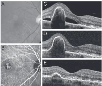
Figure 2. A representative case showing treatment outcome of polypoidal choroidal vasculopathy. (A-C) Best-corrected visual acu- ity (BCVA) at diagnosis (before treatment) was 20/25. (D) Subretinal fluid was no longer present after three consecutive ranibizumab injections. The eye underwent five ranibizumab injections during the 24-month follow-up period. (E) The BCVA was maintained over 2 years and was 20/25 at 24 months. (A) Fundus photography, (B) indo- cyanine green angiography, (C-E) optical coherence tomography.
A C
D
E B
0.6
0.4
0.2
0
500 400 300 200 100 0
BCVA (logMAR)
Baseline
p = 0.560
p = 0.015
3M 12M 24M
Central foveal thickness (µm)
Baseline
Follow-up period
3M 12M 24M
A
B
baseline and 238.1 ± 52.3 μm at the last follow-up visit.
Discussion
In the present study, eyes diagnosed with PCV that had good initial visual acuity maintained acuity up to 24 months after diagnosis. Additionlly, CFT at 24 months was significantly lower than baseline value and BCVA remained relatively stable in the majority of eyes during the follow-up period.
However, a ≥0.2 logMAR deterioration in BCVA was noted in some eyes, suggesting that a good initial visual acuity
does not guarantee a favorable visual outcome.
Monthly follow-up examinations, including OCT, are generally recommended after initial anti-VEGF treatment for exudative AMD. This approach facilitates early detection and prompt treatment of any recurrent exudation [11,12].
Delaying treatment by even one month can significant- ly affect visual prognosis [13]. One study that evaluated 12-month treatment outcomes in PCV patients with a good initial visual acuity involved monthly follow-ups and OCT examinations [8]. This allowed for prompt treatment of any recurrence. Unfortunately, a strict monthly follow-up proto- col with frequent OCT examinations is not possible in many clinical settings. It has been reported that outcomes follow- ing anti-VEGF therapy for exudative AMD are relatively unfavorable in the clinical setting [14,15]. The follow-up schedule and frequency of OCT examinations for the present study were not strictly controlled. During the 24-month fol- low-up period, the mean number of follow-up visits was only 9.5 and the mean number of OCT examinations was only 7.0.
Additionally, the mean number of anti-VEGF injections was 5.8, far less than that reported by previous well-controlled clinical trials [5,11,12]. However, the maintenance or im- provement of visual acuity observed in the majority of eyes, despite this study’s shortcoming in the follow-up schedule, Table 2. Comparison of best-corrected visual acuity (BCVA) and central foveal thickness (CFT) at 24 months in groups divided according to baseline characteristics
Characteristic 24-month BCVA p-value* 24-month CFT p-value*
Sex
Male (n = 14 patients) 0.25 ± 0.34 0.220 219.1 ± 83.6 0.370
Female (n = 7 patients) 0.06 ± 0.05 200.4 ± 62.6
Choroidal vascular hyperpermeability
Present (n = 9 eyes) 0.19 ± 0.29 0.361 193.4 ± 35.9 0.594
Absent (n = 12 eyes) 0.19 ± 0.30 227.4 ± 95.2
Location of polyps
Subfoveal/juxtafoveal (n = 10 eyes) 0.17 ± 0.31 0.262 196.1 ± 26.4 0.888
Extrafoveal (n = 11 eyes) 0.19 ± 0.28 228.1 ± 102.1
Fovea-involving PED
Presence 0.23 ± 0.37 0.689 191.6 ± 29.3 0.636
Absence 0.17 ± 0.25 223.5 ± 90.3
Data are presented as mean ± standard deviation where applicable. Visual acuities were presented as logarithm of minimal angle of resolu- tion scale.
PED = pigment epithelial detachment.
*Indicates statistical analysis performed using a Mann Whitney U-test.
Table 3. Correlation of baseline factors with best-corrected visual acuity (BCVA) and central foveal thickness (CFT) at 24 months
Characteristic p-value*
(24-month BCVA)
p-value* (24-month CFT)
Age 0.650 0.180
Baseline BCVA 0.486 0.821
Baseline CFT 0.204 0.759
Greatest linear dimension 0.660 0.756
*Indicates statistical analysis performed using Pearson’s correlation analysis.
highlights the favorable outcomes of anti-VEGF therapy in eyes with PCV and a good initial visual acuity.
Here, a deterioration in BCVA of ≥0.2 logMAR was not- ed in 4 eyes. Persistent serous subretinal fluid was noted in one of these eyes, suggesting that chronic subretinal fluid can negatively affect visual prognosis. It has been shown that photodynamic therapy alone or in combination with anti-VEGF therapy can resolve exudate in eyes with fluid that persists after anti-VEGF therapy [16]. These treatment modalities represent useful alternatives in such cases.
Two eyes exhibited signs of submacular hemorrhage, which has been associated with a poor visual prognosis in exudative AMD [17,18]. These two eyes were treated with either pneumatic displacement or intravitreal anti-VEGF monotherapy, both of which have been shown to be effective in treating submacular hemorrhage secondary to exudative AMD [19-24]. However, visual acuity failed to return to baseline values in both patients. This suggests that subma- cular hemorrhage development has a negative influence on visual prognosis in eyes with PCV. The development of a subfoveal polypoidal lesion was noted in one eye. This was not surprising because new polypoidal lesion development is not unusual in eyes with PCV [25]. What was notable was that the newly developed lesion was a cluster of grape-like
polyps [25-27]. This type of lesion has been associated with a poor prognosis after photodynamic [26] and/or anti-VEGF therapy [27], conversion to typical choroidal neovascular- ization [26] and the development of submacular hemorrhage [27]. Although our patient did not develop signs of submacu- lar hemorrhage, massive exudate from the newly developed polyps caused a marked deterioration in visual acuity that did not improve with anti-VEGF therapy. This suggests that additional studies are needed to evaluate the efficacy of oth- er treatments not involving anti-VEGF agents on eyes with similar disease.
The results of the present study show that visual prognosis was favorable, even in eyes with subfoveal/juxtafoveal pol- yps. Because recurrent fluid is associated with a poor visual prognosis in eyes with PCV [28], our result may have reflect- ed the small number eyes with recurrent fluid that required retreatment. In the study by Kang and Koh [28], subjects received an average of 5.39 ± 0.50 injections during the first year of treatment and 3.00 ± 0.53 injections during the sec- ond year of treatment. Additionally, the subjects in the study by Lee and Lee [29] received an average of 12.50 ± 2.77 in- jections over a 2 year treatment period. In the present study, only 5.6 injections were administered during the 24-month follow-up period, with 5.1 injections administered to eyes Table 4. Comparison of baseline characteristics in eyes with a stable best-corrected visual acuity (BCVA, n = 17 eyes) and eyes with a deterio- rated BCVA (n = 4 eyes)
Characteristic Stable BCVA group Deteriorated BCVA group p-value
Age (years) 64.6 ± 6.6 68.8 ± 9.7 0.462*
Sex (n, %)
Male 10 (58.8) 4 (100) 0.255†
Female 7 (41.2) 0
logMAR BCVA 0.08 ± 0.04 0.05 ± 0.06 0.484*
Central foveal thickness (µm) 344.4 ± 126.2 240.5 ± 35.9 0.172*
Greatest linear dimension (mm) 1,895.4 ± 808.3 2,566.8 ± 1,492.8 0.362*
Choroidal vascular hyperpermeability (n, %)
Present 8 (47.1) 1 (25.0) 0.603†
Absent 9 (52.9) 3 (75.5)
Location of PCV lesion (n, %)
Subfoveal 8 (47.1) 2 (50) 1.000†
Extrafoveal 9 (52.9) 2 (50)
Data are presented as mean ± standard deviation where applicable.
logMAR = logarithm of minimal angle of resolution; PCV = polypoidal choroidal vasculopathy.
*Indicates statistical analysis performed using a Mann Whitney U-test; †Indicates statistical analysis performed using a Fisher’s exact test.
with subfoveal/juxtafoveal polyps. Therefore, our subjects had a markedly lower injection frequency than that of previ- ous studies. Second, baseline visual acuity was predictive of long-term visual outcome in eyes with PCV [30,31]. Because our patients had a good baseline visual acuity, regardless of polyp location, this may have influenced our visual out- comes.
In the present study, we attempted to identify factors that were predictive of 24-month visual outcomes. However, none of the variables examined were significantly associated with 24-month visual acuity. Choroidal vascular hyper- permeability is frequently observed on ICGA in eyes with PCV [9,10,32,33] and is associated with greater choroidal thickness [10,32], bilateral neovascular membranes [10]
and a poor response to initial ranibizumab treatment [10].
Although a recent study showed that choroidal vascular hy- perpermeability is associated with a poor visual prognosis at 12 months [33], the association of this peculiar finding with the treatment outcome beyond this time point is not known.
Here, we found that 42.8% of eyes with PCV had choroidal vascular hyperpermeability. The visual acuity at 24 months was not different between eyes with and without choroidal vascular hyperpermeability, suggesting that the presence of choroidal vascular hyperpermeability may not significant- ly influence long-term visual prognosis in eyes with PCV and a good initial visual acuity. However, considering that only half of our examined patients completed 2 years of fol- low-up, further studies are needed to confirm our findings.
We also found that baseline characteristics were not signifi- cantly different between patients who had a marked visual acuity deterioration and patients who maintained stable vision. However, the number of patients ultimately included in the analyses was small, which may have influenced our results. Therefore, further studies on larger populations are needed to confirm our findings.
Our study had some limitations, including its retrospective design and relatively small sample size. The small sample size may have influenced the results of statistical analyses.
Additionally, the follow-up examination and treatment schedules were not controlled and only half of eligible pa- tients completed the required 2-year follow-up period. The exact reason for loss of follow-up was also not identified in the majority of lost patients. Therefore, a selection bias to- wards a favorable outcome may have significantly affected study results. In the present study, no common guidelines
were used to select anti-VEGF agents. Ranibizumab and/
or bevacizumab were used in each patient at the discretion of the treating physician. In addition, both ranibizumab and bevacizumab were used in the majority of eyes. Therefore, this study cannot contribute to information on the efficacy of a single anti-VEGF agent. However, both agents have com- parable long-term efficacy for treating exudative AMD and the influence of using both anti-VEGF agents may not have significantly influenced study results. Lastly, because ICGA was not routinely performed during the follow-up period, this study does not provide further insight into the influence of anti-VEGF therapy on polypoidal lesion and branching vascular network regression.
In spite of these deficiencies, our study demonstrates that visual acuity is generally maintained over a 2-year follow-up period when anti-VEGF therapy is used to treat PCV in eyes with a good initial visual acuity. However, 19% of the in- cluded eyes did have a deterioration in logMAR BCVA of 0.2 or more. This suggests that outcome expectations should be managed by informing patients that their prognosis could be unfavorable even though visual acuity is good at the time of diagnosis. Further studies on consecutive patients are needed to identify predictive factors of long-term (24 months) visual outcomes for this condition.
Conflicts of interest There are no conflicts of interest.
References
1. Brown DM, Kaiser PK, Michels M, et al. Ranibizumab versus verteporfin for neovascular age-related macular degeneration.
N Engl J Med 2006;355:1432-44.
2. Rosenfeld PJ, Brown DM, Heier JS, et al. Ranibizumab for neo- vascular age-related macular degeneration. N Engl J Med 2006;355:1419-31.
3. Rasmussen A, Bloch SB, Fuchs J, et al. A 4-Year longitudinal study of 555 patients treated with ranibizumab for neovascular age-re- lated macular degeneration. Ophthalmology 2013;120:2630-6.
4. Abraham P, Yue H, Wilson L. Randomized, double-masked, sh- am-controlled trial of ranibizumab for neovascular age-related macular degeneration: PIER study year 2. Am J Ophthalmol 2010;150:315-24.e1.
5. Comparison of Age-related Macular Degeneration Treatments
Trials (CATT) Research Group, Martin DF, Maguire MG, et al.
Ranibizumab and bevacizumab for treatment of neovascular age-related macular degeneration: two-year results. Ophthal- mology 2012;119:1388-98.
6. Jonas JB, Tao Y, Schlichtenbrede FC. Intravitreal bevacizumab for exudative age-related macular degeneration in clinical practice.
J Ocul Pharmacol Ther 2011;27:467-70.
7. Inoue M, Arakawa A, Yamane S, Kadonosono K. Long-term outcome of intravitreal ranibizumab treatment, compared with photodynamic therapy, in patients with polypoidal choroidal vasculopathy. Eye (Lond) 2013;27:1013-20; quiz 21.
8. Mori R, Yuzawa M, Akaza E, Haruyama M. Treatment results at 1 year of ranibizumab therapy for polypoidal choroidal vas- culopathy in eyes with good visual acuity. Jpn J Ophthalmol 2013;57:365-71.
9. Sasahara M, Tsujikawa A, Musashi K, et al. Polypoidal choroidal vasculopathy with choroidal vascular hyperpermeability. Am J Ophthalmol 2006;142:601-7.
10. Koizumi H, Yamagishi T, Yamazaki T, Kinoshita S. Relationship be- tween clinical characteristics of polypoidal choroidal vasculopa- thy and choroidal vascular hyperpermeability. Am J Ophthalmol 2013;155:305-13.e1.
11. Fung AE, Lalwani GA, Rosenfeld PJ, et al. An optical coherence tomography-guided, variable dosing regimen with intravitreal ranibizumab (Lucentis) for neovascular age-related macular de- generation. Am J Ophthalmol 2007;143:566-83.
12. CATT Research Group, Martin DF, Maguire MG, et al. Ranibi- zumab and bevacizumab for neovascular age-related macular degeneration. N Engl J Med 2011;364:1897-908.
13. Rauch R, Weingessel B, Maca SM, Vecsei-Marlovits PV. Time to first treatment: the significance of early treatment of exudative age-related macular degeneration. Retina 2012;32:1260-4.
14. Cohen SY, Dubois L, Tadayoni R, et al. Results of one-year’s treat- ment with ranibizumab for exudative age-related macular de- generation in a clinical setting. Am J Ophthalmol 2009;148:409- 13.
15. Bandukwala T, Muni RH, Schwartz C, et al. Effectiveness of intra- vitreal ranibizumab for the treatment of neovascular age-related macular degeneration in a Canadian retina practice: a retro- spective review. Can J Ophthalmol 2010;45:590-5.
16. Cho M, Barbazetto IA, Freund KB. Refractory neovascular age-re- lated macular degeneration secondary to polypoidal choroidal vasculopathy. Am J Ophthalmol 2009;148:70-8.e1.
17. Avery RL, Fekrat S, Hawkins BS, Bressler NM. Natural history of subfoveal subretinal hemorrhage in age-related macular de-
generation. Retina 1996;16:183-9.
18. Bennett SR, Folk JC, Blodi CF, Klugman M. Factors prognostic of visual outcome in patients with subretinal hemorrhage. Am J Ophthalmol 1990;109:33-7.
19. Shienbaum G, Garcia Filho CA, Flynn HW Jr, et al. Management of submacular hemorrhage secondary to neovascular age-relat- ed macular degeneration with anti-vascular endothelial growth factor monotherapy. Am J Ophthalmol 2013;155:1009-13.
20. Iacono P, Parodi MB, Introini U, et al. Intravitreal ranibizumab for choroidal neovascularization with large submacular hemorrhage in age-related macular degeneration. Retina 2014;34:281-7.
21. Ron Y, Ehrlich R, Axer-Siegel R, et al. Pneumatic displacement of submacular hemorrhage due to age-related macular degenera- tion. Ophthalmologica 2007;221:57-61.
22. Cakir M, Cekiç O, Yilmaz OF. Pneumatic displacement of acute submacular hemorrhage with and without the use of tissue plasminogen activator. Eur J Ophthalmol 2010;20:565-71.
23. Ohji M, Saito Y, Hayashi A, et al. Pneumatic displacement of sub- retinal hemorrhage without tissue plasminogen activator. Arch Ophthalmol 1998;116:1326-32.
24. Shin JY, Lee JM, Byeon SH. Anti-vascular endothelial growth factor with or without pneumatic displacement for submacular hemorrhage. Am J Ophthalmol 2015;159:904-14.e1.
25. Uyama M, Wada M, Nagai Y, et al. Polypoidal choroidal vascu- lopathy: natural history. Am J Ophthalmol 2002;133:639-48.
26. Lee WK, Kim KS, Kim W, et al. Responses to photodynamic ther- apy in patients with polypoidal choroidal vasculopathy con- sisting of polyps resembling grape clusters. Am J Ophthalmol 2012;154:355-65.e1.
27. Hikichi T, Higuchi M, Matsushita T, et al. Factors predictive of outcomes 1 year after 3 monthly ranibizumab injections and as-needed reinjections for polypoidal choroidal vasculopathy in Japanese patients. Retina 2013;33:1949-58.
28. Kang HM, Koh HJ. Long-term visual outcome and prognostic factors after intravitreal ranibizumab injections for polypoidal choroidal vasculopathy. Am J Ophthalmol 2013;156:652-60.
29. Lee JH, Lee WK. Anti-vascular endothelial growth factor mono- therapy for polypoidal choroidal vasculopathy with polyps resembling grape clusters. Graefes Arch Clin Exp Ophthalmol 2016;254:645-51.
30. Chang YS, Kim JH, Kim KM, et al. Long-term outcomes of an- ti-vascular endothelial growth factor therapy for polypoidal choroidal vasculopathy. J Ocul Pharmacol Ther 2016;32:219-24.
31. Hikichi T, Kitamei H, Shioya S. Prognostic factors of 2-year out- comes of ranibizumab therapy for polypoidal choroidal vascu-
lopathy. Br J Ophthalmol 2015;99:817-22.
32. Jirarattanasopa P, Ooto S, Nakata I, et al. Choroidal thickness, vascular hyperpermeability, and complement factor H in age-related macular degeneration and polypoidal choroidal vasculopathy. Invest Ophthalmol Vis Sci 2012;53:3663-72.
33. Cho HJ, Kim HS, Jang YS, et al. Effects of choroidal vascular hyperpermeability on anti-vascular endothelial growth factor treatment for polypoidal choroidal vasculopathy. Am J Ophthal- mol 2013;156:1192-200.e1.