© 2015 The Korean Ophthalmological Society
This is an Open Access article distributed under the terms of the Creative Commons Attribution Non-Commercial License (http://creativecommons.org/licenses /by-nc/3.0/) which permits unrestricted non-commercial use, distribution, and reproduction in any medium, provided the original work is properly cited.
Original Article
Aflibercept Treatment for Neovascular Age-related Macular Degeneration and Polypoidal Choroidal Vasculopathy Refractory to
Anti-vascular Endothelial Growth Factor
Da Ru Chi Moon, Dong Kyu Lee, Soon Hyun Kim, Yong Sung You, Oh Woong Kwon
Retina Center, Nune Eye Hospital, Seoul, Korea
Purpose: To report the results of switching treatment to vascular endothelial growth factor (VEGF) Trap-Eye (aflibercept) in neovascular age-related macular degeneration (AMD) and polypoidal choroidal vasculopathy (PCV) refractory to anti-VEGF (ranibizumab and bevacizumab).
Methods: This is a retrospective study involving 32 eyes from 29 patients; 18 were cases of neovascular AMD and 14 were cases of PCV. The best-corrected visual acuity (BCVA) and central macular thickness (CMT) of spectral-domain optical coherence tomography were evaluated.
Results: BCVA and CMT improved from 0.58 to 0.55 (p = 0.005) and from 404 to 321 μm (p < 0.001), respectively, after switching to aflibercept. The 14 eyes that received 6 or more aflibercept injections remained stable at 0.81 to 0.81 and 321 to 327 μm (p = 1.0, 0.29), respectively, after 3 aflibercept injections. The 10 eyes that received 3 or more bevacizumab injections after 3 or more aflibercept injections worsened, from 0.44 to 0.47 and from 332 to 346 μm (p = 0.06, 0.05), respectively. The results showed similar improvement of BCVA and CMT in neovascular AMD and PCV.
Conclusions: Aflibercept seems to be effective for improvement and maintenance of BCVA and CMT for neovascular AMD and PCV refractory to anti-VEGF. Switching from aflibercept back to bevacizumab treatment may not be a proper strategy.
Key Words: Aflibercept, Anti-vascular endothelial growth factor, Bevacizumab, Macular degeneration, Polypoi- dal choroidal vasculopathy
Neovascular age-related macular degeneration (AMD) is a leading cause of blindness in aged populations in the industrialized world [1-3]. Currently, intravitreal injection of anti-vascular endothelial growth factor (VEGF) is the standard therapy for treating neovascular AMD [4,5].
Bevacizumab and ranibizumab injections play important roles in preserving vision and preventing vision loss [6-8].
However, for approximately 10% of AMD cases, treatment with anti-VEGF has been reported to have no initial effect or induce resistance with repeated treatments [9,10].
Possible mechanisms of resistance are tolerance or tachyphylaxis, which is defined as decreased response to repeated treatment with a medication [10,11].
Polypoidal choroidal vasculopathy (PCV) is characterized by frequent recurrence, bilateral asymmetry, and serous and hemorrhagic detachment of retinal pigment
Received: October 3, 2014 Accepted: January 5, 2015
Corresponding Author: Oh Woong Kwon, MD, PhD. Retina Center, Nune Eye Hospital, #404 Seolleung-ro, Gangnam-gu, Seoul 135-841, Korea. Tel: 82-2-2086-7792, Fax: 82-2-2086-7779, E-mail: owkwon0301@
gmail.com
epithelium, which can also cause vision loss [12]. Among several treatment strategies, photodynamic therapy (PDT) and intravitreal injection of anti-VEGF have shown the most favorable results [13,14]. However, serious complications such as foveal atrophy after PDT and limited responsiveness to anti-VEGF treatment have also been reported [14,15].
Possible countermeasures include temporary cessation [10] or increasing the volume of anti-VEGF [16]. However, these measures might result in deterioration of the lesions or increase the risk of adverse effects. Another possible countermeasure is the introduction of a drug with a different mode of action [10]. In this context, aflibercept, which has a different mode of action compared to anti- VEGF, is expected to be a novel treatment option for the above cases by acting as a ‘trap system’ [17,18].
In this study, we evaluated the visual and anatomical response of intravitreal injection of aflibercept in eyes with neovascular AMD and PCV refractory to treatment with ranibizumab or bevecizumab.
Materials and Methods
Design
This study is a retrospective chart review of patients with neovascular AMD and PCV refractory to treatment with ranibizumab or bevecizumab. They were switched to aflibercept treatment with at least 3 consecutive injections at approximately one-month intervals between July 1, 2013 and June 30, 2014.
Patient selection
Inclusion criteria of this study are as follows: known choroidal neovascularization secondary to AMD or PCV diagnosed with fluorescein angiography and indocyanine green angiography, persistent or recurrent fluids on spec- tral-domain optical coherence tomography (SD-OCT), or leakage on angiography after 3 or more anti-VEGF treat- ments prior to switching to aflibercept. After switching, patients with more than three initial monthly loading doses of intravitreal aflibercept (2 mg in 0.05 mL) were enrolled.
Follow-up examination and data
A full ophthalmic examination, including best-corrected visual acuity (BCVA), intraocular pressure, slit lamp exam- ination, indirect ophthalmoscopy, fundus photograph, and SD-OCT (Heidelberg Spectralis; Heidelberg Engineering, Heidelberg, Germany) were conducted at each monthly vis- it. Demographic data and treatment history (number of in- jections and duration of follow-up) were also collected.
BCVA (Snellen) were obtained on the day of aflibercept initiation, at subsequent follow-up visits, and at the final follow-up visit. Central macular thickness (CMT) was cal- culated with custom software. Segmentation lines and cen- tral foveal location were manually adjusted when necessary.
Analysis
The primary outcomes of this study were the mean change in logarithm of the minimal angle of resolution in visual acuity and CMT after switching to aflibercept injec- tions. SD-OCT was used to analyze the fluid resolution in- cluding the intraretinal fluid, subretinal fluid, pigment epi- thelial detachments, and subretinal hemorrhage. Changes of the fluid from baseline were classified as complete reso- lution, partial resolution, unchanged or worse. Complete resolution was defined as resolution of all components of the fluid from baseline. The same analysis was conducted in each of the neovasuclar AMD (18 cases) and PCV (14 cases) patients. Further analysis was performed in patients treated with aflibercept 3 to 4 times and more than 4 times (aflibercept on-going) as well as on those who returned to bevacizumab treatment with more than 3 injections (switching-back). Statistical analysis was performed using the SPSS ver. 16.0 (SPSS Inc., Chicago, IL, USA). Wilcox- on signed rank tests were used to compare the differences in means between baseline and follow-up examinations for BCVA and CMT. Mann-Whitney U-tests were used to identify the differences in baseline characteristics between AMD and PCV patients. A p-value less than 0.05 was con- sidered to be statistically significant.
Results
patient characteristics at the time of switching to aflibercept (baseline) for 32 cases (17 refractory cases, 15 recurrent cases) of 29 pateints (16 male and 13 female) are
listed in Table 1. The mean age at baseline was 63.8 ± 11.3 years (range, 50 to 85 years). The clinical characteristics of all cases are listed in Table 2. All cases were treated three or more times with other drugs before switching to aflibercept: 12 were previously treated with bevacizumab alone, 7 with ranbizumab, 13 with both bevacizumab and ranibizumab, and 10 were treated with PDT on one or more occasions. The mean number and duration of anti- VEGF/PDT treatment prior to switching were 19.2 times (range, 3 to 64 times) / 3.0 (1 to 5) and 28.8 months (range, 4 to 78 months), respectively. The mean number and duration of aflibercept injection after switching were 4.3 times (range, 3 to 9 titmes) and 5.5 ± 1.9 months (range, 3 to 9 months), respectively. Out of all cases, 8 were treated with only 3 to 4 aflibercept injections (loading treatment), 14 were treated with more than 4 aflibercept injections and 6 month follow-up (on-going treatment) because of excellent clinical response to aflibercept or patient requests.
There were 10 cases treated with bevacizumab again (switching-back treatment) after 3 or 4 injections of aflibercept due to poor response to aflibercept or expense issues despite excellent response to aflibercept.
Changes in mean BCVA were measured (Fig. 1). BCVA significantly improved at all follow-up visits compared with baseline (p = 0.005). The mean BCVA improvement was highest at 2.7 months after switching to aflibercept (p
= 0.003). The mean BCVA improved from 0.31 to 0.24 (p = 0.06) in the loading treatment patients (8 eyes) and remained stable, changing from 0.81 to 0.81 (p = 1.0), in the on-going treatment patient group (14 cases) who were injected with aflibercept 4 or more times and received continual follow-up for 6 months. The average number of additional aflibercept injections and follow-up were 2.6 times and 3.1 months, respectively. However, the mean
BCVA deteriorated from 0.44 to 0.47 (p = 0.06) in the switching-back treatment patient group (10 cases) who were returned to bevacizumab injections administered an average of 4.7 times (range, 3 to 7 times) over 4.9 months (range, 3 to 7 months).
There was no significant difference in the change in the mean BCVA between the two patient groups (on-going and switching-back) from baseline to final follow-up period (p
= 0.102 and 0.414, respectively). However, the BCVA worsened by more than 1 line from the baseline in 4 of the switching-back patients, whereas there was no worsening in the on-going group. Demographic characteristics including BCVA, age, treatment history (anti-VEGF and PDT), and duration of disease did not show significant differences between the on-going and switching-back treatment patients (data not shown).
Anatomical changes were analyzed for all groups (Fig.
2). CMT reduction changed significantly for all follow-up visits compared with baseline measurements (p = 0.000).
The maximum mean reduction was 83 μm at 4.6 months, which corresponded to the time point just before switching back to bevacizumab in 10 of the 32 cases. Similar to the visual acuity changes, CMT improved from 341 to 291 μm ( p = 0.04) in loading treatment patients (8 eyes) and remained stable in the on-going treatment patient group (14 cases) during the treatment period (321 to 327 μm, p = 0.29). Meanwhile, CMT deteriorated in the switching-back treatment patient group (10 cases) during the switch-back period (332 to 346 μm, p = 0.05). The mean change in CMT was statistically significant only in the on-going treatment patients (14 cases, p = 0.02) from baseline to the final follow-up period.
Further analyses for SD-OCT were also conducted (Table 3) [5]. Upon qualitative analysis of OCT findings [5],
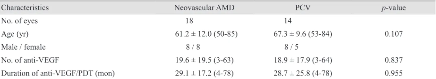
Table 1. Demographics of neovascular AMD and PCV patients
Characteristics Neovascular AMD PCV p-value
No. of eyes 18 14
Age (yr) 61.2 ± 12.0 (50-85) 67.3 ± 9.6 (53-84) 0.107
Male / female 8 / 8 8 / 5
No. of anti-VEGF 19.6 ± 19.5 (3-63) 18.9 ± 17.9 (3-64) 0.837
Duration of anti-VEGF/PDT (mon) 29.1 ± 17.2 (4-78) 28.7 ± 25.8 (4-78) 0.955
Values are presented as number or mean ± standard deviation (range).
AMD = age-related macular degeneration; PCV = polypoidal choroidal vasculopathy; VEGF = vascular endothelial growth factor; PDT
= photodynamic therapy.
6 cases with intraretinal fluid, 15 with subretinal fluid, 29 with pigment epithelial detachment, and 3 with subretinal hemorrhage were observed at the baseline. After switching to aflibercept, more than 70% of patients responded to treatment. However, 17% were unchanged and 6% had a worsening of their condition.
Subanalysis of SD-OCT findings in the on-going and switching-back groups showed maintenance of the results in 91.6% (11 / 12) of the on-going group. In the switching- back group, the SD-OCT results worsened in 35.3% (7 / 16) of cases and was maintained in 64.7% (11 / 17) (data not shown).
Another analysis of the changes in SD-OCT findings, CMT and BCVA of choroidal neovascularization (18 cases), and PCV (14 cases) patients showed similar results: overall treatment response was more than 70% in both patient groups, BCVA and CMT improved respectively from 0.54 to 0.51 (p = 0.06) and 409 to 341 (p = 0.017) in neovascular
AMD and from 0.64 to 0.61 (p = 0.06) and 398 to 308 (p = 0.005) in PCV (data not shown).
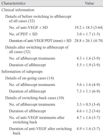
Table 2. Clinical characteristics of all 32 cases
Characteristics Value
Clinical information
Details of before switching to aflibercept of all cases (32)
No. of anti-VEGF ± SD 19.2 ± 18.5 (3-64)
No. of PDT ± SD 3.0 ± 1.7 (1-5)
Duration of anti-VEGF/PDT (mon) ± SD 28.8 ± 26.1 (4-78) Details after switching to aflibercept of
all cases (32)
No. of aflibercept treatments 4.3 ± 1.6 (3-9) Duration of aflibercept 5.5 ± 1.9 (3-9) Information of subgroups
Details of on-going cases (14)
No. of aflibercept treatments 5.6 ± 1.6 (4-9) Duration of aflibercept 7.3 ± 1.1 (6-9) Details of switching-back cases (10)
No. of aflibercept treatments 3.3 ± 0.5 (3-4) Duration of aflibercept 4.6 ± 1.2 (3-6) No. of anti-VEGF treatments after
switching back 4.7 ± 1.6 (3-7)
Duration of anti-VEGF after switching
back 4.9 ± 1.6 (3-7)
Values are presented as mean± SD (range).
VEGF = vascular endothelial growth factor; SD = standard devi- ation; PDT = photodynamic therapy.
Fig. 1. Changes in mean best-corrected visual acuity (logarithm of the minimal angle of resolution, logMAR) for all cases in- cluding on-going and switching-back cases during the entire fol- low-up period. 2.7 months was the time point after 3 initial load- ing injections of aflibercept in all cases. 4.2 months was the time point of 4 or more on-going treatment of aflibercept in 14 cases. 4.6 months was the time point of switching-back treatment of beva- cizumab in 10 cases. Improvement of best-correct visual acuity from baseline of all cases was statistically significant, but not sig- nificant in on-going or switching-back treatment cases during the entire follow-up period. Best-correct visual acuity was stable in on-going cases (p = 1.0) but deteriorated in switching-back cases (p = 0.06). VA = visual acuity. *p < 0.05.
0.5 0.58
*0.54 *0.55
0.6 0.7
0.8 0.8
0.84
0.81 0.81
0.9
0.4
0.45 0.42 0.44
*330
*327
*321
*332
*329
346 404
421 423
0.47
320 340 360 380 400 420
On-going injection
at 4.2 mon 14 Cases, 7.3 mon
Stable VA
14 Cases, 7.3 mon Stable CMT
10 Cases, 9.5 mon Decreased VA
10 Cases, 9.5 mon Increased CMT 2.7 mon
5.5 mon
Baseline:
switching time point to aflibercept
Switching-back at 4.6 mon
On-going injection at 4.2 mon
Switching-back at 4.6 mon
5.5 mon
On-going injection of aflibercept All cases
Switching and switching-back
On-going injection of aflibercept All cases
Switching and switching-back
Baseline:
switching time point to aflibercept
VA (logMAR)CMT (μm)
Fig. 2. Changes in mean central macular thickness (CMT) of all cases, on-going cases, and switching-back cases during the entire follow-up period. 4.2 months was the time point of 4 or more on-going treatments of aflibercept in 14 cases. 4.6 months was the time point of switching-back treatment of bevacizumab in 10 cases. Improvement of CMT from baseline in all cases, including on-going and switching-back cases, was statistically significant during the entire follow-up period. However, after switching back to bevacizumab, CMT increased (p = 0.05) during the switch- ing-back period. In contrast, CMT remained stable in on-going 4 or more treatment cases of aflibercept (p = 0.29) during the treat- ment period. *p < 0.05.
0.5 0.58
*0.54 *0.55
0.6 0.7
0.8 0.8
0.84
0.81 0.81
0.9
0.4
0.45 0.42 0.44
*330
*327
*321
*332
*329
346 404
421 423
0.47
320 340 360 380 400 420
On-going injection
at 4.2 mon 14 Cases, 7.3 mon
Stable VA
14 Cases, 7.3 mon Stable CMT
10 Cases, 9.5 mon Decreased VA
10 Cases, 9.5 mon Increased CMT 2.7 mon
5.5 mon
Baseline:
switching time point to aflibercept
Switching-back at 4.6 mon
On-going injection at 4.2 mon
Switching-back at 4.6 mon
5.5 mon
On-going injection of aflibercept All cases
Switching and switching-back
On-going injection of aflibercept All cases
Switching and switching-back
Baseline:
switching time point to aflibercept
VA (logMAR)CMT (μm)
Adverse events
No ocular adverse events were observed, including endophthalmitis, retinal detachment, retinal pigment epithelial tears, submacular hemorrhage, uveitis, or sustained intraocular pressure elevation requiring other treatment.
Discussion
It has been reported that aflibercept is an effective treat- ment not only for patients with naïve neovascular AMD [19] but also for those who show no response or resistance to existing anti-VEGF treatments [4,5,20,21]. In this study, switching to aflibercept resulted in significant improvement in visual acuity and CMT in AMD and PCV patients who showed no response or resistance to existing treatments. In addition, improved visual acuity and CMT were maintained for more than 6 months in the group that continued aflibercept treatment.
Notably, we analyzed the results of switching-back treatment, which involves returning to anti-VEGF treatment after switching to aflibercept. Unlike the results of the on-going treatment, there was deterioration in visual acuity and CMT in the 10 patients who were switched back to bevacizumab. The reason for switching back to bevacizumab was cost for 5 cases, even though they were responsive to aflibercept. The other 5 cases were not responsive to aflibercept. Although the numbers of cases were small, these data suggest that switching back from aflibercept to bevavizumab is not an effective strategy.
These data might be derived from the differences in the basic affinity of each drug for receptors. Aflibercept, which is a trap-eye system and works on placental growth factor as well as VEGF B, has better affinity to VEGF A than existing anti-VEFG drugs such as ranibizumab and bevacizumab, which are anti-VEFG antibodies [17,18]. In addition to potency, abrupt discontinuation of aflibercept may be another explanation. For uveitis, changes into less potent drugs deteriorated the disease progress. Thus, slow tapering of the drug may be more effective for these cases.
This observation also suggests that switching back to bevacizumab after gradual discontinuation of aflibercept using methods such as the treat and extend or capped pro re nata in the VEGF-Trap Eye: Investigation of Efficacy and Safety in Wet AMD (VIEW) study are possible solutions when switching treatment from aflibercept back to bevacizumab is inevitable [19]. Another possible solution is switching back to super-high or high dose anti- VEGF treatment. The SAVE (Super-dose Anti-VEGF) study already demonstrated the effectiveness of super-high dose anti-VEFG treatment for recalcitrant neovascular AMD [16]. Since the cost difference between aflibercept and bevacizumab may result in more switching-back treatments, f urther long-term studies on gradual discontinuation are needed. Additionally, identification of deteriorating factors in switching back as well as baseline patient demographic characteristics are required. Studies on switching to ranibizumab and switching from naïve af- libercept treatment are also needed.
In this study, 9 patients did not show appropriate responses to aflibercept. When the cases were divided into Table 3. Qualitative analysis based on spectral domain optical coherence tomography findings of all cases
OCT findings Baseline no. of cases Worse Unchanged Improvement
Partial resolution Complete resolution
IRF 6 0 (0) 1 (16.7) 3 (50) 2 (33.3)
SRF 15 2 (13.3) 2 (13.3) 9 (60) 2 (13.3)
PED 29 1 (3.4) 6 (20.7) 17 (58.7) 5 (17.2)
SRH 3 0 (0) 0 (0) 3 (100) 0 (0)
Overall 53 3 (5.7) 9 (17) 32 (60.3) 9 (17)
Values are presented as number or number (%); From Ho VY, et al. Am J Ophthalmol 2013;156:23-8.e2, with permission from Elsevier [5]; Results of all cases with refractory disease prior to anti-vascular endothelial growth factor treatments at 4.6 months after switching to aflibercept (4.6 months: time point before initiation of switching-back treatment); This trend was consistent among all 3 subgroups treated with previous bevacizumab treatment, ranibizumab treatment, or treatment with both agents (data not shown).
OCT = optical coherence tomography; IRF = intraretinal fluid; SRF = subretinal fluid; PED = pigment epithelial detachment; SRH = sub- retinal hemorrhage (additional assessment with fundus examination).
responsive and non-responsive groups, significant differences (p = 0.018, Mann-Whitney test) in the duration of disease were observed between the groups. The area under the receiver operating characteristic curve was 0.77 (p = 0.019), implying that long-duration of disease led to less effective aflibercept treatment (in refractory cases).
Limitations of this study include retrospective chart review, low case number, short-term follow-up, and non- controlled study. However, this study was not designed to test a hypothesis but to identify a trend in the management of refractory AMD and PCV cases. Thus, further prospective and controlled studies with higher case numbers and long-term follow-up are needed.
In conclusion, switching treatment to aflibercept seems to be effective for improvement and maintenance of BCVA and CMT over 6months for neovascular AMD and PCV refractory to anti-VEGF. In addition, switching back to bevacizumab is not recommended and further study of switching back the treatment to prior anti-VEGF is necessary.
Conflict of Interest
No potential conflict of interest relevant to this article was reported.
References
1. Bressler NM. Age-related macular degeneration is the lead- ing cause of blindness. JAMA 2004;291:1900-1.
2. Congdon N, O'Colmain B, Klaver CC, et al. Causes and prevalence of visual impairment among adults in the Unit- ed States. Arch Ophthalmol 2004;122:477-85.
3. Klaver CC, Wolfs RC, Vingerling JR, et al. Age-specific prevalence and causes of blindness and visual impairment in an older population: the Rotterdam Study. Arch Oph- thalmol 1998;116:653-8.
4. Chang AA, Li H, Broadhead GK, et al. Intravitreal afliber- cept for treatment-resistant neovascular age-related macu- lar degeneration. Ophthalmology 2014;121:188-92.
5. Ho VY, Yeh S, Olsen TW, et al. Short-term outcomes of af- libercept for neovascular age-related macular degeneration in eyes previously treated with other vascular endothelial growth factor inhibitors. Am J Ophthalmol 2013;156:23-8.e2.
6. Avery RL, Pieramici DJ, Rabena MD, et al. Intravitreal bevacizumab (Avastin) for neovascular age-related macular degeneration. Ophthalmology 2006;113:363-72.e5.
7. Comparison of Age-related Macular Degeneration Treat- ments Trials (CATT) Research Group, Martin DF, Maguire MG, et al. Ranibizumab and bevacizumab for treatment of neovascular age-related macular degeneration: two-year re- sults. Ophthalmology 2012;119:1388-98.
8. Folk JC, Stone EM. Ranibizumab therapy for neovascular age-related macular degeneration. N Engl J Med 2010;363:
1648-55.
9. Otsuji T, Nagai Y, Sho K, et al. Initial non-responders to ranibizumab in the treatment of age-related macular de- generation (AMD). Clin Ophthalmol 2013;7:1487-90.
10. Binder S. Loss of reactivity in intravitreal anti-VEGF ther- apy: tachyphylaxis or tolerance? Br J Ophthalmol 2012;
96:1-2.
11. Schaal S, Kaplan HJ, Tezel TH. Is there tachyphylaxis to intravitreal anti-vascular endothelial growth factor phar- macotherapy in age-related macular degeneration? Oph- thalmology 2008;115:2199-205.
12. Yannuzzi LA, Sorenson J, Spaide RF, Lipson B. Idiopathic polypoidal choroidal vasculopathy (IPCV). Retina 1990;10:1-8.
13. Gomi F, Ohji M, Sayanagi K, et al. One-year outcomes of photodynamic therapy in age-related macular degeneration and polypoidal choroidal vasculopathy in Japanese patients.
Ophthalmology 2008;115:141-6.
14. Hikichi T, Higuchi M, Matsushita T, et al. One-year results of three monthly ranibizumab injections and as-needed re- injections for polypoidal choroidal vasculopathy in Japa- nese patients. Am J Ophthalmol 2012;154:117-24.e1.
15. Akaza E, Yuzawa M, Mori R. Three-year follow-up results of photodynamic therapy for polypoidal choroidal vascu- lopathy. Jpn J Ophthalmol 2011;55:39-44.
16. Wykoff CC, Brown DM, Chen E, et al. SAVE (Super-dose anti-VEGF) trial: 2.0 mg ranibizumab for recalcitrant neo- vascular age-related macular degeneration: 1-year results.
Ophthalmic Surg Lasers Imaging Retina 2013;44:121-6.
17. Papadopoulos N, Martin J, Ruan Q, et al. Binding and neu- tralization of vascular endothelial growth factor (VEGF) and related ligands by VEGF Trap, ranibizumab and beva- cizumab. Angiogenesis 2012;15:171-85.
18. Stewart MW, Rosenfeld PJ. Predicted biological activity of intravitreal VEGF Trap. Br J Ophthalmol 2008;92:667-8.
19. Heier JS, Brown DM, Chong V, et al. Intravitreal afliber-
cept (VEGF trap-eye) in wet age-related macular degener- ation. Ophthalmology 2012;119:2537-48.
20. Cho H, Shah CP, Weber M, Heier JS. Aflibercept for exu- dative AMD with persistent fluid on ranibizumab and/or bevacizumab. Br J Ophthalmol 2013;97:1032-5.
21. Yonekawa Y, Andreoli C, Miller JB, et al. Conversion to aflibercept for chronic refractory or recurrent neovascular age-related macular degeneration. Am J Ophthalmol 2013;
156:29-35.e2.