처음 진단된 삼출 나이관련황반변성에서 애플리버셉트 치료의 임상적 결과
Clinical Outcomes of Aflibercept Treatment for Treatment-naive Exudative Age-related Macular Degeneration
최유진1, 최영제1, 조용운1,2, 유웅선1,2, 정인영1,2
Yu-Jin Choi1, Young Je Choi1, Yong Wun Cho1,2, Woong-Sun Yoo1,2, In Young Chung1,2
1경상국립대학교 의과대학 안과학교실, 2경상국립대학교 건강과학연구원
1Department of Ophthalmology, Gyeongsang National University School of Medicine, Jinju, Korea
2Gyeongsang Institute of Health Science, Gyeongsang National University, Jinju, Korea
Purpose: To evaluate the 1-year outcomes of the treat-and-extend regimen using intravitreal injection of aflibercept for treatment-naive exudative age-related macular degeneration (AMD) and the clinical results of switching to ranibizumab within the same period.
Methods: The change in best-corrected visual acuity (BCVA), central macular thickness (CMT) and number of injections was evaluated in 36 eyes first diagnosed with exudative AMD and treated with the treat-and-extend regimen using intravitreal injection of aflibercept, as well as 16 eyes switched from aflibercept to ranibizumab for improved efficacy within the same period.
Results: Mean BCVA improved significantly from 0.57 ± 0.28 logarithm of the minimum angle of resolution (logMAR) to 0.39 ± 0.29 logMAR at 12 months (p = 0.015) and mean CMT decreased significantly from 406.34 ± 69.18 μm to 269.58 ± 83.15 μm at 12 months in the 36 eyes on the treat-and-extend regimen of aflibercept (p = 0.004). The average number of injections per year was 6.8 ± 0.5. Of the 16 eyes that underwent a change in medication, seven eyes (63.6%) exhibited improvement or maintenance of initial visual acuity; mean CMT decreased significantly from 415.27 ± 73.63 μm prior to drug switch to 289.63 ± 62.78 μm after drug switch in the 11 eyes that re- ceived three injections of ranibizumab (p = 0.009).
Conclusions: A treat-and-extend regimen of intravitreal aflibercept injection was effective for exudative AMD in a clinical setting.
Switching to ranibizumab for improved efficacy led to anatomical improvement and could help stabilize vision.
Keywords: Aflibercept; Age-related macular degeneration; Treat-and-extend regimen
Address reprint requests to In Young Chung, MD, PhD
Department of Ophthalmology, Gyeongsang National University School of Medicine, #15 Jinju-daero, 816 beon- gil, Jinju 52727, Korea
Tel: 82-55-750-8171, Fax: 82-55-758-4158 E-mail: [email protected]
Received: 2021. 8. 9.
Revised: 2021. 9. 7.
Accepted: 2021. 9. 10.
서론
삼출 나이관련황반변성은 50세 이상의 환자에서 실명을 유발할 수 있는 질환으로 알려져 있다[1]. 노령인구의 증가에 따라 국내 에서의 유병률이 증가하고 있으며 나이관련황반변성에 동반된 맥락막 신생혈관 발생에 의한 부종, 출혈 등 삼출성 변화로 인 하여 시력저하가 나타나게 된다[2]. 그러나 항혈관내피성장인자 치료가 도입됨에 따라 치료 결과가 향상되어 상당수의 환자에 서 해부학적 및 기능적인 결과를 호전 및 유지시킬 수 있는 것 으로 보고되고 있다[3].
현재 삼출 나이관련황반변성의 일반적인 치료로는 유리체 내 항혈관내피성장인자 주입술이 시행되고 있으며, 베바시주맙 (AvastinⓇ, Genentech Inc., Oceanside, CA, USA and The Roche Group, Basel, Switzerland), 라니비주맙(LucentisⓇ, Genentech Inc., South San Fransisco, CA, USA and Novartis Pharma AG, Basel, Switzerland), 애플리버셉트(EyleaⓇ, Regeneron, Tarrytown, NY, USA and Bayer HealthCare, Berlin, Germany) 가 효과적인 약제로 널리 이용되고 있다. 항혈관내피성장인자 약 제들 중 특히 애플리버셉트는 약제의 지속시간이 상대적으로 길 며, VIEW (vascular endothelial growth factor [VEGF] Trap-eye:
investigation of efficacy and safety in wet age-related macular degeneration [AMD]) 1과 VIEW 2 연구에서는 3회의 로딩 주입 술 후 8주 간격으로 유리체내 애플리버셉트를 주입하는 것이 매 달 라니비주맙을 주입하는 것과 비교했을 때 유사한 시력 변화 와 해부학적 호전을 보여주었다[4].
하지만 주기적인 주사와 방문에 대한 경제적, 치료적 부담감 에 대한 불편감으로 인하여 주사와 방문 횟수를 줄이면서 치료 효과를 얻을 수 있는 방법에 대한 연구들이 진행되어 왔다. 대 표적으로 정기적인 경과 관찰을 시행하면서 재발하는 경우에 주사를 시행하는 pro ne nata (PRN) 방법이 있다. 그러나 이는 주사 횟수를 줄이면서 치료 효과를 유지할 수 있음이 보고되었 지만 정기적인 경과 관찰을 하지 못하는 경우 시력을 유지하기 가 어렵다는 부분도 알려진 바 있다[5,6]. 매번 병원 방문에 대한 환자들의 불편감을 감소시키고 반복적인 유리체내 항혈관내피 성장인자주입술 치료에 따르는 합병증을 감소시키면서 시력 개 선을 장기적으로 유지하기 위해 치료 및 연장(treat-and-extend) 방법의 치료적 접근이 제시되었다. 여러 연구들은 치료 및 연 장 방법이 fixed dosing 방법으로 주사하는 것과 유사한 수준의 기능적 해부학적 호전을 얻을 수 있다고 보고하였으며 현재 삼 출 나이관련황반변성의 치료 스케줄로 널리 사용되고 있다[7,8].
본 연구에서는 전향적 연구를 통해 이미 치료 및 연장 방법 의 장점이 많이 알려져 있기에 실제 의료현장에서 애플리버셉 트주입술의 치료 및 연장 방법에 따른 임상적 결과를 알아보고 자 하였다. 삼출 나이관련황반변성으로 처음 진단받고 유리체 내 애플리버셉트주입술을 치료 및 연장 방법의 원칙하에 시행
하고 1년 이상 경과 관찰이 가능하였던 환자들을 대상으로 치 료 효과를 분석하였고, 같은 기간 내 약제를 라니비주맙으로 변 경하여 치료한 경우의 치료 효과를 분석하였다.
대상과 방법
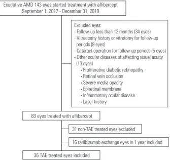
본 연구는 후향적으로 의무기록을 분석하였으며 헬싱키 선언에 입각한 생명의학 연구윤리심의위원회의 승인하에 진행되었다(승 인번호: 2020-11-005). 2017년 09월부터 2019년 12월까지 삼출 나 이관련황반변성으로 진단되어 최초 치료로 유리체내 애플리버 셉트주입술로 치료를 시작한 143안을 분석하여 12개월 이상 경 과 관찰이 불가능하였던 34안, 유리체절제술 과거력이 있거나 치 료 도중 유리체절제술을 시행한 8안, 경과 관찰 기간 동안 백내 장수술을 시행한 5안, 시력에 영향을 주는 다른 안과적 질환이 동반된 13안, 치료 및 연장 방법으로 치료하지 못한 31안을 제외 하고 연구기준에 적합한 환자들 중 애플리버셉트 치료 및 연장 방법으로 치료한 34명 36안과 치료시작 1년 내에 임상의사의 판 단에 따라 애플리버셉트가 시력이나 해부학적 반응에 효과가 부 족하다고 생각되어 라니비주맙으로 약제를 변경하였던 16안을 대상으로 분석을 시행하였다(Fig. 1).
연구에 포함된 환자는 50세 이상의 삼출 나이관련황반변성 으로 진단되고 진단 당시 최대교정시력 0.1-0.8 (decimal), 형광 안저혈관조영 및 인도시아닌그린혈관조영에서 황반하 신생혈관 과 누출이 관찰되는 경우, 빛간섭단층촬영에서 망막하액과 망
Exudative AMD 143 eyes started treatment with aflibercept September 1, 2017 - December 31, 2019
83 eyes treated with aflibercept
31 non-TAE treated eyes excluded
16 ranibizumab exchange eyes in 1 year included 36 TAE treated eyes included
Excluded eyes:
- Follow-up less than 12 months (34 eyes) - Vitrectomy history or vitretomy for follow-up periods (8 eyes)
- Cataract operation for follow-up periods (5 eyes) - Other ocular diseases of affecting visual acuity (13 eyes)
• Proliferative diabetic retinopathy • Retinal vein occlusion • Severe media opacity • Epiretinal membrane • Inflammatory ocular disease • Laser history
Figure 1. Flow chart for patients included in the study. AMD = age-related macular degeneration; TAE = treat-and-extend.
막내액이 관찰되고 1년 이상 관찰이 가능하였던 환자들을 대 상으로 하였다[9,10]. 다음과 같은 경우는 연구에서 제외하였다.
1) 증식당뇨망막병증, 망막혈관폐쇄증이 동반된 경우, 2) 심한 매체혼탁이 있는 경우, 3) 망막전막이나 염증성 질환 등 시력에 영향을 줄 수 있는 기타 다른 유리체망막병증이 동반된 경우 4) 유리체절제술 과거력이 있거나 치료 도중 유리체절제술을 시 행한 경우, 5) 치료 기간 동안 백내장수술을 시행한 경우, 6) 레 이저치료 병력이 있는 경우.
모든 환자에서 진단시 최대교정시력, 세극등검사, 안저검사, 빛 간섭단층촬영(HRA spectralis + OCT, Heidelberg Engineering, Heidelberg, Germany), 형광안저혈관조영(HRA2 FAG, Heidelberg Engineering Gmbh, Heidelberg, Germany) 및 인도시아닌그린혈 관조영(HRA2 ICG, Heidelberg Engineering Gmbh)을 시행하였
다. 형광안저혈관조영과 인도시아닌그린혈관조영 결과를 종합적 으로 판단하여 분지혈관망과 말단부의 결절 병변을 보이는 특징 적 소견이 있는 경우 결절맥락막혈관병증(polypoidal choroidal vasculopathy, PCV)으로 진단하였고, 망막내 혈관신생, 망막-망 막 혈관문합, 혹은 망막-맥락막 혈관문합이 관찰되는 경우 망 막혈관종성증식(retinal angiomatous proliferation, RAP)으로 진 단하였으며 이를 제외한 경우를 전형적 삼출 나이관련황반변 성(typical neovascular age-related macular degeneration, typical AMD)으로 분류하였다.
유리체내 주사는 수술실에서 시행되었으며 0.5% proparacaine hydrochloride (AlcaineⓇ, Alcon, Inc., Fort Worth, TX, USA)으 로 시술전 점안마취한 뒤 povidone iodine으로 속눈썹과 눈주위 를 소독하고, 개검기를 끼우고 1.25% povidone iodine으로 세척
Figure 2. Fundus photographs and optical coherence tomography (OCT) images of a case of age-related macular degeneration treated with intravitreal aflibercept treat-and-extend regimen. (A, B) At diagnosis, best-corrected visual acuity (BCVA) was 0.15. (C) Subretinal fluid (SRF) was decreased after three monthly aflibercept injections. BCVA was 0.2. (D) Eight weeks after the fourth intravitreal aflibercept injection, OCT showed persistent SRF with unchanged fluid. BCVA was 0.2. There was no visual loss. So, the patient was treated with a fifth intravitreal aflibercept injec- tion at an interval of 8 weeks to maintain the injection interval. (E) Eight weeks after the fifth intravitreal aflibercept injection, OCT showed com- plete absorption of SRF. BCVA was 0.3. The patient was treated with a sixth intravitreal aflibercept injection at an interval of 10 weeks to extend the injection interval. (F) Ten weeks after the sixth intravitreal aflibercept injection, there was no fluid on OCT. BCVA was 0.3. The patient was treated with a seventh intravitreal aflibercept injection at an interval of 12 weeks to further extend the injection interval.
A B
C
E
D
F
한 뒤 각막 윤부에서 하외측 3.0 mm 혹은 3.5 mm 부위의 섬모 체 평면부를 통해 30게이지 일회용 바늘을 이용하여 애플리버 셉트(2.0 mg/0.05 mL) 또는 라니비주맙(0.5 mg/0.05 mL)을 주 입하였다.
유리체내 애플리버셉트주입술 스케줄 방법에 따라 치료 및 연장 방법으로 치료받은 환자들은 초기 4주 간격으로 3회의 주 사가 시행되었으며 빛간섭단층촬영에서 마른 황반부 상태가 될 때까지 8주 간격으로 주사를 시행하였다. 의사의 판단에 따라 임상 상태와 환자의 필요 등을 고려하였을때 재발 유무와 관계 없이 지속적으로 주사하는 치료가 도움이 된다고 판단되어 병 변의 활동성에 따라 치료 및 연장(treat-and-extend, TAE) 방법 으로 치료한 환자들은 TAE군으로 정의하였다. TAE군은 빛간 섭단층촬영에서 마른 황반부 소견을 보이면 주사 간격을 2주씩 늘리는 것을 원칙으로 하되 환자 사정에 따라 1-3주 간격을 연 장하였으며 주사 최대 간격은 16주로 하였다. 빛간섭단층촬영 에서 망막하액과 망막내액이 증가한 경우, 망막하출혈 및 망막
내출혈이 새롭게 발생한 경우, 새로운 신생혈관이 발생한 경우, 최대교정시력이 감소한 경우에는 질환의 활동성이 재발한 것으 로 판단하여 주사 간격을 2주씩 감소시키는 것을 원칙으로 하 되 환자 사정을 고려하는 경우 1-3주씩 간격을 단축하였으며 주 사 최소 간격은 8주로 하였다(Fig. 2). 치료 및 연장 방법의 주 사 간격에 대한 단축, 유지, 연장 기준은 Table 1에 나타내었다.
최대교정시력, 세극등검사, 안저검사, 빛간섭단층촬영이 경과 관찰 때마다 시행되었으며 decimal 방식으로 측정된 최대교정 시력은 통계분석을 위해 logarithm of the minimum angle of resolution (logMAR)값으로 환산하여 분석하였다. 중심황반두 께는 빛간섭단층촬영을 이용하여 내경계막에서 브루크막까지 의 거리를 수직으로 측정하였다. TAE 방법으로 치료한 환자들 의 12개월 동안 최대교정시력 변화 정도와 중심황반두께 변화 정도를 분석하였고, 12개월에 3줄 이상 시력 호전을 보인 비율 과 주사 횟수, 주사 간격을 조사하였다. 또한 유리체내 애플리 버셉트 주입술 치료 1년 이내 애플리버셉트에 효과가 부족하여 라니비주맙으로 치료 약제를 변경한 경우 초기 진단시, 마지막 애플리버셉트 주사 후, 변경된 라니비주맙으로 3회 치료 후 4주 뒤의 최대교정시력과 중심황반두께를 비교하였다.
통계분석에는 SPSS 프로그램(SPSS version 21.0, software for windows; SPSS Inc., Chicago, IL, USA)을 이용하였다.
Kolmogorov-Smirnov test와 Shapiro-Wilk test을 통하여 정규 성 검정을 하고 repeated measures analysis of variances 방법 을 이용하여 서로 다른 시점에 측정된 값을 비교하였으며, 서 로 다른 두 시점 사이의 비교는 bonferroni method를 이용하였 다. 두 군 간의 비교는 bonferroni method 후 Student’s t-test로 분석하였다. p값이 0.05 미만인 경우에 통계적으로 유의한 것 으로 정의하였다.
결과
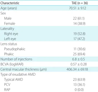
유리체내 애플리버셉트주입술을 치료 및 연장 방법으로 1년 이 상 치료하였던 34명 36안의 평균 연령은 70.51 ± 9.12세였고 남 성이 22안(66.1%), 여성이 14안(38.9%)이었다. TAE군에서 전형적 삼출 나이관련황반변성은 23안(63.9%), 결절맥락막혈관병증이 13안(36.1%), 망막혈관종성증식이 0안(0.0%)이었고 1년간 유리 체내 애플리버셉트 평균 주사 횟수는 6.8 ± 0.5회였다(Table 2).
유리체내 애플리버셉트 주입은 치료 후 유의한 시력 호전 을 보였다. 평균 최대교정시력은 TAE군에서 초기 0.57 ± 0.28 logMAR에서 3개월째 0.41 ± 0.30 logMAR, 12개월째 0.39 ± 0.29 logMAR로 주사 후 통계적으로 유의한 향상을 보였다(p
= 0.021, p = 0.015) (Fig. 3). 12개월째 초기 최대교정시력보다 TAE군에서 평균 0.18 logMAR 시력 상승이 관찰되었다. 최대교 정시력 변화 정도를 분석하였을 때, TAE군에서 유리체내 애플 Table 1. Criteria for intravitreal aflibercept injection interval in the
treat-and-extend regimen Criteria for the injection interval
Criteria for shortening the injection interval Any of following
• The presence of new IRF or SRF on OCT
• The presence of persistent IRF or SRF with increased fluid on OCT
• The presence of persistent IRF with unchanged fluid on OCT
• Loss of BCVA from the previous visit in conjunction with recurrent fluid on OCT
• Increased on CMT measured by OCT
• New neovascularization
• New macular hemorrhage
Criteria for maintaining the injection interval Any of following
• The presence of persistent IRF or SRF with decreased fluid on OCT
• The presence of persistent SRF with unchanged fluid on OCT
And
• No loss of BCVA from the previous visit
• No new neovascularization
• No new macular hemorrhage Criteria for extending the injection interval
• If none of the criteria for shortening were met and there was no fluid on OCT, the injection interval was extended IRF = intraretinal fluid; SRF = subretinal fluid; OCT = optical coher- ence tomography; BCVA = best-corrected visual acuity; CMT = central macular thickness.
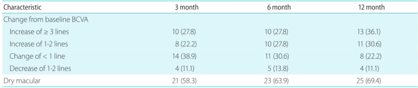
리버셉트 주사 12개월째 최대교정시력이 3줄 이상 호전된 눈은 13안(36.1%)이었으며 시력이 떨어진 눈은 4안(11.1%)이었고 나머 지 19안(52.8%)에서는 2줄 이하의 시력 상승을 보이거나 시력이 유지되었다(Table 3).
중심황반두께는 유리체내 애플리버셉트 주사 후 유의한 호 전을 보였다. TAE군에서 초기 406.34 ± 69.18 μm에서 3개월 째 279.61 ± 78.12 μm, 12개월째 269.58 ± 83.15 μm로 주사 후 통계적으로 유의한 중심황반두께 감소를 보였다(p = 0.011, p = 0.004) (Fig. 3). 마른 황반부 정도는 TAE군에서 3개월째 21안(58.3%), 6개월째 23안(63.9%), 12개월째 25안(69.4%)이었다 (Table 3).
환자군을 유형에 따라 전형적 삼출 나이관련황반변성과 결 절맥락막혈관병증으로 나누어서 분석해 본 결과 전형적 삼출 나이관련황반변성 환자군의 TAE군에서 최대교정시력은 12개 월째 초기 최대교정시력보다 0.15 logMAR 시력 상승 소견을 보 였고 결절맥락막혈관병증 환자군의 TAE군에서 최대교정시력 은 초기 최대교정시력보다 12개월째 0.19 logMAR 시력 상승 소 견을 보였으며 두 군 사이의 최대교정시력 변화 정도는 유의한 차이가 없었다(Fig. 4). 중심황반두께는 전형적 삼출 나이관련 황반변성 환자군의 TAE군에서 초기 중심황반두께보다 5개월 째 115.43 ± 94.26 μm 감소를 보였으며 결절맥락막혈관병증 환 자군의 TAE군에서 초기 중심황반두께보다 5개월째 158.41 ±
89.46 μm 감소로 결절맥락막혈관병증 환자군에서 더 큰 유의한 중심황반두께 감소 소견을 보였다(p = 0.006). 12개월째 중심황 반두께 변화는 전형적 삼출 나이관련황반변성 환자군의 TAE 군에서 132.42 ± 89.67 μm 감소, 결절맥락막혈관병증 환자군의 TAE군에서 144.21 ± 79.24 μm 감소로 결절맥락막혈관병증 환 자군에서 더 큰 감소 소견을 보였으나 5개월째를 제외한 각 시 기의 두 군 사이의 중심황반두께 변화 정도는 통계적으로 유의 한 차이를 보이지 않았다(Fig. 4). 1년간 유리체내 애플리버셉트 평균 주사 횟수는 전형적 삼출 나이관련황반변성 환자군에서 6.7 ± 0.6회, 결절맥락막혈관병증 환자군에서 7.0 ± 0.8회였다.
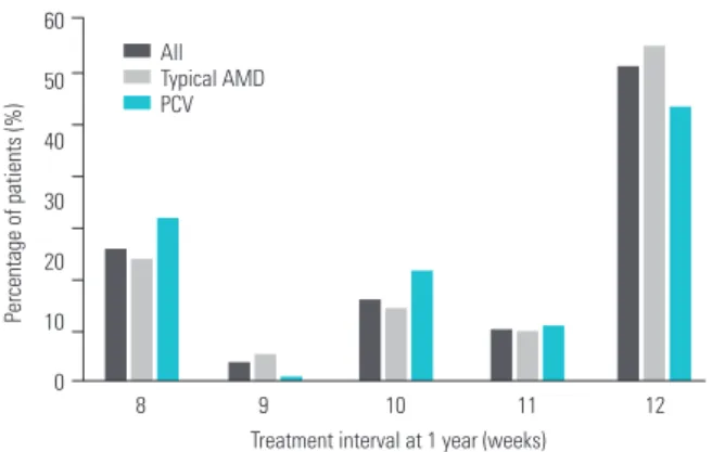
12개월 시점에서 유리체내 애플리버셉트주입술을 치료 및 연 장 방법으로 1년 이상 치료하였던 전체 36안에서 유리체내 애 플리버셉트 주사 간격이 8주인 경우가 8안(22.2%), 12주 간격으 로 늘릴수 있던 경우가 19안(52.8%)이었다. 유형별 TAE군의 전 형적 삼출 나이관련황반변성 환자군에서 유리체내 애플리버셉 트 주사 간격은 8주 간격 5안(20.0%), 12주 간격 14안(56.0%)이 었으며, 결절맥락막혈관병증 환자군에서 유리체내 애플리버셉 트 주사 간격이 8주 간격 3안(27.3%), 12주 간격 5안(45.5%)이었
Figure 3. The mean change in (A) BCVA and (B) central macular thickness (CMT) from baseline to 12 months for all patients with treat- and-extend regimen using aflibercept for exudative age-related macular degeneration. BCVA = best-corrected visual acuity; logMAR
= logarithm of the minimum angle of resolution. *p < 0.05.
Table 2. Patient baseline characteristics and demographics
Characteristic TAE (n = 36)
Age (years) 70.51 ± 9.12
Sex Male Female
22 (61.1) 14 (38.9) Laterality
Right eye 19 (52.8)
Left eye 17 (47.2)
Lens status
Pseudophakic 11 (30.6)
Phakic 25 (69.4)
Number of injections 6.8 ± 0.5
BCVA (logMAR) 0.57 ± 0.28
Central macular thickness (μm) 406.34 ± 69.18 Type of exudative AMD
Typical AMD 23 (63.9)
PCV 13 (36.1)
RAP 0 (0.0)
Values are presented as mean ± standard deviation or number (%).
TAE = treat-and-extend; BCVA = best-corrected visual acuity;
logMAR = logarithm of the minimum angle of resolution; AMD
= age-related macular degeneration; PCV = polypoidal choroidal vasculopathy; RAP = retinal angiomatous proliferation.
Mean logMAR BCVAMean CMT,µm
Follow-up period (months)
Follow-up period (months) 1.0
0.9 0.8 0.7 0.6 0.5 0.4 0.3 0.2 0.1 0
12 6
3 Baseline
600 550 500 450 400 350 300 250 200 150
12 6
3 Baseline
*
*
*
* *
A
B
다(Fig. 5). 12개월간 유리체내 애플리버셉트주입술 시행으로 인 한 혈관색전성질환, 안내염, 망막박리와 같은 심각한 합병증은 발생하지 않았다.
유리체내 애플리버셉트주입술을 치료 및 연장 방법으로 치료 시작하고 1년 이내에 약제를 라니비주맙으로 변경한 눈은 16안 이었고 이를 분석해 보았다. 평균 연령은 71.83 ± 8.94세였으 며 남성이 10안(62.5%), 여성이 6안(37.5%)이었다. 라니비주맙으 로 변경한 군에서 전형적 삼출 나이관련황반변성은 12안(75.0%), 결절맥락막혈관병증이 4안(25.0%)이었다. 라니비주맙으로 약제 를 변경하기 전 애플리버셉트 주사 횟수는 3회 9안, 4회 3안, 5회 2안, 6회 2안이었다. 약제를 변경한 16안 중 라니비주맙 3회 이 상의 주사를 시행한 11안을 대상으로 초기 진단시, 마지막 애 플리버셉트 주사 후, 변경된 라니비주맙으로 3회 치료 후 4주 뒤의 최대교정시력과 중심황반두께를 비교해 보았다. 초기 진 단시, 마지막 애플리버셉트 주사 후, 변경된 라니비주맙으로 3회 치료 후 4주 뒤의 평균 최대교정시력은 각각 0.51 ± 0.30 logMAR, 0.68 ± 0.26 logMAR, 0.61 ± 0.27 logMAR이었다.
각 시점에서 통계적으로 유의한 차이는 관찰되지 않았지만 초 기 진단시와 비교하여 라니비주맙 약제로 변경 후 4안(36.4%)에 서 시력호전을 보였으며 3안(27.3%)에서 시력이 유지되어 총 7안 (63.6%)에서 시력을 보존할 수 있었다. 초기 진단시, 마지막 애플 리버셉트 주사 후, 변경된 라니비주맙으로 3회 치료 후 4주 뒤의 평균 중심황반두께는 각각 394.49 ± 92.31 μm, 415.27 ± 73.63 μm, 289.63 ± 62.78 μm이였으며 마지막 애플리버셉트 주사 후 와 비교하여 라니비주맙 약제 변경후 통계적으로 유의한 중심황 반두께 감소를 보였다(p = 0.009) (Fig. 6).
고찰
혈관내피성장인자는 신생혈관 증식을 유발하는 중요한 역할을 Table 3. Changes in the visual acuity and retinal fluid component of patients with exudative age-related macular degeneration treated with a treat-and-extend regimen using intravitreal aflibercept injections
Characteristic 3 month 6 month 12 month
Change from baseline BCVA
Increase of ≥ 3 lines 10 (27.8) 10 (27.8) 13 (36.1)
Increase of 1-2 lines 8 (22.2) 10 (27.8) 11 (30.6)
Change of < 1 line 14 (38.9) 11 (30.6) 8 (22.2)
Decrease of 1-2 lines 4 (11.1) 5 (13.8) 4 (11.1)
Dry macular 21 (58.3) 23 (63.9) 25 (69.4)
Values are presented as number (%).
BCVA = best-corrected visual acuity.
Figure 4. The mean change in (A) BCVA and (B) CMT from baseline to 12 months for patients with typical neovascular age-related macu- lar degeneration (AMD) and with polypoidal choroidal vasculopathy (PCV). (A) There was no significant difference in BCVA changes be- tween the typical AMD group and PCV group. (B) At month 5, CMT change showed a significantly greater decrease in the PCV group than the typical AMD group. BCVA = best-corrected visual acuity;
logMAR = logarithm of the minimum angle of resolution; CMT = central macular thickness. *p < 0.05.
Mean change from baseline BCVA (logMAR)
Follow-up period (months)
Typical AMD PCV
p = 0.326 p = 0.341 p = 0.813 p = 0.792 p = 0.195 p = 0.429 p = 0.528
3 4 5 6 12
1 2
0.1
0
-0.1
-0.2
-0.3
-0.4
Mean change from baseline CMT, µm
Follow-up period (months)
Typical AMD PCV
p = 0.942 p = 0.818 p = 0.458 p = 0.291 p = 0.006 p = 0.245
*
p = 0.103
3 4 5 6 12
1 2
0 -20 -40 -60 -80 -100 -120 -140 -160 -180
A
B
하는 인자로 밝혀지면서 이에 대한 길항치료인 항혈관내피성장 인자가 나이관련황반변성에서 맥락막신생혈관의 치료에 널리 이용되고 있다[11]. 애플리버셉트는 베바시주맙, 라니비주맙과 달리 혈관내피성장인자 A (vascular endothelial growth factor A)뿐 아니라 혈관내피성장인자 B (vascular endothelial growth factor B)와 태반성장인자(placental growth factor)에도 결합하 여 더 높은 결합 친화도를 보인다[12-14]. Zhang et al. [15]은 나 이관련황반변성 환자에서 애플리버셉트가 라니비주맙에 비해 좋은 효과를 보였음을 보고하였고, Kim et al. [16]은 베바시주 맙과 라니비주맙에 반응하지 않은 나이관련황반변성 환자에서 중심망막두께가 유의하게 감소하는 것을 보고하였다.
애플리버셉트는 초기 4주마다 3회의 로딩 후 일반적으로 8주 간격으로 주기적으로 유리체내 주입술을 시행한다. 하지만 나
이관련황반변성은 만성적인 경과로 장기적인 치료와 관리가 필 요하고 이에 임상적으로 일정 간격으로 유리체내 주입술을 시 행하는 치료방법은 환자와 치료자 모두에게 비용, 빈번한 외래 내원, 시간, 주사치료에 따른 부작용 발생 가능성과 같은 부담 감이 따를 수 있게 된다. 따라서 실제적으로 많은 임상의사들은 treat-and-extend 방법이나 PRN 방법과 같이 치료에 따른 부담 감을 덜 수 있는 치료법을 택하기도 한다[17].이러한 treat-and- extend 치료는 혈관내피성장인자 A의 억제 기간이 환자들마다 다를 수 있다는 원리에 기반하여 가능할 수 있다[18,19]. 따라서 환자의 주입술 간의 최대 주사 허용 간격이 달라지고 횟수에도 환자의 상태에 따라 달라질 수 있는 것이다.
Treat-and-extend 방법의 가장 큰 장점은 PRN 방법에 비하여 방문 횟수를 줄이면서 Fixed dosing 방법과 유사한 만족할만한 시력 결과를 얻을 수 있다는 점이다. Fixed dosing 방법처럼 일 정한 간격을 두고 지속적으로 주사하여 고가의 약제를 자주 주 입하는 것에 뒤따르는 의료비 상승의 문제를 개선할 수 있고, 매달 경과 관찰하고 재발하는 경우에만 치료하는 방식인 PRN 방법에서 발생하는 매달 방문에 따른 경제적 부담과 경과 관찰 이 되지 않을 경우 예후가 좋지 못할 가능성에 대한 부분을 감 소시킬 수 있다.
이러한 treat-and-extend 치료 방법의 효용성에 대하여 외국 의 연구들이 진행되고 있고 애플리버셉트를 대상으로 이루어 진 연구들의 임상 특징을 Table 4에 정리해 보았다[7,10,20-22].
이와 같은 연구들은 유리체내 애플리버셉트주입술 treat-and- extend 치료로 기능적 및 해부학적 호전이 되는 것을 보여준다.
ALTAIR 연구[20]를 포함한 대부분의 연구들에서 최대교정시 력의 호전과 중심망막두께의 감소를 보였으며, 12주 간격 이상 주사 간격을 늘릴 수 있었던 환자는 Ohji et al. [20]의 연구에서 42.3% (IVT-AFL-2W), 49.6% (IVT-AFL-4W), Gillies et al. [21]
에서 16.0%, Decroos et al. [10]의 연구에서 35.0%, Yamamoto et al. [22]에서 17.9%였다.
본 연구에서 나이관련황반변성에서 treat-and-extend 방법으 로 유리체내 애플리버셉트주입술을 시행하면서 1년 동안 평균 6.8회의 주사를 시행하였으며 평균 0.18 logMAR의 시력 상승을 보였다. 중심황반두께는 주사 12개월 후 유의한 두께 감소를 유 지할 수 있었으며 많은 환자들에서 마른 황반부를 보존할 수 있 었다. 본 연구에서는 주사치료 간격을 12주 이상 늘릴수 있었던 환자군은 19안(52.8%)으로 다소 높은 경향을 보였다. 이는 후향 적 연구로 담당의사의 판단에 따라 본 연구의 treat-and-extend 주사 간격을 2주씩 늘리는 것을 원칙으로 하되 환자 사정에 따 라 최대 3주까지 간격이 연장되는 것을 허용하였고 애플리버셉 트 치료 도중 라니비주맙으로 변경된 환자들을 제외하고 분석 하였기에 결과에 영향을 미쳤을 것으로 생각된다.
Park et al. [23]의 국내의 나이관련황반변성에 대한 기초역학 조사에 따르면 전형적 삼출 나이관련황반변성이 62.1%, 결절맥 Figure 5. Distribution of the treatment intervals at 1 year in treat-
and-extend groups. AMD = age-related macular degeneration; PCV
= polypoidal choroidal vasculopathy.
Treatment interval at 1 year (weeks)
Percentage of patients (%)
AllTypical AMD PCV 60
50
40
30
20 10
0 8 9 10 11 12
Figure 6. Mean change in best-corrected visual acuity and central macular thickness at baseline, after last aflibercept injection(A), and after switching to three injections of ranibizumab(R). CMT = central macular thickness; BCVA = best-corrected visual acuity; logMAR = logarithm of the minimum angle of resolution. *p < 0.05.
Mean CMT,µm Mean BCVA (logMAR)
900 800 700 600 500 400 300 200
0.2 0.4 0.6 0.8 1.0 1.2 1.4 Initial After injection(A) After injection(R) 1.6
CMT BCVA
*
락막혈관병증이 31.7%, 망막혈관종성증식이 6.2%로 분포한다 고 보고하고 있다. 본 연구에서도 전형적 삼출 나이관련황반변 성 환자가 63.9%, 결절맥락막혈관병증 환자의 비율이 36.1%로 국내의 보고와 비슷한 분포를 보였다. 본 연구에서 질환의 유형 에 따른 시력 변화는 12개월 경과 관찰 기간 동안 전형적 삼출 나이관련황반변성 환자에서 0.15 logMAR 시력 상승, 결절맥락 막혈관병증 환자에서 0.19 logMAR 시력 상승을 보였고 중심황 반두께는 경과 관찰 기간 동안 전형적 삼출 나이관련황반변성 환자에서 132.4 μm, 결절맥락막혈관병증 환자에서 144.8 μm 감 소를 보였다. 5개월 시점의 중심황반두께를 제외하고 두 군 간 의 통계적 유의성을 보이지는 않았지만 결절맥락막혈관병증에 서 전형적 삼출 나이관련황반변성에 비하여 기능적 및 해부학 적 호전이 더 큰 경향을 보였다. 애플리버셉트는 결절맥락막혈 관병증에서 결절 병변과 망막색소상피박리를 소실시키고 다른 항혈관내피성장인자에 비하여 더 강한 효과를 보이는 경우가 있는 것으로 알려져 있는데 본 연구에서도 결절맥락막혈관병증 환자에서의 치료 효과는 애플리버셉트의 유용성을 뒷받침해 줄 수 있을 것으로 생각된다[24].
본 연구에서 애플리버셉트에서 라니비주맙으로 약제를 변경 한 환자들을 분석한 결과 56.3% (9안)가 라니비주맙으로 변경 전 애플리버셉트 주사 횟수가 3회로, 초기에 약제 변경을 시행 하는 경향을 보였다. 약제 변경의 효과를 분석한 결과 초기 진 단시 시력과 라니비주맙 3회 주사 후 4주 뒤의 시력 비교에서 통 계적으로 유의한 차이를 보이지는 않았으나 63.6% (7안)에서 시 력을 보존할 수 있었으며, 해부학적으로는 마지막 애플리버셉트 주사 후와 비교하여 라니비주맙 약제 변경 후 통계적으로 유의 한 중심황반두께 감소를 보임을 알 수 있었다.
라니비주맙에서 애플리버셉트으로 변경한 경우 구조적 반응 의 향상을 보였다는 다수의 보고들에 비해 애플리버셉트에서 라니비주맙으로 약제 변경에 대한 치료 효과에 대한 연구들은 아직까지는 드물게 보고하고 있다[25,26]. Marquis and Mantel
[27]은 애플리버셉트에서 라니비주맙 약제 변경 후 시력과 중심 망막두께의 유의한 차이는 없었지만 망막색소상피박리의 높이 와 망막하액 높이의 구조적 변화에 유의한 감소를 보임을 보고 하였고, Despreaux et al. [28]의 연구에서는 과거 라니비주맙에 서 애플리버셉트로 변경하였던 환자에서 다시 라니비주맙으로 재변경 후 유의한 시력 호전의 결과를 보였고 중심망막두께는 라니비주맙에서 애플리버셉트로 변경 후 시점과 애플리버셉트 에서 라니비주맙으로 재변경 후 시점의 비교에는 유의한 차이 를 보이지는 않았지만 최초 약제 변경 이전 초기 시점과 애플 리버셉트에서 라니비주맙으로 재변경 후 시점을 비교하였을때 중심망막두께의 유의한 감소를 보였다. 애플리버셉트에서 라니 비주맙으로 변경한 경우에 대한 대부분의 연구들은 과거 라니 비주맙에서 애플리버셉트로 변경 후 다시 이전 약제인 라니비 주맙으로 돌아가는 환자들을 연구 대상에 포함하고 있지만 본 연구는 초기 애플리버셉트에서 라니비주맙으로 변경한 환자들 을 대상으로 연구가 시행되었다는 점에서 의의가 있으며 단기간 의 결과 해부학적 호전과 시력 안정화에 도움을 줄 수 있었다.
본 연구에는 다음과 같은 제한점이 있다. 본 연구는 첫째, 의 무기록을 후향적으로 분석한 연구로 전향적 연구에서처럼 환자 마다의 질병의 경중에 따른 여러 종류의 항혈관내피성장인자 약제 선택의 엄격한 기준을 적용하지 못하였고 둘째, 1년 이상 관찰 가능하였던 환자들만을 대상으로 하였으며 셋째, 애플리 버셉트에 효과가 적다고 생각되어 라니비주맙으로 약제를 변경 한 경우를 라니비주맙 주입군으로 분류하였다. 이는 본 연구 결 과가 타 연구 결과와 비교했을 때 총 주사 횟수가 적고 12주의 최대 주사 간격을 유지하는 비율이 높게 나타나는 것의 한 원인 으로 작용했을 가능성이 있어 이를 염두에 두고 결과를 해석해 야 할 것이다. 또한 전체 대상안이 적고 아류형별 분석시 비교 적 표본수가 적다는 점, 애플리버셉트에서 라니비주맙으로 약 제 변경 후 단기간의 치료 결과를 분석하였으나 장기간의 예후 를 보여주지 못한 한계점이 있다. 이러한 한계점에도 불구하고 Table 4. Review of treat-and-extend regimens of intravitreal aflibercept injection in exudative age-related macular degeneration at 1 year
Study N Mean BCVA
gain
Mean CMT change (μm)
Mean injections
≥ 12 weeks injection interval (%)
Ohji et al. [20] (2020) 123 (IVT-AFL-2W) 9.0 letters (ETDRS) -130 7.2 42.3
123 (IVT-AFL-4W) 8.4 letters (ETDRS) -126 6.9 49.6
Gillies et al. [21] (2019) 281 4.9 letters (ETDRS) 9.7 16.0
Haga et al. [7] (2018) 41 -0.32 logMAR -161 7.5
Decroos et al. [10] (2017) 31 7.2 letters (ETDRS) -143 8.0 35.0
Yamamoto et al. [22] (2017) 67 -0.15 logMAR -194 8.3 17.9
Our study 36 -0.18 logMAR -136 6.8 52.8
BCVA = best-corrected visual acuity; CMT = central macular thickness; IVT-AFL = intravitreal aflibercept; 2W = 2 weeks adjustment; ETDRS
= early treatment diabetic retinopathy study letter score; 4W = 4 weeks adjustment; log MAR = logarithm of the minimal angle of resolu- tion.
실제 의료 현장에서 삼출 나이관련황반변성으로 진단받고 최초 치료로 유리체내 애플리버셉트주입술을 시작한 환자들의 임상 적 결과를 분석하고 치료 도중 라니비주맙으로 약제를 변경하 였던 환자들의 단기간의 치료 효과를 분석한 보고로서 본 연구 의 의의가 있다고 생각된다.
결론적으로 유리체내 애플리버셉트주입술 treat-and-extend 방법 치료는 실제 임상에서 삼출 나이관련황반변성에서 기능 적 및 구조적 개선을 보이며 방문 및 주사 회수를 최소화하여 치료 순응도를 높일 수 있는 효과적인 방법임을 알 수 있었다.
또한 애플리버셉트 치료 도중 치료에 대한 반응이 제한적인 환 자에서 라니비주맙으로 약제를 변경하는 것은 단기적인 해부학 적 호전과 시력보존을 유지하는 데 있어 대안 치료제로 유용하 며 치료 계획 수립에 도움을 줄 수 있을 것으로 생각한다. 향 후 유리체내 애플리버셉트주입술의 treat-and-extend를 하지 못 한 환자군의 비교에 대한 추가적인 연구가 필요할 것으로 보이 며 추후 장기적인 치료 경과에 대한 추가 연구가 의미가 있을 것으로 생각된다.
References
1. Congdon N, O’Colmain B, Klaver CC, et al. Causes and preva- lence of visual impairment among adults in the United States.
Arch Ophthalmol 2004;122:477-85.
2. Park SJ, Lee JH, Woo SJ, et al. Age-related macular degeneration:
prevalence and risk factors from Korean National Health and Nutrition Examination Survey, 2008 through 2011. Ophthalmol- ogy 2014;121:1756-65.
3. Rofagha S, Bhisitkul RB, Boyer DS, et al. Seven-year outcomes in ranibizumab-treated patients in ANCHOR, MARINA, and HORI- ZON: a multicenter cohort study (SEVEN-UP). Ophthalmology 2013;120:2292-9.
4. Heier JS, Brown DM, Chong V, et al. Intravitreal aflibercept (VEGF trap-eye) in wet age-related macular degeneration. Ophthal- mology 2012;119:2537-48.
5. Muftuoglu IK, Arcinue CA, Tsai FF, et al. Long-term results of pro re nata regimen of aflibercept treatment in persistent neo- vascular age-related macular degeneration. Am J Ophthalmol 2016;167:1-9.
6. Hufendiek K, Pielen A, Framme C. Strategies of intravitreal injec- tions with anti-VEGF: “Pro re nata versus treat and extend”. Klin Monbl Augenheilkd 2018;235:930-9.
7. Haga A, Kawaji T, Ideta R, et al. Treat-and-extend versus ev- ery-other-month regimens with aflibercept in age-related mac- ular degeneration. Acta Ophthalmol 2018;96:e393-8.
8. Figueras-Roca M, Parrado-Carrillo A, Nguyen V, et al. Treat-and- extend versus fixed bimonthly treatment regimens for treat- ment-naive neovascular age-related macular degeneration: real world data from the Fight Retinal Blindness registry. Graefes Arch Clin Exp Ophthalmol 2021;259:1463-70.
9. Ito A, Matsumoto H, Morimoto M, et al. Two-year outcomes of a treat-and-extend regimen using intravitreal aflibercept injec- tions for typical age-related macular degeneration. Ophthalmo- logica 2017;238:236-42.
10. Decroos FC, Reed D, Adam MK, et al. Treat-and-extend therapy using aflibercept for neovascular age-related macular degenera- tion: a prospective clinical trial. Am J Ophthalmol 2017;180:142-50.
11. Ferrara N. Vascular endothelial growth factor: basic science and clinical progress. Endocr Rev 2004;25:581-611.
12. Papadopoulos N, Martin J, Ruan Q, et al. Binding and neutral- ization of vascular endothelial growth factor (VEGF) and related ligands by VEGF Trap, ranibizumab and bevacizumab. Angio- genesis 2012;15:171-85.
13. Stewart MW, Rosenfeld PJ. Predicted biological activity of intrav- itreal VEGF Trap. Br J Ophthalmol 2008;92:667-8.
14. Rudge JS, Thurston G, Davis S, et al. VEGF trap as a novel antian- giogenic treatment currently in clinical trials for cancer and eye diseases, and VelociGene- based discovery of the next genera- tion of angiogenesis targets. Cold Spring Harb Symp Quant Biol 2005;70:411-8.
15. Zhang Y, Chioreso C, Schweizer ML, Abràmoff MD. Effects of aflibercept for neovascular age-related macular degeneration: a systematic review and meta-analysis of observational compara- tive studies. Invest Ophthalmol Vis Sci 2017;58:5616-27.
16. Kim JH, Cho NC, Kim WJ. Intravitreal aflibercept for neovascular age-related macular degeneration resistant to bevacizumab and ranibizumab. J Korean Ophthalmol Soc 2015;56:1359-64.
17. Lanzetta P, Loewenstein A; Vision Academy Steering Commit- tee. Fundamental principles of an anti-VEGF treatment regimen:
optimal application of intravitreal anti-vascular endothelial growth factor therapy of macular diseases. Graefes Arch Clin Exp Ophthalmol 2017;255:1259-73.
18. Fauser S, Muether PS. Clinical correlation to differences in ran- ibizumab and aflibercept vascular endothelial growth factor suppression times. Br J Ophthalmol 2016;100:1494-8.
19. Fauser S, Schwabecker V, Muether PS. Suppression of intraocular vascular endothelial growth factor during aflibercept treat- ment of age-related macular degeneration. Am J Ophthalmol 2014;158:532-6.
20. Ohji M, Takahashi K, Okada AA, et al. Efficacy and safety of intra-
vitreal aflibercept treat-and-extend regimens in exudative age- related macular degeneration: 52- and 96-week findings from ALTAIR : a randomized controlled trial. Adv Ther 2020;37:1173-87.
21. Gillies MC, Hunyor AP, Arnold JJ, et al. Effect of ranibizumab and aflibercept on best-corrected visual acuity in treat-and-extend for neovascular age-related macular degeneration: a random- ized clinical trial. JAMA Ophthalmol 2019;137:372-9.
22. Yamamoto A, Okada AA, Nakayama M, et al. One-year out- comes of a treat-and-extend regimen of aflibercept for exu- dative age-related macular degeneration. Ophthalmologica 2017;237:139-44.
23. Park KH, Song SJ, Lee WK, et al. The results of nation-wide reg- istry of age-related macular degeneration in Korea. J Korean Ophthalmol Soc 2010;51:516-23.
24. Cho HJ, Kim KM, Kim HS, et al. Intravitreal aflibercept and ranibi- zumab injections for polypoidal choroidal vasculopathy. Am J Ophthalmol 2016;165:1-6.
25. Kumar N, Marsiglia M, Mrejen S, et al. Visual and anatomical outcomes of intravitreal aflibercept in eyes with persistent sub- foveal fluid despite previous treatments with ranibizumab in patients with neovascular age-related macular degeneration.
Retina 2013;33:1605-12.
26. Slean GR, Hemarat K, Khurana RN, Stewart JM. Conversion back to bevacizumab or ranibizumab for recurrent neovascular activ- ity with aflibercept in age-related macular degeneration: a case series. Int J Retina Vitreous 2016;2:2.
27. Marquis LM, Mantel I. Beneficial switch from aflibercept to ranibizumab for the treatment of refractory neovascular age-re- lated macular degeneration. Graefes Arch Clin Exp Ophthalmol 2020;258:1591-6.
28. Despreaux R, Cohen SY, Semoun O, et al. Short-term results of switchback from aflibercept to ranibizumab in neovascular age-related macular degeneration in clinical practice. Graefes Arch Clin Exp Ophthalmol 2016;254:639-44.
처음 진단된 삼출 나이관련황반변성에서 애플리버셉트 치료의 임상적 결과
목적: 처음 진단된 삼출 나이관련황반변성에서 유리체내 애플리버셉트 치료 및 연장 방법의 1년 결과와 같은 기간 내 라니비주맙으로 변 경한 경우의 임상 양상의 변화를 알아보고자 하였다.
대상과 방법: 삼출 나이관련황반변성으로 처음 진단되고 유리체내 애플리버셉트주입술을 치료 및 연장 방법으로 치료하였던 36안과 같은 기간 내 효과가 부족하여 라니비주맙으로 변경하였던 16안을 대상으로 최대교정시력 변화, 중심황반두께 변화, 주사 횟수를 분석 하였다
결과: 애플리버셉트 치료 및 연장 방법으로 치료한 36안은 평균 최대교정시력 초기 0.57 ± 0.28 logarithm of the minimum angle of resolution (logMAR)에서 12개월째 0.39 ± 0.29 logMAR로 유의한 시력 호전을 보였으며(p = 0.015), 평균 중심황반두께는 초 기 406.34 ± 69.18 μm에서 12개월째 269.58 ± 83.15 μm로 유의한 중심황반두께 감소를 보였다(p = 0.004). 1년간 평균 주사 횟 수는 6.8 ± 0.5회였다. 약제를 변경한 16안 중 라니비주맙 3회 이상 주사를 시행한 11안에서 초기 시력이 호전되거나 유지할 수 있었 던 경우는 7안(63.6%)이었으며 평균 중심황반두께는 약제 전환 전 415.27 ± 73.63 μm에서 전환 후 289.63 ± 62.78 μm로 유의 한 감소를 보였다(p = 0.009).
결론: 삼출 나이관련황반변성에서 유리체내 애플리버셉트 치료 및 연장 방법은 실제 임상에서 효과적이었고, 또한 애플리버셉트에 효 과가 부족하여 라니비주맙으로 변경한 경우 해부학적 호전을 보였으며 시력 안정화에 도움을 줄 수 있었다.
국문초록