Usefulness of Artificial Jump Graft to Portal Vein Thrombosis in Deceased Donor Liver Transplantation
Hong Pil Hwang,
1,2Jae Do Yang,
1,2Sang In Bae,
1,2Si Eun Hwang,
3Baik Hwan Cho,
1,2,4and Hee Chul Yu
1,2,41Department of Surgery, Chonbuk National University Medical School and Hospital, Jeonju;
2Biomedical Research Institute, Chonbuk National University Hospital, Jeonju;
3Department of Surgery, Daejeon Sun Hospital, Daejeon;
4Research Institute of Clinical Medicine, Chonbuk National University, Jeonju, Korea.
Received: July 17, 2014 Revised: November 12, 2014 Accepted: December 10, 2014 Corresponding author: Dr. Hee Chul Yu, Department of Surgery, Chonbuk National University Medical School and Hospital, 20 Geonji-ro, Deokjin-gu,
Jeonju 561-712, Korea.
Tel: 82-63-250-1576, Fax: 82-63-271-6197 E-mail: [email protected]
∙ The authors have no financial conflicts of interest.
© Copyright:
Yonsei University College of Medicine 2015 This is an Open Access article distributed under the terms of the Creative Commons Attribution Non- Commercial License (http://creativecommons.org/
licenses/by-nc/3.0) which permits unrestricted non- commercial use, distribution, and reproduction in any medium, provided the original work is properly cited.
Severe portal vein thrombosis (PVT) is often considered a relative contraindica- tion for living donor liver transplantation due to high associated risks and morbidi- ty. Meanwhile, improvement in operative techniques, resulting in higher success rates has removed PVT from the list of contraindications in deceased donor liver transplantation (DDLT). In this report, we describe a surgical technique for DDLT using polytetrafluoroethylene graft from the inferior mesenteric vein for portal in- flow in patient with portomesenteric thrombosis.
Key Words: Liver transplantation, portal vein thrombosis, artificial graft, inferior mesenteric vein, polytetrafluoroethylene
INTRODUCTION
Portal vein thrombosis (PVT) is a common complication of end-stage liver disease with an incidence of 0.6‒16% in patients with well-compensated disease, increas- ing up to 35% in cirrhotic patients with hepatocellular carcinoma.1 As recently as the 1990s, PVT was associated with an increased incidence of complications and a higher mortality rate.2 Additionally, blood loss volumes in patients with complete thrombosis were reported to be significantly higher than those in patients without PVT or with partial PVT.3 Nevertheless, the presence of PVT is no longer consid- ered an absolute contraindication for liver transplantation, as many improvements have been made in surgical technique and perioperative management.2 In this re- port, we describe using polytetrafluoroethylene (PTFE) jump graft from the inferi- or mesenteric vein (IMV) for portal inflow in a patient with severe portomesenter- ic venous thrombosis.
CASE REPORT
A 64-year-old male patient was admitted to our hospital for deceased donor liver
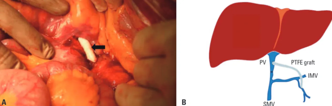
enteric vein. However, the peripancreatic area showed se- vere adhesion with superior mesenteric vessels. We had no choice except to perform a bypass from the IMV to the por- tal vein using PTFE (diameter: 7 mm) graft since the length of the iliac vessel of the donor was not sufficient for bypass and cryopreserved vein. After IMV was exposed below the ligament of Treitz, end_ to_ side anastomosis was performed using polypropylene 6-0 continuous running sutures be- tween PTFE with IMV and end_ to_ side anastomosis be- tween PTFE and the graft’s portal vein (Fig. 2). The PTFE graft was passed through the mesentery above the pancreas, and anastomosis was performed in the same pattern be- tween PTFE with distal portal vein. Intraoperative Doppler assessment after bypass showed a mean portal vein velocity 48.4 cm/second. We performed gauze packing with exter- nalization at the mesentery of the pancreatic head area be- cause of uncontrolled oozing-like bleeding. The weight of the graft was 1799 gram, the cold ischemic time was 318 minutes, the warm ischemic time was 40 minutes, and the anhepatic time was 194 minutes. Intraoperative transfu- sions were performed with 50 packs of packed red blood cells, 20 packs of fresh frozen plasma, and 20 packs of platelet concentrate. The total operative time was 14 hours and 5 minutes.
On the first postoperative day, portal vein Doppler ultra- sound revealed normal waveform (mean flow velocity of 34.9 cm/second) without thrombus, and the vital signs and laboratory findings were relatively stable. The patient un- derwent general anesthesia again on the 16th day after transplantation for perihepatic hematoma removal, but the portal vein and the PTFE graft were intact.
After the transplant operation, the amount of ascites was 500‒1500 cc/day until post-transplant 3 weeks, and we com- pensated daily ascites by 5% albumin and Hartmann solu- transplantation. The patient’s previous medical history re-
vealed alcoholic liver cirrhosis and diabetes mellitus. Also, he had undergone transarterial chemoembolization with adriamycin three times in the past because of hepatocellular carcinoma in segment 8 with minimal portal vein thrombo- sis, as well as balloon occluded retrograde transvenous obliteration and/or endoscopic variceal band ligation six times, because of gastric and esophageal variceal bleeding.
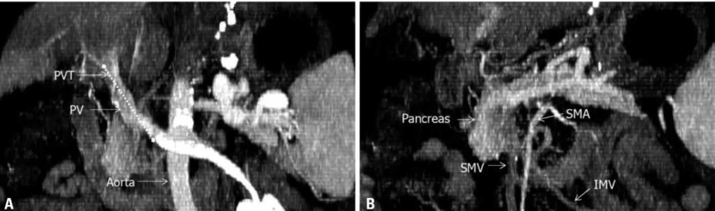
At the time of admission, his Child-Turcotte-Pugh score was 10 points (grade C), and his model for end-stage liver disease score was 9.0 points. Preoperative computed to- mography revealed portomesenteric thrombosis from the portal vein to the proximal superior mesenteric vein with complete luminal obstruction, and venous collateral flow was not prominent (Fig. 1). The segment of portomesenter- ic thrombus was about 6.8 cm and the IMV had no throm- bosis. The donor was a 56-year-old male who had brain death due to traumatic cerebral hemorrhage.
During exploration for recipient hepatectomy, portal ve- nous flow was not detected by intraoperative doppler ultra- sound and venous collateral flow was not prominent. Me- chanical thrombectomy and low dissection of the portal vein was performed, and the portal venous inflow was im- proved. After the liver graft was placed in the recipient’s abdomen, the donor’s inferior vena cava was anastomosed to the recipient’s inferior vena cava by applying the piggy- back technique. Then, the portal vein of the recipient was anastomosed to the graft portal vein in an end_ to_ end fash- ion. Although the graft was recovered from ischemia after reperfusion, portal blood flow decreased gradually over a few minutes. Although repeated mechanical thrombectomy with portal venotomy was executed, the portal inflow did not improve. Thus we attempted to search for other area for portal inflow along more distal regions of the superior mes-
Fig. 1. Portal venous thrombosis is shown in preoperative computed tomography (A and B, maximum intensity projection of serial coronal view). Thrombosis was extended to the proximal superior mesenteric vein (dot line; length, 6.8 cm) for a Yedel classification Grade 3. PV, portal vein; PVT, portal vein thrombo- sis; SMV, superior mesenteric vein; SMA, superior mesenteric artery; IMV, inferior mesenteric vein.
A B
flow without thrombus on the 7th day and 27 months after liver transplantation (Fig. 3). Now, he is healthy without transplant-related complication, although he still sometimes reports paralytic ileus.
DISCUSSION
PVT is caused by decreased portal flow from progressive liver cirrhosis and the development of periportal lymphangi- tis and fibrosis.1 PVT is more frequent in patients with auto- immune, cryptogenic, and alcoholic cirrhosis than in patients with hepatitis C.2 The pathophysiology of PVT is complex, but related to liver cirrhosis, which causes the elevation of portal pressure in relation to endothelial injury and thrombus formation.4 Enhanced coagulability as a result of decreased levels of natural anticoagulants, such as protein C, protein S, and antithrombin III, as well as coagulation factors are also observed in patients with PVT.1
Yerdel, et al.5 classified PVT into four grades according to its extent and the severity of luminal occlusion by the throm- tion. The patient needed continuous renal replacement thera-
py for oliguria for 3 days, and we transported the patient to a general ward at post-transplant 10 days. After hematoma re- moval at post-transplant 16 days, the patient was admitted again to the surgical intensive care unit. At this time, the pa- tient’s mental status was deep drowsy for 3 weeks, and we performed tracheostomy at post-transplant 27 days for con- tinuous ventilation. Brain magnetic resonance imaging re- vealed an acute ischemic lesion in the mid pons. At post- transplant 56 days, the patient was transported to a general ward; however, his lower extremities revealed muscle atro- phy, which required rehabilitation for about 4 months. The patient stayed in the intensive care unit for 50 days, and the total hospital stay was 179 days. The patient was managed with prostaglandin E1 (0.01 lg/kg/h) and a protease inhibi- tor (mesilate gabexate; 1 mg/kg/h) for 7 days after transplan- tation. Immunosuppressive agents included a triple therapy of calcineurin inhibitor (tacrolimus), mycophenolate mofetil, and steroid. Anti-platelet agent (aspirin 100 mg/day) was prescribed at post-transplant 30 days, and is still being used.
Follow-up computed tomography showed normal portal
Fig. 3. Postoperative computed tomography at post operative 7 days (A) and 27 months (B) shows that the polytetrafluoroethylene graft has no thrombosis and no anastomotic stricture. PTFE, polytetrafluoroethylene; IMV, inferior mesenteric vein.
Fig. 2. After thrombectomy and lower dissection, portal vein anastomosis with polytetrafluoroethylene (PTFE; arrow) vascular artificial graft was performed with continuous running suture using polypropylene (A). Since massive bleeding and adhesion precluded the possibility of distal superior mesenteric vein interposition, we performed anastomosis between the proximal inferior mesenteric vein and distal portal vein using PTFE (B; schematic representation). PV, portal vein; SMV, superior mesenteric vein; IMV, inferior mesenteric vein.
A A
B B
PV
SMV
IMV PTFE graft
The use of a temporary porto-caval shunt is associated with better hemodynamic stability and improved renal func- tion, as well as decreased transfusion requirements in piggy back liver transplantation.10,11 In our case, we performed by- pass from the IMV to the graft portal vein after reperfusion of the liver graft. In fact, preoperative CT scan did not clear- ly show the range of portomesenteric thrombosis. More- over, we thought that first mechanical thrombectomy was done successfully because of sufficient portal back flow. If the porto-caval shunt was performed before portal recon- struction and reperfusion, the patient may have shown bet- ter progression during hospitalization.
There are only a few reports associated with IMV for portal vein inflow. Kobayashi, et al. reported on IMV to the left gonadal vein shunt for gastroesophageal varices and ex- trahepatic PVT after liver transplantation.12 In our case, the SMV was difficult to use for portal vein reconstruction be- cause of severe adhesions and friable vessel walls. After the successful portal vein reconstruction using the IMV with PTFE, the patient had no vascular problems for 27 months.
In conclusion, PTFE jump graft from the IMV to the por- tal vein may be a feasible option in patients with portomes- enteric thrombosis, and the IMV can be a potential source of portal inflow in liver transplants.
REFERENCES
1. Kim SJ, Kim DG, Park JH, Moon IS, Lee MD, Kim JI, et al. Clin- ical analysis of living donor liver transplantation in patients with portal vein thrombosis. Clin Transplant 2011;25:111-8.
2. Lladó L, Fabregat J, Castellote J, Ramos E, Torras J, Jorba R, et al.
Management of portal vein thrombosis in liver transplantation: in- fluence on morbidity and mortality. Clin Transplant 2007;21:716- 3. Iyer SG, Lau CL, Chang KY, Mak SW, Madhavan KK. Success-21.
ful living donor liver transplantation in portomesenteric thrombo- sis. Am J Transplant 2010;10:1483-5.
4. Wu TH, Lin YS, Lee CF, Wu TJ, Yu MC, Chan KM, et al. Clinical analysis and strategy for liver transplantation in patients with pre- existing portal vein thrombosis. Chang Gung Med J 2011;34:426- 34.
5. Yerdel MA, Gunson B, Mirza D, Karayalçin K, Olliff S, Buckels J, et al. Portal vein thrombosis in adults undergoing liver trans- plantation: risk factors, screening, management, and outcome.
Transplantation 2000;69:1873-81.
6. Sugawara Y, Makuuchi M, Tamura S, Matsui Y, Kaneko J, Hasegawa K, et al. Portal vein reconstruction in adult living donor liver transplantation using cryopreserved vein grafts. Liver Transpl 2006;12:1233-6.
7. Hwang S, Jung DH, Ha TY, Ahn CS, Moon DB, Kim KH, et al.
Usability of ringed polytetrafluoroethylene grafts for middle he-
bus. Our case, exhibited grade 3 PVT, showing complete thrombosis on both the PV and the proximal superior mes- enteric vein (SMV).
Although an optimal technique for managing PVT dur- ing liver transplantation does not exist, the most preferred method of portal vein reconstruction is eversion thrombecto- my, especially in grade 1 or 2 PVT.1,2,5,6 Grade 2 and 3 PVT can be managed with either thrombectomy or anastomosis to the splenomesenteric confluence with or without graft, or anastomosis onto a varix, depending on the anatomy and thrombus severity, as well as extent in the patient.2 Lladó, et al. recommend a venous graft or anastomosis to a collateral vein for the patients with grade 3 thrombosis and involve- ment of the SMV.2,5 Especially, a jump venous graft from the SMV is indicated only when the portal flow can be re- stored in cases of extensive PVT or when there is no suit- able engorged collateral coronary vein available.4 However, prosthetic vessel grafts are the only choice if a venous con- duit is not possible. The indications for prosthetic vessel grafts for the splanchnic venous system have been limited because the blood flow in this system is relatively slow, that is, highly thrombogenic. Moreover, little clinical experience with the use of PTFE grafts in the splanchnic venous sys- tem has been accumulated.7 PTFE seems to be a good alter- native for outflow venoplasty, when autologous or allogeneic vascular patch is not available or when in critical situations.8 The hematological condition of patients undergoing liver transplantation is less thrombogenic than that in canine mod- els, as shown in the impairment of coagulation profiles, such as moderate-to-severe thrombocytopenia, the low viscosity from lowered hematocrits, and the concurrent use of anti- platelet agents.7 Luminal thrombus formation is uncommon when a prosthetic graft is used as a conduit for large arter- ies, such as the inferior vena cava, or even the portal vein.
These vessels can be classified as high-flow vessels regard- less of the pressure, so that there is a low risk of luminal thrombus formation.7 Another problem with the use of PTFE is infection. Pomposelli, et al.9 reported no infection com- plications cases when PTFE was used for venous recon- struction. Due to the inert nature of PTFE, the result of any infection is likely to be due to early conduit thrombosis and eventual encapsulation of the graft. Therefore, decisions re- garding the type of conduit can be made based on availabil- ity of resources and preference of the surgeon. Moreover, one advantage of the synthetic graft material is reduced cost, which is approximately 10% less than that of cryopreserved vessels.9
shunt in orthotopic liver transplantation: a single center analysis.
Transpl Int 2011;24:243-50.
11. Pratschke S, Meimarakis G, Bruns CJ, Kaspar M, Prix N, Zacho- val R, et al. Temporary intraoperative porto-caval shunt: useless or beneficial in piggy back liver transplantation? Transpl Int 2013;
26:90-8.
12. Kobayashi T, Sato Y, Yamamoto S, Oya H, Kokai H, Hatakeyama K. The inferior mesenteric vein to the left gonadal vein shunt for gastroesophageal varices and extrahepatic portal vein thrombosis after living donor liver transplantation: a case report. Transplant Proc 2012;44:591-3.
patic vein reconstruction during living donor liver transplantation.
Liver Transpl 2012;18:955-65.
8. Chen TH, Jeng LB, Lee CC, Li PC, Huang JC, Hsu CH, et al.
Polytetrafluoroethylene patch venoplasty for outflow reconstruc- tion in living donor liver transplantation: two case reports. Trans- plant Proc 2008;40:2529-30.
9. Pomposelli JJ, Akoad M, Khwaja K, Lewis WD, Cheah YL, Ver- besey J, et al. Evolution of anterior segment reconstruction after live donor adult liver transplantation: a single-center experience.
Clin Transplant 2012;26:470-5.
10. Ghinolfi D, Martí J, Rodríguez-Laiz G, Sturdevant M, Iyer K, Bassi D, et al. The beneficial impact of temporary porto-caval