Long-term results of conjoined unification venoplasty for multiple portal vein branches of the right liver graft in living donor liver transplantations
6
0
0
전체 글
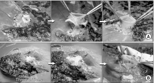
(2) Yoo SY, et al. Conjoined unification venoplasty. 24 months from July 2014 to December 2015. During this. HIGHLIGHTS. study period, 21 patients were selected. Recipients show-. ∙ Portal vein reconstruction using the conjoined unification venoplasty technique appears to be effective in preventing portal vein complications. ∙ It is a useful technique to reconstruct right liver grafts with multiple portal vein orifices.. ing hepatocellular recurrence were excluded to avoid unnecessary bias. The participants underwent regular follow-ups at our institution’s outpatient clinic. We collected follow-up data until December 2018 or until their deaths making the observation period >3 years. This study protocol was approved by the Institutional Review. of the PV branch. To resolve this issue, we developed. Board of our institution (IRB No. 2018-1386).. a conjoined unification venoplasty (CUV) method which is a technical modification of conventional autologous PYG. Surgical Techniques of CUV. interpositions [7,8]. This study presents the long-term. The surgical techniques for CUV included excising a. outcomes of CUVs for reconstructions of double PV or-. 5-mm-long segment of the autologous sectional PV. ifice grafts in a high-volume LDLT center.. branch creating a 5×12-mm-sized venous patch. When a PV patch was not available, an autologous greater sa-. METHODS. phenous venous patch was used. This patch was inserted as a central intervening venous patch between the two. Study Design. sectional PV orifices with small niches. Crutch-opened. This was a retrospective single-arm study regarding the. autologous PYGs were anastomosed to the unified PV or-. outcomes of CUV for reconstruction of double PV orifice. ifice grafts creating a potbelly-shaped PV confluence. grafts. The primary purpose of this study was to present. (Fig. 1). Details of these procedures have been pre-. the 3-year actual patency rates of PV reconstructions. sented elsewhere [7,8].. using CUV. The secondary purpose was to present the long-term morphometric changes in PV reconstructions. Morphometric Evaluations of PV Reconstructions. using CUVs.. by CUVs. CUVs were first applied to LDLTs in July 2014 at Asan. According to our LDLT management protocol, Doppler. Medical Center. Therefore, the study period was set to. ultrasonography was performed each day during the first. Fig. 1. Illustration of the conjoined unification venoplasty technique. (A) First, the recipient’s autologous portal vein (PV) Y-graft was harvested and the crutch of the PV Y-graft was opened (arrows) creating a funnel-shaped vessel graft. (B) A central vein patch is attached between the two sectional PV orifices of the right liver graft, which converts the two PV orifices to one large PV orifice. The crutch-opened PV graft is anastomosed to the unified PV graft. Dotted lines indicate the approximation points for end-to-end anastomosis. (C) The final configuration of the unified PV makes a potbelly-shaped PV confluence. This conjoined PV confluence provides a wider range of tolerance for malalignment (bidirectional arrow) and PV size mismatches.. 107.
(3) Korean J TransplantㆍDecember 2019ㆍVolume 33ㆍIssue 4. week after LDLT, and once or twice per week thereafter. tients; model for end-stage liver disease scores, 15.3±. during hospitalization. Post-transplant dynamic com-. 6.4; ABO blood-incompatible LDLTs, 2; and graft-re-. puted tomography (CT) scans were routinely performed. cipient weight ratio, 1.12±0.21.. every week while the patients were in the hospital, and. Recipient PYGs were harvested in all cases. For the. at 1, 3, 6, and 12 months after LDLTs. Thereafter, fol-. central intervening vein patch, a PV segment was used. low-up abdominal CT scans were repeated annually for. in six cases and an autologous greater saphenous vein. 5 years and biannually after 5 years. In this study, we. patch was used in the remaining 15 cases (Figs. 2 and 3).. reconstructed the shape of the PV system using portal. All living donors were blood-relatives or relatives. phase CT images in a three-dimensional mode and com-. through marriage with type III PV anomalies, in which. pared their serial changes for the first 3 years after. the right anterior and posterior PV branches were sepa-. LDLTs.. rated by a rectangular gap [4,5]. The number of right liver graft PV orifices was two in 19 cases and three in. Statistical Analysis. two cases. All of the PV orifices were unified into one. All numerical data were presented as mean values with. orifice using the CUV technique.. standard deviations. The patency rates were determined using the Kaplan-Meier method. Statistical analyses. Long-Term Patency and Morphometric Changes of. were performed using IBM SPSS ver. 22.0 (IBM Corp.,. PV Reconstructions by CUV. Armonk, NY, USA).. During a mean follow-up of 4 years, the 21 patient cohort displayed a 100% 4-year patient survival rate. None of. RESULTS. them underwent any PV interventions including interventional stenting. Three-dimensional reconstruction of the. Patient Profiles. recipient PVs using serial follow-up CT scans showed. The clinical profiles of 21 patients who underwent LDLTs. progressive streamlined reshaping (Fig. 3). The PV con-. using a right liver graft were as follows: mean age,. fluence portion was over-expanded at portal reperfusion;. 51.7±4.9 years; male to female ratio, 15:6; primary di-. however, within a few weeks, it showed streamlined con-. agnoses: hepatitis B viral infections in 14 patients, hep-. figuration with the entire reshaping process occurring over. atitis C viral infections in two patients, alcoholic liver. several months. Thereafter, the stable reshaped config-. disease in three patients, and other diseases in two pa-. uration of the reconstructed PV maintained for 3 years.. Fig. 2. Operative photographs of the conjoined unification venoplasty technique applied to the two and three portal vein (PV) orifices. (A) The two PV orifices were unified by a central vein patch from a short PV segment. The crutch-opened autologous PV Y-graft is anastomosed to make a single PV orifice. (B) The three PV orifices were unified by a Yshaped incised central vein patch from an autologous greater saphenous vein segment. The crutch-opened autologous PV Y-graft is attached to anastomose this enlarged PV graft orifice.. 108.
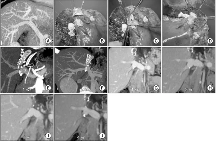
(4) Yoo SY, et al. Conjoined unification venoplasty. Fig. 3. Sequential changes of the recipient portal vein (PV) reconstructed by the conjoined unification venoplasty technique. (A) The donor PV showed a type III anomaly. (B-D) Two graft PV orifices were unified by a conjoined unification venoplasty. (E-J) Computed tomography portal phase follow-up images showed slight expansion of the anastomotic confluence during the first week, which was reshaped at 1 month. There was no noticeable configurational changes in the streamlined reshaped PV after 1 year.. DISCUSSION. depending on the surgeons’ preference and donor PV anatomies. Based on our experience, primary indications. Following its first introduction in 2001, an autologous. for CUVs appear to be a combination of one small and. PYG interposition has been the standard procedure for. one large sectional PV orifice, the presence of a small. grafts with double PV orifices because it is technically. accessory PV branch, a long extrahepatic PV branch, and. simplistic and has excellent long-term outcomes [1,5].. the poor conditions of autologous PYGs. In cases with. An autologous PYG interposition is simple and intuitive.. multiple PV grafts (>2) or severely damaged recipient. However, in practice, it is often technically demanding. PYGs, CUV should be adopted preferentially because. and has occasionally resulted in stenoses because of ana-. these conditions are highly disadvantageous if conven-. tomical variations and discrepancies between the recipi-. tional autologous PYG interposition is considered [9].. ent and graft PVs. To resolve such issues, we developed. Typically, we performed intraoperative cine-portograms. a CUV method to improve the technique of conventional. to determine the shape of the reconstructed PV when. PYG interposition. Long-term outcomes of PV re-. PYG interpositions were undertaken. It also enabled us. constructions by CUV were excellent and showed no. to detect and manage the spontaneous portosystemic col-. complications for >3 years in this study.. laterals [10]. We believe that CUVs or similar techniques. In our institution, conventional autologous PYG inter-. are important components of standardized modified right. positions and CUVs have been concurrently performed. lobe grafts in order to minimize vascular complications in. 109.
(5) Korean J TransplantㆍDecember 2019ㆍVolume 33ㆍIssue 4. ACKNOWLEDGMENTS. adult LDLTs [11,12]. The only disadvantage of CUVs compared with conventional PYG interpositions is the complexity of the. Conflict of Interest. bench work. If surgeons fully understand the primary. No potential conflict of interest relevant to this article. mechanisms of CUV reconstruction, the surgical design. was reported.. for CUV may not be difficult. The most critical technical point is to ensure that the PV confluence portion is suffi-. Funding/Support. ciently large, which automatically resolves anatomical. This study was supported by the intramural research fund. variations and discrepancies between the recipient and. of Asan Medical Center Organ Transplantation Center. graft PVs. The technical principles of CUV are markedly. (2018-01).. similar to those of the quilt venoplasty technique used for. This study was supported by research grant from the. outflow vein reconstructions of the extended right lobe. Korean Society for Transplantation (2019-04-01002-. grafts [13,14]. PV reconstructions with CUVs do not re-. 005).. quire any anticoagulant or antiplatelet agents after LDLTs.. ORCID Sung Yeon Yoo. https://orcid.org/0000-0002-3145-9354. autologous PYGs, such as long autologous greater saphe-. Shin Hwang. https://orcid.org/0000-0002-9045-2531. nous venous patches or fresh cold-stored homologous. Tae-Yong Ha. https://orcid.org/0000-0001-9932-0212. venous patches. We do not recommend using cry-. Gi-Won Song. https://orcid.org/0000-0002-4235-0434. opreserved venous grafts for PV replacements because. Dong-Hwan Jung. https://orcid.org/0000-0001-5984-023X. they can induce aneurysmal dilatation or shrinkage [15].. Gil-Chun Park. https://orcid.org/0000-0003-1631-3258. The primary mechanism of CUV reconstruction is a. CUVs can be performed using vein sources other than. Chul-Soo Ahn. https://orcid.org/0000-0002-3844-3646. gradual reshaping of the dual PVs into one single PV ac-. Deok-Bog Moon. https://orcid.org/0000-0002-8209-3540. cording to hemodynamic principles. Follow-up CT im-. Ki-Hun Kim. https://orcid.org/0000-0002-4016-0995. ages revealed that the luminal configuration of the unified. Young-In Yoon. https://orcid.org/0000-0002-9308-0366. PV reconstruction with CUV was markedly similar to that. Yo-Han Park. https://orcid.org/0000-0002-2242-0968. obtained using a single PV reconstruction. This indicates. Hui-Dong Cho. https://orcid.org/0000-0001-8501-3385. that the PV confluence portion usually over-expands at. Yong-Kyu Chung. https://orcid.org/0000-0002-2132-2450. the time of portal reperfusion, but shortly thereafter, re-. Sang-Hyun Kang. https://orcid.org/0000-0002-8518-1941. shapes and streamlines according to the principles of. Jin-Uk Choi. https://orcid.org/0000-0001-8078-0593. hemodynamics. This reshaping process began within a few. Sung-Gyu Lee. https://orcid.org/0000-0001-9161-3491. weeks, stabilized within several months, and continued for >3 years. We expect that this reshaped configuration. Author Contributions. will continue to be stable over the life of the patient.. Conceptualization: SH. Data curation: SYY, TYH, GWS,. In conclusion, PV reconstructions using the CUV tech-. DHJ, GCP, CSA, DBM, KHK, YIY, YHP, HDC, YKC,. nique appear to be significantly effective in preventing PV. SHK, JUC. Formal analysis: SH, TYH. Funding acquis-. complications. We believe that the CUV method is a use-. ition: SH. Methodology: SH, DHJ. Project admin-. ful technical option for reconstruction of right liver grafts. istration: SH, SGL. Visualization: SH. Writing - original. with multiple PV orifice grafts during LDLTs because it. draft: SH, SYY. Writing - review & editing: SH.. can overcome the disadvantages of conventional autologous PYG interpositions.. REFERENCES 1. Hwang S, Lee SG, Lee YJ, Sung KB, Park KM, Kim. 110.
(6) Yoo SY, et al. Conjoined unification venoplasty. KH, et al. Lessons learned from 1,000 living donor liver. 9. Kwon JH, Hwang S, Song GW, Moon DB, Park GC,. transplantations in a single center: how to make living. Kim SH, et al. Conjoined unification venoplasty for triple. donations safe. Liver Transpl 2006;12:920-7.. portal vein branches of right liver graft: a case report. 2. Marcos A, Orloff M, Mieles L, Olzinski A, Sitzmann J. Reconstruction of double hepatic arterial and portal venous branches for right-lobe living donor liver transplantation. Liver Transpl 2001;7:673-9.. and technical refinement. Korean J Hepatobiliary Pancreat Surg 2016;20:61-5. 10. Moon DB, Lee SG, Ahn C, Hwang S, Kim KH, Ha T, et al. Application of intraoperative cine-portogram to de-. 3. Thayer WP, Claridge JA, Pelletier SJ, Oh CK, Sanfey. tect spontaneous portosystemic collaterals missed by in-. HA, Sawyer RG, et al. Portal vein reconstruction in right. traoperative doppler exam in adult living donor liver. lobe living-donor liver transplantation. J Am Coll Surg 2002;194:96-8.. transplantation. Liver Transpl 2007;13:1279-84. 11. Hwang S, Ahn CS, Kim KH, Moon DB, Ha TY, Song. 4. Lee SG, Hwang S, Kim KH, Ahn CS, Park KM, Lee. GW, et al. Standardization of modified right lobe grafts. YJ, et al. Approach to anatomic variations of the graft. to minimize vascular outflow complications for adult living. portal vein in right lobe living-donor liver transplantation.. donor liver transplantation. Transplant Proc 2012;44:. Transplantation 2003;75(3 Suppl):S28-32.. 457-9.. 5. Hwang S, Lee SG, Ahn CS, Kim KH, Moon DB, Ha. 12. Hwang S, Ha TY, Ahn CS, Moon DB, Kim KH, Song. TY, et al. Technique and outcome of autologous portal. GW, et al. Standardized surgical techniques for adult living. Y-graft interposition for anomalous right portal veins in. donor liver transplantation using a modified right lobe. living. graft: a video presentation from bench to reperfusion.. donor. liver. transplantation.. Liver. Transpl. 2009;15:427-34.. Korean J Hepatobiliary Pancreat Surg 2016;20:97-101.. 6. Lee HJ, Hwang S, Ahn CS, Kim KH, Moon DB, Ha TY,. 13. Hwang S, Lee SG, Ahn CS, Moon DB, Kim KH, Ha TY,. et al. Long-term outcomes of portal Y-graft interposition. et al. Outflow vein reconstruction of extended right lobe. for anomalous right portal veins in living donor liver. graft using quilt venoplasty technique. Liver Transpl. transplantation. Transplant Proc 2012;44:454-6.. 2006;12:156-8.. 7. Ha TY, Hwang S, Moon DB, Ahn CS, Kim KH, Song. 14. Hwang S, Ha TY, Ahn CS, Moon DB, Song GW, Kim. GW, et al. Conjoined unification venoplasty for graft dou-. KH, et al. Hemodynamics-compliant reconstruction of. ble portal vein branches as a modification of autologous. the right hepatic vein for adult living donor liver trans-. Y-graft interposition. Liver Transpl 2015;21:707-10.. plantation with a right liver graft. Liver Transpl 2012;. 8. Hwang S, Ha TY, Song GW, Jung DH, Moon DB, Ahn. 18:858-66.. CS, et al. Conjoined unification venoplasty for double por-. 15. Sugawara Y, Makuuchi M, Tamura S, Matsui Y, Kaneko. tal vein branches of right liver graft: 1-year experience. J, Hasegawa K, et al. Portal vein reconstruction in adult. at a high-volume living donor liver transplantation center.. living donor liver transplantation using cryopreserved vein. J Gastrointest Surg 2016;20:199-205.. grafts. Liver Transpl 2006;12:1233-6.. 111.
(7)
수치

관련 문서
• We report a case of successful pediatric deceased do- nor liver transplantation for symptomatic congenital absence of the portal vein with whole liver graft from a hepatitis
Funneling venoplasty for anomalous graft left hepatic vein in living donor liver transplantation using a split left lateral section graft for an infant patient.. Jung-Man Namgoong
Introduction: The anastomosis between the left renal vein and graft portal vein is one of the crucial options in patients with end-stage liver disease with portal venous thrombosis
Middle hepatic vein (MHV) reconstruction with vas- cular graft interposition has been accepted as a standard procedure for living donor liver transplantation (LDLT) using
Surgical complications after living donor liver transplantation in patients with biliary atresia: a relatively high incidence of portal vein complications. Ueda M, Egawa H, Ogawa
During operation, portal vein anastomosis of the right lobe graft was performed using an interposing cadaveric iliac vein graft and the right gastroepiploic artery was anastomosed
This study aimed to evaluate donor safety in LDLT using caudal middle hepatic vein trunk preserved right lobe (CMPRL) graft.. Methods: LDLT using MRL grafts were performed on
Single center experience of 39 patients with pre- operative portal vein thrombosis among 404 adult living donor liver transplantations.. Selection of donors and recipients for
