• 검색 결과가 없습니다.

Living donor liver transplantation in a pediatric patient with congenital absence of the portal vein

N/A
N/A
Protected

Academic year: 2021

Share "Living donor liver transplantation in a pediatric patient with congenital absence of the portal vein"

Copied!
7
0
0

로드 중.... (전체 텍스트 보기)

전체 글

(1)

Living donor liver transplantation in a pediatric patient with congenital absence of the portal vein

Jung-Man Namgoong

1

, Shin Hwang

1

, Gil-Chun Park

1

, Hyunhee Kwon

1

, Kyung Mo Kim

2

, Seak Hee Oh

2

1

Department of Surgery, Asan Medical Center, University of Ulsan College of Medicine, Seoul, Korea,

2

Department of Pediatrics, Asan Medical Center, University of Ulsan College of Medicine, Seoul, Korea

Case Report

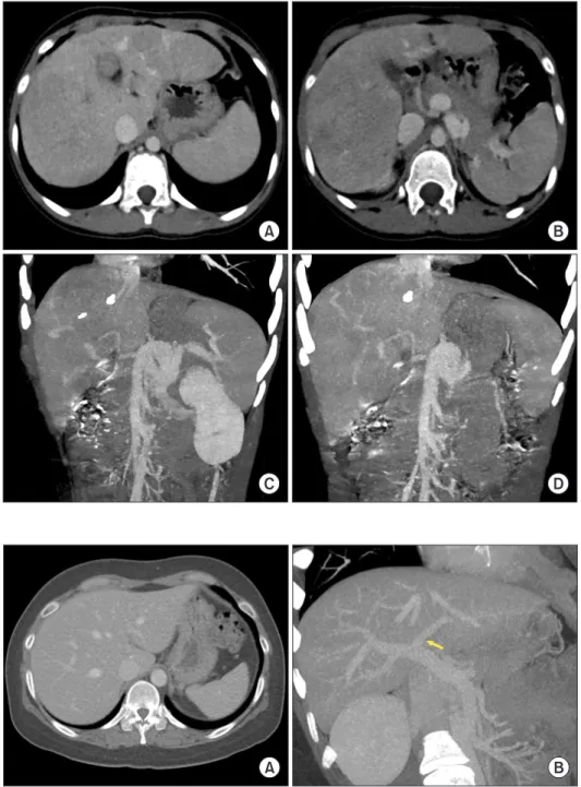
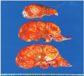
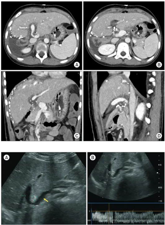
Congenital absence of the portal vein (CAPV) is a rare venous malformation in which mesenteric venous blood drains directly into the systemic circulation. We report a case of pediatric living donor liver transplantation (LDLT) for CAPV combined with focal nodular hyperplasia (FNH) and hepatocellular adenoma. A 9-year-old girl who had been diagnosed with multiple FNH had CAPV. Her blood ammonia level was raised to 137 µg/dL. However, she did not complain of any symptoms. To treat CAPV and FNH, we decided to per- form LDLT. The graft was a left liver graft from 39-year-old mother of the patient. Recipient hepatectomy was performed according to standard procedures of pediatric LDLT. Portal vein reconstruction was performed using interposition of an iliac vein homograft conduit to the superior mesenteric vein-splenic vein confluence. The CAPV-associated congenital splenorenal shunt was securely li- gated. The pathology report of the explant liver showed a 2 cm-sized hepatocellular adenoma and multiple FNH lesions measuring up to 7.1 cm. The patient recovered uneventfully from the LDLT operation. The reconstructed portal vein was maintained well without any hemodynamic abnormalities. In conclusion, as CAPV patients can have various vascular anomalies, combined vascular anomalies should be thoroughly assessed before and during liver transplantation operation. The most effective reconstruction techniques should be used to achieve satisfactory results following liver transplantation.

Key Words: Portal vein agenesis; Splenorenal shunt; Interposition graft; Iliac vein homograft; Liver tumor

INTRODUCTION

Congenital absence of the portal vein (CAPV) is a rare ve- nous malformation in which the mesenteric venous blood drains directly into the systemic circulation. The majority of patients with CAPV show no signs or symptoms of porto- systemic encephalopathy. They only show slightly abnormal results in liver function tests. Liver transplantation (LT) is indicated for patients with symptomatic CAPV refractory to medical treatments [1-4].

The congenital portocaval shunt (PCS) drains the entire

mesenteric venous blood either directly into the inferior vena cava (IVC) or the left renal vein. Thus, theoretically, there is no portal hypertension or collateral circulation [3-5]. As the liver with CAPV does not have sufficient portal inflow, the hepatic arterial flow is the main inflow of blood. If a patient cannot tolerate a medical treatment, LT should be taken into account for such a case. For patients with CAPV, their liver function profiles are not severely impaired with resultant low Pediatric End-stage Liver Disease scores. Because of the low chance for deceased donor liver transplantation in the current Korean setting, patients with CAPV need to consider living donor liver transplantation (LDLT). We herein present a case of pediatric LDLT using a left liver graft for CAPV and liver tumors.

CASE

A 9-year-old girl was referred to our hospital due to the diag- nosis of multiple liver mass. These liver lesions were diagnosed with focal nodular hyperplasia (FNH) on imaging studies and percutaneous liver biopsy. There was no abnormal finding on the diagnostic exome sequencing. The patient was prematurely

Received: February 2, 2021, Revised: February 11, 2021, Accepted: February 14, 2021

Corresponding author: Shin Hwang

Department of Surgery, Asan Medical Center, University of Ulsan College of Medicine, Olympic-ro 43-gil 88, Songpa-gu, Seoul 05505, Korea

Tel: +82-2-3010-3930, Fax: +82-2-3010-6701, E-mail: shwang@amc.seoul.kr ORCID: https://orcid.org/0000-0002-9045-2531

Copyright Ⓒ The Korean Association of Hepato-Biliary-Pancreatic Surgery

This is an Open Access article distributed under the terms of the Creative Commons Attri- bution Non-Commercial License (http://creativecommons.org/licenses/by-nc/4.0) which permits unrestricted non-commercial use, distribution, and reproduction in any medium, provided the original work is properly cited.

(2)

procedures of pediatric LDLT. As the recipient’s native portal measuring 400 g at the back table was recovered, yielding a

Fig. 1. Pretransplant computed tomography findings of the recipient. (A, B) Multiple liver tumors can be seen. (C, D) The portal vein is absent, which is compatible with the congenital absence of the portal vein.

A B

C D

Fig. 2. Preoperative computed tomography findings of the living donor. (A) The volume of the left liver is measured to be 508 mL by volumetry. (B) The diameter of the left portal vein appears unusually small (arrow).

A B

(3)

graft-to-recipient ratio of 1.7%. The graft left portal vein was unusually small, measuring only 7 mm in diameter. The graft hepatic vein was separated into three openings, which were unified into a 3 cm-wide single orifice through unification venoplasty (Fig. 3D).

After the native liver was removed, the hepatic vein orifices at the recipient IVC were unified with venoplasty using a cryo- preserved saphenous vein homograft patch because the right hepatic vein trunk was hypoplastic (Fig. 4A). The vein branch- es at the SMV-SV confluence portion were securely clamped (Fig. 4B) and a 1.5 cm-long longitudinal incision was made at the confluence portion (Fig. 4C). A 4 cm-long cold-stored iliac vein homograft conduit was anastomosed to the SMV-SV confluence in an end-to-side fashion using 6-0 Prolene (Fig.

4D–4F).

The graft hepatic vein orifice was anastomosed with the size- matched recipient IVC orifice (Fig. 5A, 5B). The interposed ili- ac vein conduit was anastomosed with the graft portal vein (Fig.

5C, 5D). Because the graft portal vein was much smaller than the conduit, a small niche was made each at the anterior and posterior walls of the graft portal vein to facilitate wide anasto- mosis. Thereafter, graft reperfusion was initiated. The spleno- renal shunt was securely ligated (Fig. 5E), and then marked in- crease in the portal blood flow was observed (Fig. 5F). Surgical microscopy was used for graft hepatic artery reconstruction.

Intraoperative direct portography was performed through a jejunal vein branch, in which the portal blood flow was well

maintained (Fig. 6A). An enlarged collateral vein through the inferior mesenteric vein and the lumbar vein was identified (Fig. 6B) and ligated. Roux-en-Y hepaticojejunostomy was used for biliary reconstruction.

The pathology report of the explant liver showed two types of tumors. The first was a 2 cm-sized hepatocellular adenoma of beta-catenin mutated subtype at segment IV. Another one comprised of multiple (> 10) FNH lesions measuring up to 7.1 cm in size that scattered over both lobes, indicating multiple FNH syndrome. There were also multiple regenerative nodules measuring up to 2.7 cm in size in both lobes (Fig. 7).

The patient recovered uneventfully from the LDLT operation.

The reconstructed portal vein was maintained well without any hemodynamic abnormalities (Fig. 8, 9). This patient has been doing well for the past 6 months after the LDLT.

DISCUSSION

CAPV is a rare venous malformation in which mesenteric ve- nous blood drains directly into the systemic circulation. There are two types of congenital PCS: intrahepatic PCS and extra- hepatic PCS. Intrahepatic PCS is localized between the portal and hepatic veins [6]. Extrahepatic PCS is divided into type I and type II according to intrahepatic portal venous supply [7].

Type I PCS is an extrahepatic shunt without patent intrahepat- ic portal vein. Thus, the entire mesenteric venous blood drains directly into the systemic vein such as the IVC or the left renal Fig. 3. Intraoperative photographs for recipient hepatectomy and graft bench work. (A) The hepatic artery branches are meticulously dissected after transection of the common bile duct. (B) The retropancrea- tic space is dissected and the left renal vein (yellow vessel loop) and the insertion site of the splenorenal shunt (black silk sling) are isolated. (C) The confluence portion of the mesentero-splenic vein is meticulously dissected. (D) The graft hepatic veins are separated into three openings, which are unified to make a wide single orifice.

A B

C D

(4)

vein. This type is called CAPV. Type II PCS is an extrahepatic shunt with patent intrahepatic portal vein. Thus, the patent portal vein perfuses the liver and the shunt vessel drains some mesenteric venous blood into the systemic circulation. Our patient had type I PCS because there was no portal venous structure and all splanchnic blood flow was drained through the splenorenal shunt.

The standard treatment for CAPV has not been established yet. Although PCS can be accompanied by hyperammonemia, the majority of patients with PCS have no signs of encepha- lopathy, as shown in the present case. Such patients only show slightly abnormal results in liver function tests. The majority of patients with CAPV receive conservative medical treatment for hyperammonemia, whereas only a small portion of patients with CAPV require surgical treatments including LT. Surgi- cal treatment is indicated when hyperammonemia or porto- systemic encephalopathy is refractory to medical treatment.

CAPV is a venous malformation in which mesenteric venous blood drains directly into the systemic circulation. It might be accompanied by hepatopulmonary syndrome. Surgical treat- ment for CAPV can also be indicated for hepatopulmonary syndrome [8,9].

Pretransplant imaging studies in CAPV patients have usually demonstrated a large communication vein to the IVC through the splenorenal shunt, thus no evidence of portal hypertension has been observed in the imaging studies or intraoperative findings. However, we have previously reported an atypical case of CAPV showing portal hypertension with gastric and esophageal varix, which was the primary cause that led to LT [10].

Most patients with type I PCS are indicated for LT as surgi- cal reconstruction of portal vein structures of the native liver is impossible, as in our patient. Although LT for symptomatic CAPV has been reported in the literature [1-3,5,9,11-13], tech- niques for portal vein reconstruction have not been well estab- lished yet. There are two methods of portal vein reconstruction in LT for CAPV. The first method is to anastomose the PCS directly to the graft portal vein in an end-to-end fashion [2,11].

The second method is to use a venous interposition graft through an end-to-side anastomosis to the PCS [3,5]. As the present case lacked any portal vein stump, direct anastomosis was technically impossible. Thus, we used vein conduit inter- position as an end-to-side anastomosis to the PCS. The prereq- uisite for reconstruction with a vein conduit is the availability Fig. 4. Intraoperative photographs of the hepatic vein and portal vein venoplasty. (A) The hepatic vein orifices at the recipient inferior vena cava are unified with venoplasty using a cryopreserved saphenous vein patch. (B) The vein branches at the mesentero-splenic confluence portion are securely clamped. (C) A 1.5 cm-long longitudinal incision is made at the confluence portion. (D–F) A 4 cm-long fresh-stored iliac vein conduit is anastomosed to the confluence portion in an end-to-side fashion.

A B C

D E F

(5)

of an adequate vein allograft. To obtain suitable vein homo- graft, we had to wait for one month before the LDLT operation.

There is a strong association between the development of liver masses such as FNH and hepatocellular adenomas in patients with CAPV. This association has been hypothesized to be related to the lack of portal vein flow, which can lead to compensatory arterial hyperperfusion and the formation of regenerative nodules. Although rare, these neoplasms may degenerate to hepatocellular carcinoma [14-18]. Imaging char- acteristics of these hepatic neoplasms can be challenging due to the absence of intrahepatic portal systems and their associ-

Fig. 5. Intraoperative photographs of graft implantation. (A, B) The graft hepatic vein orifice is anastomosed with the size-matched recipient hepatic vein orifice. (C, D) The portal iliac vein conduit is anastomosed with the graft portal vein. (E) The congenital splenorenal shunt is securely ligated. (F) Portal blood flow is increased after ligation of the splenorenal shunt.

A B C

D E F

Fig. 6. Intraoperative direct portography findings. (A) Portal blood flow is well maintained. (B) A collateral vein through the inferior mesenteric vein and the lumbar vein is identified and ligated.

A B

Fig. 7. Gross photographs of the explant liver. There are three types of

liver nodules. A 2 cm-sized hepatocellular adenoma of beta-catenin

mutated subtype is located at segment IV. Multiple focal nodular

hyperplasia nodules measuring up to 7.1 cm in size are scattered over

both lobes. Multiple regenerative nodules measuring up to 2.7 cm in

size are also present in both lobes.

(6)

ated portal venous or delayed imaging patterns. Apparently, a biopsy of these lesions is critical in cases showing an interval growth or a change in enhancement pattern [14-18].

In conclusion, as CAPV patients can have various vascular anomalies, such combined vascular anomalies should be thor- oughly assessed before and during the LT operation. The most effective reconstruction techniques should be used to achieve satisfactory results following LT.

CONFLICT OF INTEREST

No potential conflict of interest relevant to this article was reported.

ORCID

Jung-Man Namgoong, https://orcid.org/0000-0002-9237-7440 Shin Hwang, https://orcid.org/0000-0002-9045-2531 Gil-Chun Park, https://orcid.org/0000-0003-1631-3258 Hyunhee Kwon, https://orcid.org/0000-0001-6647-9155

A B

C D

Fig. 8. Posttransplant computed tomo- graphy scan taken at four days after the transplantation. (A, B) The graft portal vein reconstruction appears smooth and streamlined. (C, D) The iliac vein conduit from the mesentero-splenic vein junction is well visualized as a newly made main portal vein.

A B

Fig. 9. Posttransplant Doppler ultrasono- graphy at 21 days after transplantation.

(A) The contour of portal vein anastomosis appears smooth and streamlined with slight stenosis at the anastomosis site. Arrow indicates the interposed iliac vein conduit.

(B) The portal vein blood flow is well

maintained.

(7)

Kyung Mo Kim, https://orcid.org/0000-0001-7896-6751 Seak Hee Oh, https://orcid.org/0000-0002-9672-8877

AUTHOR CONTRIBUTIONS

Conceptualization: JMN, SH. Data curation: KMK, SHO.

Methodology: JMN, SH, GCP, HK. Visualization: SH. Writing - original draft: JMN, SH. Writing - review & editing: All au- thors.

REFERENCES

1. Sanada Y, Mizuta K, Kawano Y, Egami S, Hayashida M, Wakiya T, et al. Living donor liver transplantation for congenital absence of the portal vein. Transplant Proc 2009;41:4214-4219.

2. Shinkai M, Ohhama Y, Nishi T, Yamamoto H, Fujita S, Take H, et al.

Congenital absence of the portal vein and role of liver transplantation in children. J Pediatr Surg 2001;36:1026-1031.

3. Wojcicki M, Haagsma EB, Gouw AS, Slooff MJ, Porte RJ. Orthot- opic liver transplantation for portosystemic encephalopathy in an adult with congenital absence of the portal vein. Liver Transpl 2004;10:1203-1207.

4. Ikeda S, Sera Y, Ohshiro H, Uchino S, Uchino T, Endo F. Surgical indications for patients with hyperammonemia. J Pediatr Surg 1999;34:1012-1015.

5. Sumida W, Kaneko K, Ogura Y, Tainaka T, Ono Y, Seo T, et al. Living donor liver transplantation for congenital absence of the portal vein in a child with cardiac failure. J Pediatr Surg 2006;41:e9-e12.

6. Watanabe A. Portal-systemic encephalopathy in non-cirrhotic pa- tients: classification of clinical types, diagnosis and treatment. J Gas- troenterol Hepatol 2000;15:969-979.

7. Morgan G, Superina R. Congenital absence of the portal vein: two cases and a proposed classification system for portasystemic vascular anomalies. J Pediatr Surg 1994;29:1239-1241.

8. Emre S, Arnon R, Cohen E, Morotti RA, Vaysman D, Shneider BL.

Resolution of hepatopulmonary syndrome after auxiliary partial orthotopic liver transplantation in Abernethy malformation. A case

report. Liver Transpl 2007;13:1662-1668.

9. Soejima Y, Taguchi T, Ogita K, Taketomi A, Yoshizumi T, Uchiyama H, et al. Auxiliary partial orthotopic living donor liver transplan- tation for a child with congenital absence of the portal vein. Liver Transpl 2006;12:845-849.

10. Namgoong JM, Hwang S, Kim DY, Ha TY, Song GW, Jung DH, et al. Pediatric liver transplantation using a hepatitis B surface anti- gen-positive donor liver graft for congenital absence of the portal vein. Korean J Transplant 2021;35:59-65.

11. Woodle ES, Thistlethwaite JR, Emond JC, Whitington PF, Vogelbach P, Yousefzadeh DK, et al. Successful hepatic transplantation in con- genital absence of recipient portal vein. Surgery 1990;107:475-479.

12. Ohnishi Y, Ueda M, Doi H, Kasahara M, Haga H, Kamei H, et al. Suc- cessful liver transplantation for congenital absence of the portal vein complicated by intrapulmonary shunt and brain abscess. J Pediatr Surg 2005;40:e1-e3.

13. Charre L, Roggen F, Lemaire J, Mathijs J, Goffette P, Danse E, et al.

Hematochezia and congenital extrahepatic portocaval shunt with ab- sent portal vein: successful treatment by liver transplantation. Trans- plantation 2004;78:1404-1406.

14. Benedict M, Rodriguez-Davalos M, Emre S, Walther Z, Morotti R.

Congenital extrahepatic portosystemic shunt (Abernethy malforma- tion type Ib) with associated hepatocellular carcinoma: case report and literature review. Pediatr Dev Pathol 2017;20:354-362.

15. Virdis M, Monteleone M, Sposito C, Cascella T, Pellegrinelli A, Maz- zaferro V. Hepatocellular carcinoma in Abernethy malformation: a rare occurrence of congenital complete portosystemic shunt. J Vasc Interv Radiol 2018;29:1775-1778.

16. Christou N, Dib N, Chuffart E, Taibi A, Durand-Fontanier S, Valleix D, et al. Stepwise management of hepatocellular carcinoma associat- ed with Abernethy syndrome. Clin Case Rep 2018;6:930-934.

17. Chiang J, Chiu HK, Moriarty JM, McWilliams JP. Hyperandrogenism and malignant degeneration of hepatic adenomas in the setting of Abernethy malformation. Radiol Case Rep 2020;15:2701-2705.

18. Rajeswaran S, Johnston A, Green J, Riaz A, Thornburg B, Mouli S, et

al. Abernethy malformations: evaluation and management of con-

genital portosystemic shunts. J Vasc Interv Radiol 2020;31:788-794.

수치

Fig. 1. Pretransplant computed tomography  findings of the recipient. (A, B) Multiple  liver tumors can be seen
Fig. 6. Intraoperative direct portography findings. (A) Portal blood flow  is well maintained
Fig. 8. Posttransplant computed tomo- tomo-graphy scan taken at four days after the  transplantation

참조

관련 문서

(1) Infuse fluid through a large vein: subclavian vein (high osmolarity) (2) Ensure proper placement of the line (chest X-ray after line insertion) (3) Precisely regulate

The index is calculated with the latest 5-year auction data of 400 selected Classic, Modern, and Contemporary Chinese painting artists from major auction houses..

1 John Owen, Justification by Faith Alone, in The Works of John Owen, ed. John Bolt, trans. Scott Clark, "Do This and Live: Christ's Active Obedience as the

Note that the boundary with the smallest misorientation is made up of a row of dislocations, whereas the high-angle boundaries have a disordered structure in

As the manufacturing technology of a laser welding equipment supplying directly a high density heat energy in the joining part, which is different from

Purpose: Calcaneal fracture is a rare fracture, which accounts for about 2% of all fractures, but is one of the most common fractures in the ankle bone.. There is

systemic circulation, in the right ventricle and oxygenated blood from the lungs, or pulmonary circulation, in the left ventricle, as in birds and mammals.. Two vessels,

• directly ancestral to the earliest members of living species Homo sapiens.. • probably the first human to live in