ORIGINAL ARTICLE
DOI: 10.4174/jkss.2011.81.1.35
JKSS
Journal of the Korean Surgical Society pISSN 2233-7903ㆍeISSN 2093-0488
Received October 29, 2010, Accepted May 17, 2011 Correspondence to: Dong Lak Choi
Division of Hepatobiliary and Transplantation Surgery, Department of Surgery, Catholic University of Daegu School of Medicine, 3056-6 Daemyeong 4-dong, Nam-gu, Daegu 705-718, Korea
Tel: +82-53-650-4063, Fax: +82-53-650-4950, E-mail: [email protected]
cc Journal of the Korean Surgical Society is an Open Access Journal. All articles are distributed under the terms of the Creative Commons Attribution Non-Commercial License (http://creativecommons.org/licenses/by-nc/3.0/) which permits unrestricted non-commercial use, distribution, and reproduction in any medium, provided the original work is properly cited.
An early single-center experience of portal vein thrombosis in living donor liver transplantation:
clinical feature, management and outcome
Joo Dong Kim, Dong Lak Choi, Young Seok Han
Division of Hepatobiliary and Transplantation Surgery, Department of Surgery, Catholic University of Daegu School of Medicine, Daegu, Korea
Purpose: Portal vein thrombosis (PVT) has been considered a relative contraindication for living donor liver transplantation (LDLT). However, it is no longer a contraindication of LDLT due to improvement in surgical techniques and approaches to PVT. The aim of this study was to assess the impact of PVT on outcomes in LDLT patients. Methods: We retrospectively ana- lyzed the data from 97 adult patients undergoing LDLT in our center from July 2008 to June 2010. Intraoperative findings and preoperative imaging results were reviewed for PVT grading (Yerdel grading). We analyzed the technical aspects and com- parisons of risk factors, perioperative variables, and survivals between patients with and without PVT based on the grades.
Results: In the 97 LDLT patients, 18 patients were confirmed to have PVT (18.5%) including grade I cases (n = 8), grade II (n = 7), and grade III (n = 3). Prior treatment of portal hypertension was found to be an independent risk factor for PVT (P = 0.001).
The comparisons between PVT and no PVT groups showed no significant difference in intraoperative and postoperative var- iables except for postoperative bleeding (P = 0.036). The short-term portal vein patency, in-hospital mortality and survival rates were not significantly different between the PVT and control groups. Conclusion: The outcomes are similar to non-PVT group in terms of in-hospital mortality, survival rates, and postoperative complications. Therefore, our study suggests that PVT cannot be considered to be a contraindication for LDLT and LDLT could be undertaken without increased morbidity and mortality in patients with PVT, in spite of operative complexity.
Key Words: Portal vein, Thrombosis, Liver transplantation, Outcome assessment
INTRODUCTION
In the early period of liver transplantation (LT), portal vein thrombosis (PVT) was considered a contraindication for operation because of the technical difficulties it en- tailed, especially the inability to gain an adequate portal supply [1-3]. In 1985, Shaw et al. [4] reported the first suc-
cessful deceased donor liver transplantation (DDLT) for a PVT patient and since then, many innovative surgical techniques have been introduced such as thrombectomy, portal vein (PV) reconstruction using vein grafts, and cav- oportal hemitransposition [5-9]. Current PVT patients are no longer regarded to be contraindicated for LT and the re- sults for patients with PVT is now comparable to that of
patients without PVT [1,2,9,10]. Nevertheless, PVT is con- sidered to add high risk to LT because of the complexity of LT procedures, resulting in significant surgical morbidity and mortality [10,11].
In living donor liver transplantation (LDLT), the issue of subjecting a healthy donor to potentially significant morbidity and mortality has led to a critical reassessment of the recipient selection criteria that are considered ac- ceptable in DDLT [12]. In addition, there are some techni- cal difficulties due to these innovations for preexisting PVT patients; necessity of distal dissection of vascular pedicle of the hilum and restricted availability of deceased donor vein graft [10,11].
Therefore, based on greater technical difficulties and the results from DDLT in this group of patients, the pres- ence of PVT has often been considered to be a relative or absolute contraindication in LDLT [13,14].
From this point of view, the purpose of this report is to analyze single-center experiences in management of PVT, and to assess the impact of PVT on the outcomes in adult LDLT patients.
METHODS
We retrospectively studied the records of 97 LDLT pa- tients using data collected prospectively among 109 cases of consecutive adult LDLT performed at our center from July 2008 to June 2010. In our center, PVT has not been a contraindication for LDLT except where the preoperative radiologic studies demonstrate a gross tumor thrombus in the main portal vein. All PVT patients were diagnosed us- ing abdominal computed tomography (CT) performed within 3 months before transplantation, and intra- operative detection. For standardization purposes, 12 pa- tients were excluded for the following reasons: 1) no avail- ability of preoperative and postoperative CT scans, 2) in- sufficient records of the intraoperative findings, or 3) tu- mor thrombus confirmed by postoperative pathology.
PV flow was monitored routinely after transplantation by Doppler ultrasound at post-transplant days 1,4, and 7, and dynamic CT scan at days 14 and 21.
PVT was diagnosed preoperatively and/or intra-
operatively in 18 cases (18.5%). These patients with pre- operatively and/or intraoperatively confirmed PVT formed the study group. Patients without PVT and transplanted during the same period were used as the control group.
All patients with confirmed PVT were retrospectively classified into four grades according to the extent of thromboses, as described by Yerdel et al. [15]: Grade I:
minimally or partially thrombosed PV, in which the thrombus is mild or, at the most, confined to <50% of the vessel lumen with or without minimal extension into the superior mesenteric vein (SMV). Grade II showed >50%
occlusion of the PV, including total occlusion with or with- out minimal extension into the SMV. Grade III were com- plete thromboses of both PV and proximal SMV with an open distal SMV. Grade IV was complete thrombosis of the portal vein as well as the proximal and distal SMV.
The preoperative, intraoperative, and postoperative pa- rameters in these two groups were compared. The patients with PVT and those with non-PVT were followed up for a median time of 15.3 months (range, 1.2 to 36.5 months) and 14.9 months (range, 2.4 to 36.3 months).
Assessment of risk factors for PVT and outcomes analysis
Analyzed potential risk factors for PVT included; age, sex, primary disease or Child-Turcott-Pugh (CTP) score, the average model for end-stage liver disease (MELD) sore, preoperative platelet count, preoperative pro- thrombin time, living donor characteristics, quality of do- nated liver, previous treatment for portal hypertension (splenectomy, shunt operation, transjugular intrahepatic portosystemic shunt [TIPS], or sclerotherapy), and pres- ence of malignancy.
Surgery time, amount of operative red blood cell (RBC) transfusion, duration of hospital and intensive care unit (ICU) stays were analyzed. Postoperative complications (postoperative bleeding, biliary complication, PVT or stenosis, hepatic artery complication, infection, rejection) were compared. In-hospital mortality and patient survival were compared according to the presence of PVT.
Statistical analysis
Continuous variables are represented as a mean ± SD
Table 1. Recipient, donor, and graft characteristics of LDLT in patients with and without PVT
PVT (n = 18) Non-PVT (n = 79) P-value
Age (yr) 50.8 ± 7.3 50.5 ± 8.2 0.791
Recipient gender (M:F) 14:4 57:22 0.627
Primary disease
HBV/HCV related liver cirrhosis without HCC 9 (50.0) 28 (35.4) 0.289
HBV/HCV related liver cirrhosis with HCC 5 (27.8) 31 (39.2) 0.428
Alcoholic liver cirrhosis 2 (11.1) 12 (15.2) 0.99
Othersa) 2 (11.1) 8 (10.2) 0.99
CTP score 9.1 ± 1.8 8.7 ± 2.8 0.620
MELD score 17.3 ± 8.1 17.7 ± 10.3 0.831
Ascites (mL) 1,261.1 ± 1,440.2 1,383.5 ± 1,892.8 0.964
Preoperative platelet (×103/μL) 55.1 ± 31.3 68.9 ± 27.7 0.243
Preoperative prothrombin time (INR) 1.88 ± 0.63 1.92 ± 0.88 0.639
Previous treatment of portal HTN (Y:N) 6:12 5:74 0.014
Donor age (yr) 30.0 ± 7.7 27.7 ± 7.9 0.238
Donor gender (M:F) 10:9 58:22 0.159
Graft fatty change (%) 1.11 ± 3.66 2.72 ± 5.23 0.111
Graft-to-recipient weight ratio (%) 0.98 ± 0.19 1.06 ± 0.25 0.151
Type of graft
Single (right/left liver) 13:4 70:8 0.129
Dual 1 1 0.338
Values are presented as mean ± SD or number (%).
LDLT, living donor liver transplantation; PVT, portal vein thrombosis; HBV, hepatitis B virus; HCV, hepatitis C virus; HCC, hepatocellular carcinoma; CTP, Child-Turcott-Pugh; MELD, the average model for end-stage liver disease; INR, international normalized ratio; HTN, hypertension; Y, yes; N, no.
a)Cryptogenic liver cirrhosis, fulminent hepatic failure, primary biliary cirrhosis.
and categorical data were analyzed using the Fisher’s ex- act test. A Mann-Whitney U test was used to compare the group means. The Kaplan-Meier method was used to es- timate survival curves, and survival curves were com- pared by means of the log-rank test. All analyses were carried out using SPSS ver. 14.0 (SPSS Inc., Chicago, IL, USA). A P-value less than 0.05 was considered significant.
RESULTS
Incidence and grading of PVT
PVT was diagnosed in 18 of the total 97 adult patients (18.5%) who underwent LDLT including 8 cases (44.4%) of grade I; 7 (38.9%) of grade II; 3 (16.7%) of grade III.
Preoperative risk factors for PVT (Table 1)
Age, gender, primary disease, CTP score, MELD score, presence of hepatocellular carcinoma (HCC), ascites, pre-
operative platelet count, and prothrombin time showed no relationship to PVT (P > 0.05). Living donor character- istics and quality of donated liver had no significant differ- ences between the two groups. However, previous treat- ment of portal hypertension (splenectomy, shunt oper- ation, TIPS, sclerotherapy) was associated with PVT (P = 0.014).
Intraoperative and postoperative variables be- tween PVT and non-PVT groups (Table 2)
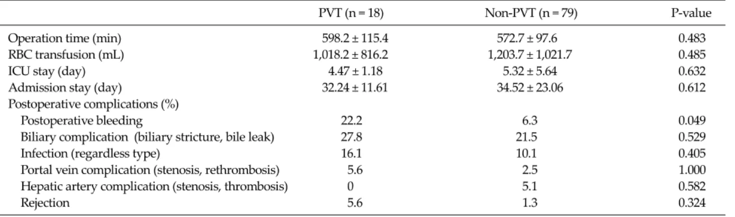
The mean duration of operation in the patients with PVT (598.2 ± 115.4 minutes) was longer than in those with- out PVT (572.6 ± 97.6 minutes), but this was not statisti- cally significant (P = 0.483). The overall mean RBC trans- fusion requirement in the patients with PVT (1,018.2 ± 816.2 mL) was not significantly different to that in those without PVT (1,203.7 ± 1,021.7 mL, P = 0.485). The mean ICU stay was similar in both groups (4.47 ± 1.12 days vs.
5.32 ± 5.6 days, P = 0.632). The mean hospital stay was also similar (32.2 ± 11.6 days vs. 34.5 ± 23.1 days, P = 0.612).
Table 2. Comparative analysis of operative and postoperative variables with or without PVT
PVT (n = 18) Non-PVT (n = 79) P-value
Operation time (min) 598.2 ± 115.4 572.7 ± 97.6 0.483
RBC transfusion (mL) 1,018.2 ± 816.2 1,203.7 ± 1,021.7 0.485
ICU stay (day) 4.47 ± 1.18 5.32 ± 5.64 0.632
Admission stay (day) 32.24 ± 11.61 34.52 ± 23.06 0.612
Postoperative complications (%)
Postoperative bleeding 22.2 6.3 0.049
Biliary complication (biliary stricture, bile leak) 27.8 21.5 0.529
Infection (regardless type) 16.1 10.1 0.405
Portal vein complication (stenosis, rethrombosis) 5.6 2.5 1.000
Hepatic artery complication (stenosis, thrombosis) 0 5.1 0.582
Rejection 5.6 1.3 0.324
PVT, portal vein thrombosis; RBC, red blood cell; ICU, intensive care unit.
Fig. 1. Survival curves in the portal vein thrombosis (PVT) and non-PVT groups (A), and in the PVT and non-PVT groups after excluding malignancy (B).
Postoperative complications of PVT patients (Table 2)
The incidence of postoperative bleeding in the patients with PVT was significant higher than in those without PVT (22.2% vs. 6.3%, P = 0.049). But, the rate of portal vein rethrombosis or stenosis, hepatic artery complication, in- fection, biliary complication (bile leak, biliary stricture), and rejection were similar in the PVT and non-PVT groups.PV patency and patient’s survival of PVT group
The PV patency of the PVT group in their follow-up pe- riod was similar to the non-PVT group. Only one of the pa- tients with PVT developed a rethrombosis. A relapar- otomy and intraoperative PV stenting were performed atthe 13th day after transplantation.
The in-hospital mortality for patients with PVT was similar to that of patients without PVT (5.6% vs. 3.8%, P
= 0.548). The 1-and 3-year actuarial survival rate in the PVT group were 87.7% and 75.2%, respectively, but there was no statistical difference between the PVT group and the non-PVT groups (log-rank test, P = 0.357) (Fig. 1A).
Considering the influence of HCC on survival, we ex- cluded patients with cancer from both groups, leaving 36 patients in the PVT group and the controls, still there was no significant difference between the two groups (log-rank test, P = 0.979) (Fig. 1B).
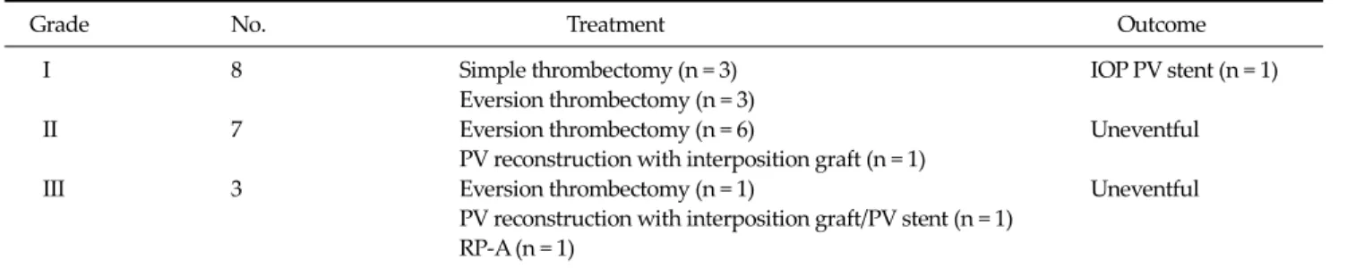
Table 3. Surgical procedures for thrombectomy and reconstruction as grade
Grade No. Treatment Outcome
I 8 Simple thrombectomy (n = 3) IOP PV stent (n = 1)
Eversion thrombectomy (n = 3)
II 7 Eversion thrombectomy (n = 6) Uneventful
PV reconstruction with interposition graft (n = 1)
III 3 Eversion thrombectomy (n = 1) Uneventful
PV reconstruction with interposition graft/PV stent (n = 1) RP-A (n = 1)
IOP, intraoperative; PV, portal vein; RP-A, renoportal anastomosis.
Fig. 2. Portal vein (PV) reconstruction with interposition vein graft in case of failed eversion thrombectomy. Preoperative computed tomography (CT) scan of this case showed that portal vein is completely obstructed (white arrow) and PV thrombosis propagated to portomesenteric junction (A). Interposition vein graft was used for portal vein reconstruction (B) and intraoperative PV stenting (black arrow) was performed due to residual thrombus (C). Patency of interposition graft was demonstrated by postoperative CT scan (D).
Operative management
Surgical management of PVT was dependent on the characteristics of the thrombus; its size (partial or com-
plete) or extension degree through the portal venous system. Surgical procedures for PVT are shown in Table 3.
Most patients with grade I and II thrombosis were man-
Fig. 3. Portal vein (PV) reconstruction with renoportal anas- tomosis. Abdominal computed tomography (CT) scan showed complete portal vein thrombosis extended to proximal superior mesenteric vein (arrow) and marked dilated splenorenal shunt drained into the left renal vein (arrow head) (A). Interposition graft was anastomosed to upper border of left renal vein and the graft PV is anastomosed to proximal end of the interposition graft (B). No signs of portal vein system stenosis were visible in postoperative abdominal CT scan (C).
aged with classic PV-PV anastomosis with or without sim- ple thrombectomy or eversion thrombectomy. In only one patient with grade II, an interposition iliac vein graft was used as anastomosis between the donor’s PV to the recipi- ent’s proximal portal vein nearby the spleno-portal junc- tion because the hilum had severe fibrotic change due to a previous hepatectomy where the thromboses were not completely removed.
One patient with grade III thrombosis was successfully treated with eversion thrombectomy after lower dis- section and classic PV-PV anastomosis. However, the oth- er two cases failed complete eversion thrombectomy, and had no suitable PV to perform the classic anastomosis. In one of these cases, interposition iliac vein graft was used for PV reconstruction. And, in addition, intraoperative PV stenting was performed because there was residual
thrombus at the spleno-mesenteric junction and proximal SMV (Fig. 2).
In the other case with complete and extended occlusion of the SMA and a large splenorenal shunt, renoportal anas- tomosis was performed in a side-to-end fashion (Fig. 3).
DISCUSSION
The PVT is a well-established complication among pa- tients with end-stage liver disease, and its incidence rang- es from 2 to 26% in various centers [8,14], reaching as high as 39% in certain patient populations [16]. The incidence of PVT in LDLT patients at our center was 18.5%.
The etiology is not completely understood, but is be- lieved to be related to anatomic change in the liver owing
to the cirrhotic process, increased portal pressure, endo- thelial injury or coagulation changes [1,15,17,18]. In past studies, high-risk factors for developing PVT included au- toimmune hepatitis, cryptogenic cirrhosis, chronic active hepatitis, Budd-Chiari syndrome, male gender, increased age, trauma, hypercoagulative states, Child-Pugh C, and treatment of portal hypertension (splenectomy, shunt op- eration, TIPS) [4,14,15,19]. But, this study found that pre- vious treatment of portal hypertension was an in- dependent risk factor for PVT prior to LDLT.
PVT was considered for 2 decades to be an absolute con- traindication for LT [17]. However, in 1985, two successful liver transplantations were reported despite PVT [4]. Since then, progress in LT has allowed surgeons to utilize multi- ple techniques including thrombectomy of native veins, extensive thromboendovenectomy up to splenomesen- teric confluence, venous interposition graft, renoportal anastomosis, cavoportal hemitransposition for over- coming this major problem and restoring PV flow [6,15,20,21]. But, there are some problems with technical difficulties and restricted availability of vein graft in LDLT as of yet [10,11,18]. The ideal surgical technique to resolve PVT during LT is not defined. The treatment depends on the characteristics of thrombosis (whether acute or chron- ic), degree (partial or complete), and especially, degree of extension to the splanchnic venous system [1,17].
In our study, 93.3% (n = 14) of patients with grade I and II PVT were successfully managed by classic PV-PV anas- tomosis with or without simple or eversion throm- bectomy. In only one patient with grade II PVT, we per- formed thrombosed PV resection and portal vein re- construction using interposition vein graft between the donor’s PV and the recipient’s proximal PV close to sple- no-portal junction because of severe porta hepatis fibrotic change and incomplete eversion thrombectomy.
In one grade III PVT patient eversion thrombectomy failed, interposition vein graft was used and intra- operative PV stenting was performed due to residual thrombus at portomesenteric confluence level. And in the other case of grade III PVT with large splenorenal shunt, we experienced portal vein reconstruction using an inter- position iliac vein graft connected to the left renal vein in a side-to-end fashion.
Regarding to blood transfusion, operation time, and in-hospital stay including ICU stay, no statistical differ- ence was observed between the PVT and non-PVT groups in our study, indicating that proper management of PVT is a controllable procedure of LDLT.
The greater technical difficulty in patients with pre-ex- isting PVT has demonstrated an increased risk of compli- cations like hepatic artery thrombosis, relaparotomy, post- operative pancreatitis, sepsis, and renal failure in the dif- ferent studies [9,15,22]. In our center, comparisons of the PVT patients and controls showed no statistical differ- ences except postoperative bleeding. This increased risk is related to pre-existing PV pathology, high blood loss, the development of coagulopathy and severe acidosis [23].
Therefore, meticulous operative procedures and close monitoring for postoperative bleeding are required.
Initially, patients with PVT undergoing LT showed a high mortality rate in some papers [24]. But, recent studies such as that of Lladó et al. [25] have described similar sur- vival in patients with PVT compared to patients without PVT. In our study, the results (in-hospital mortality, 1-and 3-year survival rates) obtained in patients undergoing LDLT with preoperative PVT are not significantly differ- ent to patients without PVT, despite short-term follow-up.
Moreover, PVT of grade II to III can be managed with dif- ferent techniques, with good postoperative results. The re- sults suggest that accurate preoperative evaluation and detailed surgical planning are essential for restoring PV patency in LDLT patients.
In conclusion, the results are similar to non-PVT group in terms of in-hospital mortality, survival rates, and post- operative complications, and PV patency. Therefore, our experience suggests that PVT cannot be considered to be a contraindication for LDLT in spite of operative com- plexity.
In PVT of grade II and III, LDLT could be undertaken successfully with accurate preoperative diagnosis and proper surgical techniques and good postoperative out- comes can be obtained. Furthermore, innovations of ther- apeutic approaches and accumulation of experience could be required to improve the outcomes in LDLT with the more extensive PVT patients.
CONFLICTS OF INTEREST
No potential conflict of interest relevant to this article was reported.
REFERENCES
1. Tao YF, Teng F, Wang ZX, Guo WY, Shi XM, Wang GH, et al. Liver transplant recipients with portal vein thrombosis:
a single center retrospective study. Hepatobiliary Pancreat Dis Int 2009;8:34-9.
2. Pan C, Shi Y, Zhang JJ, Deng YL, Zheng H, Zhu ZJ, et al.
Single-center experience of 253 portal vein thrombosis pa- tients undergoing liver transplantation in China. Trans- plant Proc 2009;41:3761-5.
3. Van Thiel DH, Schade RR, Starzl TE, Iwatsuki S, Shaw BW Jr, Gavaler JS, et al. Liver transplantation in adults.
Hepatology 1982;2:637-40.
4. Shaw BW Jr, Iwatsuki S, Bron K, Starzl TE. Portal vein grafts in hepatic transplantation. Surg Gynecol Obstet 1985;161:66-8.
5. Orlando G, De Luca L, Toti L, Zazza S, Angelico M, Casciani CU, et al. Liver transplantation in the presence of portal vein thrombosis: report from a single center.
Transplant Proc 2004;36:199-202.
6. Molmenti EP, Roodhouse TW, Molmenti H, Jaiswal K, Jung G, Marubashi S, et al. Thrombendvenectomy for organized portal vein thrombosis at the time of liver transplantation.
Ann Surg 2002;235:292-6.
7. Manzanet G, Sanjuán F, Orbis P, López R, Moya A, Juan M, et al. Liver transplantation in patients with portal vein thrombosis. Liver Transpl 2001;7:125-31.
8. Cherqui D, Duvoux C, Rahmouni A, Rotman N, Dhu- meaux D, Julien M, et al. Orthotopic liver transplantation in the presence of partial or total portal vein thrombosis:
problems in diagnosis and management. World J Surg 1993;17:669-74.
9. Dumortier J, Czyglik O, Poncet G, Blanchet MC, Boucaud C, Henry L, et al. Eversion thrombectomy for portal vein thrombosis during liver transplantation. Am J Transplant 2002;2:934-8.
10. Cho JY, Suh KS, Shin WY, Lee HW, Yi NJ, Lee KU.
Thrombosis confined to the portal vein is not a contra- indication for living donor liver transplantation. World J Surg 2008;32:1731-7.
11. Egawa H, Tanaka K, Kasahara M, Takada Y, Oike F, Ogawa K, et al. Single center experience of 39 patients with pre- operative portal vein thrombosis among 404 adult living donor liver transplantations. Liver Transpl 2006;12:1512-8.
12. Trotter JF. Selection of donors and recipients for living do- nor liver transplantation. Liver Transpl 2000;6(6 Suppl 2):S52-8.
13. Kadry Z, Selzner N, Handschin A, Müllhaupt B, Renner EL, Clavien PA. Living donor liver transplantation in pa- tients with portal vein thrombosis: a survey and review of technical issues. Transplantation 2002;74:696-701.
14. Davidson BR, Gibson M, Dick R, Burroughs A, Rolles K.
Incidence, risk factors, management, and outcome of por- tal vein abnormalities at orthotopic liver transplantation.
Transplantation 1994;57:1174-7.
15. Yerdel MA, Gunson B, Mirza D, Karayalçin K, Olliff S, Buckels J, et al. Portal vein thrombosis in adults under- going liver transplantation: risk factors, screening, man- agement, and outcome. Transplantation 2000;69:1873-81.
16. Gayowski TJ, Marino IR, Doyle HR, Echeverri L, Mieles L, Todo S, et al. A high incidence of native portal vein throm- bosis in veterans undergoing liver transplantation. J Surg Res 1996;60:333-8.
17. Ramos AP, Reigada CP, Ataíde EC, Almeida JR, Cardoso AR, Caruy CA, et al. Portal vein thrombosis and liver trans- plantation: long term. Transplant Proc 2010;42:498-501.
18. Kim SJ, Moon IS, Lee MD, Kim DG. Clinical outcome of preoperative portal vein thrombosis in living donor liver transplantation. J Korean Soc Transplant 2007;21:282-90.
19. Nonami T, Yokoyama I, Iwatsuki S, Starzl TE. The in- cidence of portal vein thrombosis at liver transplantation.
Hepatology 1992;16:1195-8.
20. Maluf D, Shim I, Posner M, Cotterell AH, Fisher RA.
Salvage procedure for unexpected portal vein thrombosis in living donor liver transplantation. Transplant Proc 2006;38:1422-4.
21. Paskonis M, Jurgaitis J, Mehrabi A, Kashfi A, Fonouni H, Strupas K, et al. Surgical strategies for liver transplantation in the case of portal vein thrombosis: current role of cav- oportal hemitransposition and renoportal anastomosis.
Clin Transplant 2006;20:551-62.
22. Bertelli R, Nardo B, Montalti R, Beltempo P, Puviani L, Cavallari A. Liver transplantation in recipients with portal vein thrombosis: experience of a single transplant center.
Transplant Proc 2005;37:1119-21.
23. Shaked A, Busuttil RW. Liver transplantation in patients with portal vein thrombosis and central portacaval shunts.
Ann Surg 1991;214:696-702.
24. Seu P, Shackleton CR, Shaked A, Imagawa DK, Olthoff KM, Rudich SR, et al. Improved results of liver trans- plantation in patients with portal vein thrombosis. Arch Surg 1996;131:840-4.
25. Lladó L, Fabregat J, Castellote J, Ramos E, Torras J, Jorba R, et al. Management of portal vein thrombosis in liver trans- plantation: influence on morbidity and mortality. Clin Transplant 2007;21:716-21.