INTRODUCTION
Congenital absence of the portal vein (CAPV) is a rare ve- nous malformation in which the mesenteric venous blood drains directly into the systemic circulation. The majority of patients with CAPV show no signs or symptoms of portosystemic encephalopathy. They only show slightly abnormal liver function tests. Liver transplantation (LT) is
indicated for patients with symptomatic CAPV refractory to medical treatments [1], especially those with hyperam- monemia, portosystemic encephalopathy, hepatopulmo- nary syndrome, hepatic tumors, or intractable complica- tions.
The congenital portocaval shunt (PCS) drains all mes- enteric venous blood either directly into the inferior vena cava (IVC) or the left renal vein. Thus, theoretically there is
Pediatric liver transplantation using a
hepatitis B surface antigen-positive donor liver graft for congenital absence of the portal vein
Jung-Man Namgoong
1, Shin Hwang
1, Dae-Yeon Kim
1, Tae-Yong Ha
1, Gi-Won Song
1, Dong-Hwan Jung
1, Kyung Mo Kim
2, Seak Hee Oh
21Department of Surgery, Asan Medical Center, University of Ulsan College of Medicine, Seoul, Korea
2Department of Pediatrics, Asan Medical Center, University of Ulsan College of Medicine, Seoul, Korea
Congenital absence of the portal vein (CAPV) is a rare venous malformation in which mesenteric venous blood drains directly into systemic circulation. Herein, we report a case of pediatric deceased donor liver transplantation (DDLT) for symptomatic CAPV with whole liver graft from a hepatitis B surface antigen (HBsAg)-positive donor. A 4-year-old boy suffered from CAPV and secondary portal hypertension. He was also diagnosed with DiGeorge syndrome and heart anomalies. After waiting for 4 months, a 5-year-old donor weighing 19 kg with positive HBsAg was allocated to this 4-year-old pa- tient weighing 15 kg. Recipient operation was performed according to the standard pro- cedures of pediatric DDLT. Portal vein reconstruction was performed using interposition of a vascular homograft conduit to the superior mesenteric vein-splenic vein confluence.
The patient recovered uneventfully from DDLT. He has been administered with lamivu- dine to prevent hepatitis B virus infection. This patient has been doing well for 5 years after DDLT without growth retardation. In conclusion, CAPV patients can have various vascular anomalies, thus combined vascular anomalies should be thoroughly assessed before and during liver transplantation operation. The most effective reconstruction techniques should be used to achieve satisfactory results following liver transplantation.
Keywords: Portal vein agenesis; Portacaval shunt; Portal hypertension; Hepatitis B virus;
Preemptive therapy
Received September 4, 2020 Revised October 1, 2020 Accepted October 2, 2020 Corresponding author: Shin Hwang Department of Surgery, Asan Medical Center, University of Ulsan College of Medicine, 88 Olympic-ro 43-gil, Songpa-gu, Seoul 05505, Korea Tel: +82-2-3010-3930 Fax: +82-2-3010-6701 E-mail: [email protected]
© The Korean Society for Transplantation This is an Open Access article distributed under the terms of the Creative Commons Attribution Non-Commercial License (http://creativecommons.org/licenses/
by-nc/4.0/) which permits unrestricted non-commercial use, distribution, and reproduction in any medium, provided the original work is properly cited.
pISSN 2671-8790
eISSN 2671-8804
no portal hypertension or collateral circulation [2]. The liver with CAPV does not have sufficient portal inflow, thus the hepatic arterial flow is the main inflow blood. If a patient cannot tolerate a medical treatment, LT should be taken into account. Although CAPV induces various clinical man- ifestations, liver function profiles of patients with CAPV are not severely impaired. Their Pediatric End-stage Liver Disease scores remain low. Since pediatric patients with CAPV do not have any priority in the waiting list, they have a very low chance of receiving deceased donor liver trans- plantation (DDLT). Herein, we present a case of pediatric DDLT for symptomatic CAPV with a whole liver graft from a hepatitis B surface antigen (HBsAg)-positive pediatric donor.
CASE REPORT
The study protocol was approved by of the Institutional Re- view Board at Asan Medical Center (IRB No. 2020-0836), which waived the requirement for informed consent due to the retrospective nature of this study.
A 4-year-old boy was referred due to melena and hema- tochezia. He was born through a normal full-term delivery.
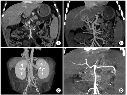
At birth, cardiac murmur was identified. His ventricular and atrial septal defects were increased during a short-term ob- servation. Thus, repair operation was performed at 40 days after birth. Seven days later, he underwent bilateral inguinal hernia operation. At 1 year of age, he underwent anoplasty for abnormal location of anus to the rectum. At 3 years of age, he received supportive care for esophageal and gas- tric varix bleeding. Serum ammonia level was within the normal limit. At 4 years of age, he underwent endoscopic varix ligation to control melena and hematochezia. Com- puted tomography showed agenesis of the portal vein with cavernous transformation and secondary portal hyperten- sion with gastric and esophageal varix (Fig. 1). Liver func- tion test findings were still normal. He was diagnosed with mild tricuspid regurgitation. He was also diagnosed with DiGeorge syndrome (microdeletion 22q11.2). However, oth- er significant anomalies were absent except heart disease and CAPV. This patient was not indicated for Rex shunt HIGHLIGHTS
• We report a case of successful pediatric deceased do- nor liver transplantation for symptomatic congenital absence of the portal vein with whole liver graft from a hepatitis B surface antigen-positive donor.
• This patient has been doing well for 5 years after trans- plantation without growth retardation.
Fig. 1. Pretransplant computed tomography
scan. (A-C) There is agenesis of the portal vein with cavernous transformation and sec- ondary portal hypertension with gastric and esophageal varix. Any large communication vein to the inferior vena cava or left renal vein is not visible. (D) The anatomy of the hepatic artery appears to be normal.
A B
C D
operation because of poor development of the intrahepatic portal vein system. Thus, we decided to perform LT.
After waiting for 4 months in the pediatric waiting list, a 5-year-old donor weighing 19 kg with positive HBsAg was allocated to this 4-year-old patient weighing 15 kg. Liver function test findings of the deceased donor were not highly impaired and significant pathological abnormality was not present on frozen-section liver biopsy. Thus, we decided to use this marginal liver graft because the patient had a very low chance to be allocated for DDLT later. The whole liver graft weighed 580 g and the graft-recipient weight ratio was 3.9%.
Recipient operation was performed according to the standard procedures of pediatric DDLT. The recipient’s native portal vein was absent. Thus, the portal collateral branches at the hepatoduodenal ligament were deeply dis- sected to fully expose the superior mesenteric vein-splenic vein confluence. After deep clamping of this confluence portion with a Satinsky clamp, an external iliac vein seg- ment harvested from the liver donor was anastomosed to the confluence in an end-to-side fashion (Fig. 2).
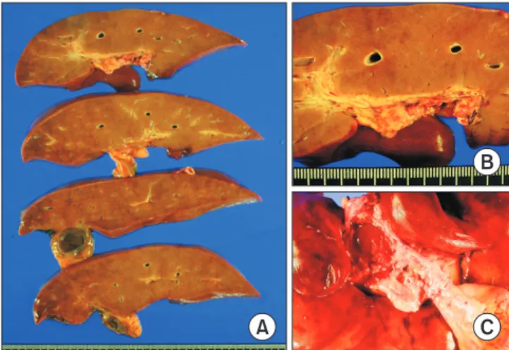
We used a modified piggyback technique making a large cavocaval anastomosis to secure graft outflow vein reconstruction. Graft portal vein was reconstructed with the interposed vein conduit. Surgical microscopy was used for hepatic artery reconstruction. Roux-en-Y hepaticoje- junostomy was used for biliary reconstruction. Coronary collaterals inducing gastric and esophageal varices were ligated. The pathology report of the explant liver showed increased vascularity with variable shapes of portal ve- nous structures and intimal fibrosis, which were compat- ible with the characteristics of CAPV (Fig. 3). Permanent wedge biopsy of the graft liver showed subcapsular and perivenular hepatic necrosis with minimal fatty change and absence of portal inflammation and cholestasis.
The patient recovered uneventfully from the DDLT
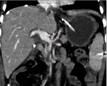
operation. The reconstructed portal vein maintained well without any hemodynamic abnormalities (Figs. 4 and 5).
Clinically suspected acute cellular rejection occurred at 2 weeks after transplantation. It responded well to a steroid pulse therapy. The patient had antibody to HBs through scheduled hepatitis B virus (HBV) vaccination before LT.
During the early posttransplant period, a combination ther- apy of hepatitis B immunoglobulin (HBIG) and lamivudine was used to prevent reactivation of HBV. HBIG 10,000 IU was administered every week during the first month and every month during the next 4 months with a target trough level of 1,000 IU/mL. After 6 months after LT, lamivudine monotherapy was used to prevent HBV infection because of high maintenance of anti-HBs titers. His HBsAg and blood HBV DNA were negative up to date. This patient has been doing well for 5 years after LT without any growth re- tardation.
A B C
Fig. 2. Surgical technique of end-to-side
conduit vein interposition. (A) Anatomy of the superior mesenteric vein-splenic vein confluence is visualized. (B) An external iliac vein graft (cylinder) was anastomosed to the superior mesenteric vein-splenic vein confluence after deep clamping of this confluence portion (blue line). (C) The inter- posed vascular conduit (cylinder) is located between the superior mesenteric vein-splen- ic vein confluence and graft portal vein.
A
B
C
Fig. 3. Gross photographs of the explant liver. (A) The liver parenchymadoes not show cirrhotic changes. (B, C) Magnification of the portal triad
area shows increased vascularity of portal venous structures with vari-
able shapes and intimal fibrosis.
DISCUSSION
CAPV is a rare venous malformation in which mesenteric venous blood drains directly into the systemic circula- tion. There are two types of congenital PCS: intrahepatic PCS and extrahepatic PCS. Intrahepatic PCS is detected between portal and hepatic veins [3]. Extrahepatic PCS is divided into type I and type II according to intrahepatic portal venous supply [4]. Type I PCS is an extrahepatic shunt without patent intrahepatic portal vein. Thus, the entire mesenteric venous blood drains directly into a sys- temic vein such as the IVC or the left renal vein. This type is called CAPV. CAPV can be further subclassified into type Ia (superior mesenteric vein and splenic vein do not
join to form confluence) and type Ib (superior mesenteric vein and splenic vein join to form confluence). If the portal vein presents a lack of complete development, the PCS is either the result of the persistence of the right vitelline vein (in which case the shunt connects to the retrohepatic IVC), or of the persistence of the left vitelline vein (the shunt is connected to the IVC or the right atrium above the conflu- ence of the hepatic veins) [5]. Type II PCS is an extrahe- patic shunt with patent intrahepatic portal vein. Thus, the patent portal vein perfuses the liver and the shunt vessel drains some mesenteric venous blood into the systemic circulation. Our patient had type Ia PCS because no portal venous structures were observed in portal triads.
The standard treatment for CAPV has not been estab- lished yet. Although PCS can be accompanied by hyper- ammonemia, the majority of patients with PCS have no signs of encephalopathy. They only show slightly abnor- mal results in liver function tests. These findings suggest that the development of portosystemic encephalopathy depends on the presence of portosystemic shunting as well as other additional factors, including insufficient re- serve of the liver to detoxify ammonia and/or a decline in neurologic tolerance to hyperammonemia. Therefore, many patients with CAPV receive conservative medical treatment for hyperammonemia. Only a small portion of patients with CAPV require surgical treatments including LT. Surgical treatment is indicated when hyperammonemia or portosystemic encephalopathy is refractory to medi- cal treatment. Surgical treatment for CAPV can also be indicated for hepatopulmonary syndrome [6]. CAPV is a venous malformation in which mesenteric venous blood drains directly into the systemic circulation, thus it might be accompanied by hepatopulmonary syndrome.
Unlike usual symptomatic cases with CAPV in the liter- ature, our patient had portal hypertension with gastric and esophageal varix that might be associated with poor devel-
Fig. 5. Posttransplant computed tomog-
raphy scan taken at 1 month after trans- plantation. (A) The intrahepatic portal vein appears normal. (B) The extrahepatic portal vein is fully expanded with collapse of the collateral veins.
A B
Fig. 4. Posttransplant computed tomography scan taken at 7 days after
transplantation. The portal vein reconstruction appears to be smooth
streamlined with resolution of variceal collaterals. An arrow indicates
the anastomosis site of the interposed vascular conduit and the superior
mesenteric vein-splenic vein confluence.
opment of congenital PCS. Pretransplant imaging studies showed absence of large communication vein to the IVC or the left renal vein. Variceal bleeding secondary to portal hypertension was the primary cause for our patient to un- dergo LT.
The strategy of surgical treatment for CAPV depends on the types of PCS. Most patients with type II PCS can be treated by ligating or banding shunt vessels while moni- toring the portal pressure to avoid the induction of portal hypertension. In contrast, if the intrahepatic portal vein is immature, LT may be required. In other words, most pa- tients with type I PCS are indicated for LT because surgical reconstruction of portal vein structures of the native liver is impossible, like in our patient.
Although LT for symptomatic CAPV has been reported in the literature, the techniques for portal vein reconstruc- tion have not been well established yet. There are two methods of portal vein reconstruction in LT for CAPV. The
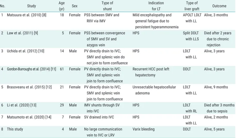
first method is to anastomose the PCS directly to the graft portal vein in an end-to-end fashion [1]. The second meth- od is to use a venous interposition graft through an end-to- side anastomosis to the PCS [2]. Our patient did not have large shunt vein to directly use it for portal vein reconstruc- tion. Thus, the first method was technically impossible. We have accumulated experience of using the second method for pediatric patients with biliary atresia and portal vein hypoplasia [7]. Thus, we applied the second method to our patient. We summarized the reported cases of LT for CAPV in Table 1 [8-14].
A congenital PCS drains all mesenteric venous blood either directly into the IVC or into the left renal vein. Thus, there is no portal hypertension or collateral circulation.
Significant splanchnic congestion may occur when a PCS is totally clamped during portal vein reconstruction, result- ing in severe bowel edema and hemodynamic instability.
We have previously experienced a similar situation during
Table 1. Summary of the reported cases of liver transplantation for congenital absence of the portal vein