108
pISSN 2288-6575 • eISSN 2288-6796 http://dx.doi.org/10.4174/astr.2014.87.2.108 Annals of Surgical Treatment and Research
TECHNICAL ADVANCE
Salvage dual graft living donor liver transplantation after major hepatectomy
Joo Dong Kim, Dong Lak Choi, Young Seok Han
Division of Hepatobiliary Pancreas Surgery and Abdominal Organ Transplantation, Department of Surgery, Catholic University of Daegu School of Medicine, Daegu, Korea
INTRODUCTION
Salvage liver transplantation (LT) has been proposed for patients who undergo primary liver resection for hepatocellular carcinoma (HCC), due to recurrence of HCC, or deterioration in liver function [1,2]. However, the technical feasibility of salvage LT must be considered in patients who have undergone prior liver resection. In particular, prior major hepatectomy such as right hepatectomy makes subsequent recipient hepatectomy and vascular graft reconstruction technically difficult [1,3].
As a result, a few salvage LTs after major hepatectomy have been reported and furthermore, few reports are available on salvage living donor liver transplantation (LDLT) after major hepatectomy due to obvious technical difficulties [1,3]. Herein, we describe a successful case of salvage dual graft LDLT after
major hepatectomy.
SURGICAL TECHNIQUE
A 48-year old man was transferred to Daegu Catholic University Medical Center because of hematemesis. He underwent right hepatectomy due to HCC 4 years ago. Gastroduodenoscopy and CT showed vari ceal bleeding at the third portion of the duodenum and newly developed HCC at segment IV. Thus, we performed a transjugular intra he patic portosystemic shunt and coil embolization for iden tified bleeding duodenal varices fed from the superior pan creaticoduodenal vein. After his condition recovered, we planned LDLT as treatment for intrahepatic recurrence and poor liver function.
The patient’s height was 172 cm and body weight was 65 kg.
Received Janaury 16, 2014, Revised February 20, 2014, Accepted February 20, 2014
Corresponding Author: Dong Lak Choi
Division of Hepatobiliary Pancreas Surgery and Abdominal Organ Transplantation, Department of Surgery, Catholic University of Daegu School of Medicine, 33 Duryugongwon-ro 17-gil, Nam-gu, Daegu 705- 718, Korea
Tel: +82-53-650-4063, Fax: +82-53-650-4950 E-mail: [email protected]
Copyright ⓒ 2014, the Korean Surgical Society
cc Annals of Surgical Treatment and Research is an Open Access Journal. All articles are distributed under the terms of the Creative Commons Attribution Non- Commercial License (http://creativecommons.org/licenses/by-nc/3.0/) which permits unrestricted non-commercial use, distribution, and reproduction in any medium, provided the original work is properly cited.
Salvage living donor liver transplantation (LDLT) after major hepatectomy has been considered a challenging procedure due to operative complexity. We report a successful case of salvage dual graft LDLT after right hepatectomy. A 48-year-old male was transferred to Daegu Catholic University Medical Center because of duodenal variceal bleeding. He underwent right hepatectomy due to hepatocellular carcinoma four years prior. We performed LDLT with dual graft from his wife and sister. During operation, portal vein anastomosis of the right lobe graft was performed using an interposing cadaveric iliac vein graft and the right gastroepiploic artery was anastomosed to the hepatic artery of the left lobe graft. Adequate graft inflow was demonstrated by postoperative imaging studies. He has been doing well with normal graft function for 31 months. Salvage dual graft LDLT could be undertaken successfully in patients with prior major hepatectomy under accurate preoperative planning and proper surgical techniques.
[Ann Surg Treat Res 2014;87(2):108-111]
Key Words: Salvage therapy, Liver transplantation, Living donors, Dual, Hepatectomy
Annals of Surgical Treatment and Research 109 His 41-year-old wife initially volunteered to be a living donor.
But, preoperative 3-dimensional CT estimated right and left lobe volume to be 630 and 200 mL (76% and 24% volume ratio), respectively, which did not meet our criteria for right lobe donation (remnant liver volume > 30% of the entire liver).
Therefore, the other donor candidate, his 44-year-old younger sister, was evaluated. CT-volumetric analysis showed that the right and left lobe volumes were 470 and 240 mL, respectively (66% and 34% volume ratio), which was safe by our criteria.
However, this donor’s right lobe had insufficient safe volume for the recipient: graft-to-recipient weight ratio (GRWR), 0.72%.
Thus, neither of the two available donors was suitable and the decision was made to perform LDLT using dual grafts. We planned to harvest the right lobe of second donor and the left lobe of first donor to ensure donor safety and adequate total graft volume.
Heavy adhesions were encountered between the cut surface and the omentum and intestine due to prior liver resection during the recipient’s operation (Fig. 1A). Thus, sharp dissection and meticulous bleeding control were required to avoid massive bleeding. Mobilizing the remaining liver was the same as that for primary LDLT after fully dissecting the surface adhesions.
The hepatic hilum had severe adhesions and fibrotic changes due to hilar dissection during the prior right hepatectomy. After the hilar dissection, we isolated the only remnant left hepatic artery, which was a very short stump of the right portal vein (PV), and the bile duct (Fig. 1B).
The first liver graft during the donor operations (right lobe:
actual graft weight, 460 g; GRWR, 0.71%) was harvested from the younger sister, and the second liver graft (left lobe: actual graft weight, 211 g; GRWR, 0.32%) was harvested from his wife. The total graft weight was 617 g, and GRWR was 0.95%.
A cryopreserved iliac vein graft was prepared to produce a portal venous conduit for PV reconstruction to the right lobe graft, because there was insufficient right PV to secure a classic anastomosis. This venous conduit was interposed to the PV orifice of the right graft to substitute for the recipient’s right PV on the back table. The reconstruction procedures for dual LDLT with right and left lobes have been described in various reports in detail, including hepatic vein reconstruction for dual grafts [4].
The interposition vein of the right graft PV was anastomosed to the recipient’s right portal stump without kinking (Fig. 2), followed by an end-to-end anastomosis of the left graft PV. The only remnant left hepatic artery was used for hepatic artery reconstruction of the right graft. However, additional inflow was needed for the other graft, so the right gastroepiploic artery (RGEA) was used as arterial inflow for the left graft (Fig. 2). The bile duct of the right graft was reconstructed with duct-to-duct anastomosis, and the bile duct of the left graft was reconstructed with hepaticojejunostomy. Intraoperative and postoperative Doppler ultrasound studies and three- dimensional CT revealed good hepatic artery, PV, and hepatic vein flow to both grafts (Fig. 3). The patient has recovered well with normal graft function and has been doing well for 31 Joo Dong Kim, et al: Dual living donor liver transplantation after major hepatectomy
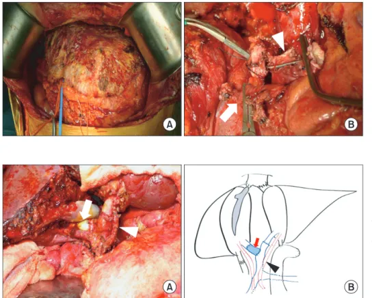
Fig. 1. Intraoperative photographs showing salvage living donor liver transplantation with dual graft.
(A) Severe perihepatic adhesion was exposed after complete mobilization of remnant liver. (B) We isolated the only remnant left hepatic artery (arrow) and short stump of right portal vein above bifurcation (arrowhead) after hilar dissection.
Fig. 2. Intraoperative photograph (A) and illustration (B) of vascular and biliary reconstruction of sal
vage dual graft living donor liver transplantation. Cryo pre served iliac vein conduit was inter posed from portal vein of rightsided graft to right por tal vein stump of recipient (arrow) and recipient’s right gastro epi ploic artery (arrow
head) was ana stomosed to left hepatic artery of leftsided graft.
110
Annals of Surgical Treatment and Research 2014;87(2):108-111
months after LDLT.
DISCUSSION
Salvage LT has been performed as a life-saving treatment for patients with intrahepatic recurrence of HCC or liver function deterioration after primary liver resection [1,5]. However, the technical difficulty during salvage LT and the risk for postoperative complications make most surgeons hesitate to perform LT. Initial clinical studies reported that salvage LT after liver resection is associated with higher operative mortality, increased risk of recurrence, and poorer outcome than those of primary LT [6]. However, recent clinical studies have indicated that salvage LT is a technically feasible procedure [1,3]. Belghiti et al. [7] reported that liver resection prior to LT does not significantly increase the technical difficulty or impair survival after LT. Hwang et al. [1] also concluded that combinations of recipients with a prior hepatectomy and living donor liver grafts for salvage LT are feasible, suggesting that salvage procedures should be extended to LDLT. Although the most recent studies have shown that salvage does not increase the difficulty of surgery, the salvage LT group had a longer operative time, more intraoperative bleeding, and increased transfusion volume, particularly in cases of a prior major hepatectomy [5,8].
Moreover, several technical aspects should be considered when performing a recipient hepatectomy, particularly in cases involving a prior major hepatectomy. The first is in regard to heavy adhesions. Some recipients may have vigorous portal collaterals, which lead to massive intraoperative bleeding and unstable condition [1,3,5]. The second technical concern is for hilar dissection, particularly when hilar dissection has been conducted extensively during major hepatectomy [5]. The third is securing vascular reconstruction for graft inflow and outflow.
A remnant right hepatic artery and PV are usually short or do not exist in cases of prior right hepatectomy [1.3]. Thus, only prior minor hepatectomy has been acceptable for salvage LDLT
in many centers, and few reports have described salvage LDLT after major hepatectomy [1,3]. Furthermore, transplant surgeons might be confronted with more serious technical obstacles during salvage LDLT with dual grafts in patients who had undergone prior major hepatectomy and so there have been only a few reports of dual graft LDLT with prior minor liver resection [1]. Several serious difficulties were encountered in the present case to secure PV and hepatic artery reconstruction because only the left hepatic artery was available, as the native recipient’s hepatic arteries and the very short right PV stump remained after a prior right hepatectomy. We utilized a cryopreserved cadaveric iliac vein graft as a substitute for the lost right PV to secure PV reconstruction to the right graft.
When the native hepatic artery is unavailable, various arterial sources such as the RGEA, the splenic artery, the left gastric artery, the gastroduodenal artery, and other interposition graft has been introduced as good alternative inflow to the graft during LDLT [9,10]. In this case, we performed hepatic artery reconstruction with the RGEA because it provided sufficient length to reconstruct the left graft arterial stumps during tran- splantation.
CONCLUSION
Salvage dual graft LDLT can be a feasible option for a patient with prior major hepatectomy in a situation with inadequate graft size for sufficient graft volume. However, accu rate preoperative planning and experienced surgical techniques lead to a successful operation. In addition, further accumulation of cases is warranted to evaluate the efficacy of complicated procedures like this case.
CONFLICTS OF INTEREST
No potential conflict of interest relevant to this article was reported.
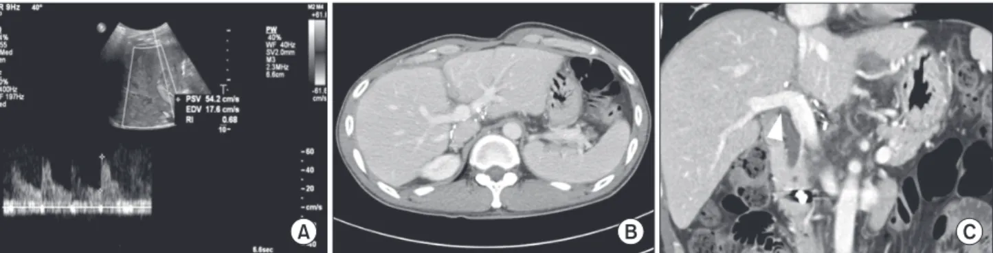
Fig. 3. Postoperative Doppler ultrasonography and CT scan. (A) Doppler ultrasonography showed good hepatic artery flow to leftsided graft using right gastroepiploic artery. (B) Dynamic CT scan on postoperative day 14 demonstrated good portal flow to both grafts. (C) The CT scan on 2 years after transplantation also showed good patency of interposed vein graft (arrowhead) used for portal vein reconstruction.
Annals of Surgical Treatment and Research 111 1. Hwang S, Lee SG, Moon DB, Ahn CS, Kim
KH, Lee YJ, et al. Salvage living donor liver transplantation after prior liver resection for hepatocellular carcinoma. Liver Transpl 2007;13:741-6.
2. Li HY, Wei YG, Yan LN, Li B. Salvage liver transplantation in the treatment of hepatocellular carcinoma: a meta-analysis.
World J Gastroenterol 2012;18:2415-22.
3. Kim BW, Park YK, Kim YB, Wang HJ, Kim MW. Salvage liver transplantation for recurrent hepatocellular carcinoma after liver resection: feasibility of the Milan criteria and operative risk. Transplant Proc 2008;40:3558-61.
4. Moon D, Lee S, Hwang S, Park K, Kim K, Ahn C, et al. Umbilical portion of reci pient's left portal vein: a useful vas-
cular conduit in dual living donor liver transplantation for the thrombosed portal vein. Liver Transpl 2004;10:802-6.
5. Wu L, Hu A, Tam N, Zhang J, Lin M, Guo Z, et al. Salvage liver transplantation for patients with recurrent hepatocellular carcinoma after curative resection. PLoS One 2012;7:e41820.
6. Adam R, Azoulay D, Castaing D, Eshke- nazy R, Pascal G, Hashizume K, et al. Liver resection as a bridge to transplan ta tion for hepatocellular carcinoma on cirrhosis:
a reasonable strategy? Ann Surg 2003;
238:508-18.
7. Belghiti J, Cortes A, Abdalla EK, Regim- beau JM, Prakash K, Durand F, et al. Re- section prior to liver transplantation for hepatocellular carcinoma. Ann Surg 2003;
238:885-92.
8. Hu Z, Wang W, Li Z, Ye S, Zheng SS. Reci- pient outcomes of salvage liver trans- plantation versus primary liver trans- plantation: a systematic review and meta- analysis. Liver Transpl 2012;18:1316-23.
9. Lee JH, Oh DY, Seo JW, Moon SH, Rhie JW, Ahn ST. Versatility of right gastroepiploic and gastroduodenal arteries for arterial reconstruction in adult living donor liver transplantation. Transplant Proc 2011;
43:1716-9.
10. Ahn CS, Hwang S, Moon DB, Song GW, Ha TY, Park GC, et al. Right gastroepiploic artery is the first alternative inflow source for hepatic arterial reconstruction in living donor liver transplantation.
Tran s plant Proc 2012;44:451-3.
REFERENCES
Joo Dong Kim, et al: Dual living donor liver transplantation after major hepatectomy