Cumulative survival rate and associated risk factors of Implantium implants: A 10-year retrospective clinical study
Jin-Hong Park1, Young-Soo Kim2, Jae-Jun Ryu1, Sang-Wan Shin1, Jeong-Yol Lee1*
1Department of Prosthodontics, Institute for Clinical Dental Research, Korea University Medical Center, Korea University, Seoul, Republic of Korea
2Department of Preventive Dentistry, Institute for Clinical Dental Research, Korea University Medical Center, Korea University, Seoul, Republic of Korea
PURPOSE. The objective of this study was to determine the cumulative survival rate (CSR) and associated risk factors of Implantium implants by retrospective clinical study. MATERIALS AND METHODS. Patients who received Implantium implants (Dentium Co., Seoul, Korea) at Korea University Guro Hospital from 2004 to 2011 were included. The period between the first surgery and the last hospital visit until December 2015 was set as the observation period for this study. Clinical and radiographic data were collected from patient records, including all complications observed during the follow-up period. Kaplan-Meier analysis was performed to examine CSR.
Multiple Cox proportional hazard model was employed to assess the associations between potential risk factors and CSR. RESULTS. A total of 370 implants were placed in 121 patients (mean age, 56.1 years; range, 19 to 75 years). Of the 370 implants, 13 failed, including 7 implants that were lost before loading. The 10-year cumulative survival rate of implants was 94.8%. The multiple Cox proportional hazard model revealed that significant risk factor of implant failure were smoking and maxillary implant (P<.05). CONCLUSION. The 10-year CSR of Implantium implants was 94.8%. Risk factors of implant failure were smoking and maxillary implant. [J Adv Prosthodont 2017;9:195-9]
KEYWORDS: Dental implant; Survival rate; Risk factors; Smoking; Arch
INTRODUCTION
To obtain favorable long-term clinical treatment outcomes of implant approach, fast and firm osseointegration with stable alveolar bone must be achieved, even after functional loading. Current implant surface treatment trends aim to
develop an implant surface that is more bioactive and pro- angiogenic in order to achieve better osseointegration.1 As a result of these efforts, a variety of new commercial implant systems have been introduced to the market. Although a large number of clinical studies have reported successful treatment outcomes of dental implants, most long-term (over 10 years) studies have used several common brands of implant systems.2-6 Additionally conflicting data have been reported about the risk factors of implant failure.
Therefore, clinicians are unable to provide concrete answers for patients who underwent failed implant treatments.
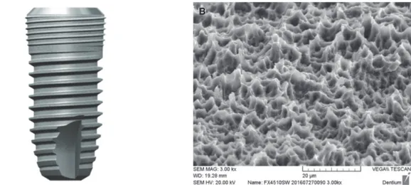
Implantium implants (Dentium Co., Seoul, Korea) is an internal connection type of conical hex with a surface sand- blasted with large grit and acid etched (SLA) (Fig. 1). Since FDA approval of this product in 2004, it has been one of the most widely used implants in South Korea. However, there are only two clinical studies on the product with fol- low-up longer than five years. Lee et al. have reported that the 5-year cumulative survival rate (CSR) of this implant is 97.37%.7 Park et al. have reported that the 10-year CSR of
Corresponding author:
Jeong-Yol Lee
Department of Prosthodontics, Institute for Clinical Dental Research, Korea University Medical Center, Korea University,
148 Gurodong-ro, Guro-gu, Seoul 08308, Republic of Korea Tel: +82226261922: e-mail, wddc@korea.ac.kr
Received August 29, 2016 / Last Revision February 9, 2017 / Accepted March 21, 2017
© 2017 The Korean Academy of Prosthodontics
This is an Open Access article distributed under the terms of the Creative Commons Attribution Non-Commercial License (http://creativecommons.
org/licenses/by-nc/3.0) which permits unrestricted non-commercial use, distribution, and reproduction in any medium, provided the original work is properly cited.
This study was supported by the Korea Health Industry Development Institute (2015-2017 Project No. HI15C0620).
this implant is 97.9% without assessing risk factors associat- ed with implant failure.8
Therefore, the objective of this retrospective study was to determine the 10-year CSR of Implantium implant and to assess the association between implant failure and related risk factors.
MATERIALS AND METHODS
This retrospective study was conducted based on review of the clinical records of all patients who received Implantium implants at the implant clinic of Korea University Guro Hospital Dental Center from June, 2004 to May, 2011.
Implants that underwent splinting with other implant sys- tems were excluded. Records which provided insufficient data presented in clinical charts were excluded as well. As a result, 370 implants in a total of 121 patients were evaluated in this study. The period between the first surgery and the last hospital visit up to December, 2015 was set as the observation period for this study.
All patients were subjected to full thickness flap eleva- tion under local anesthesia. The same surgical procedures were performed for all patients according to the manufac- turer’s recommendations. In 93 of 370 implants, ancillary surgical procedures (such as guided bone regeneration and sinus augmentation) were performed. The final prosthesis was fabricated with the conventional methods depending on the case and was loaded at least two months after the implant placement. For maintenance, regular follow-up vis- its (at least once a year) were scheduled after delivery of definitive prosthesis.
Implant survival was assessed with Albrektsson and Zarb’s criteria.9 In particular, an implant whose function was stably maintained until final observation was considered as survived, whereas removal and sleeping of the implant were
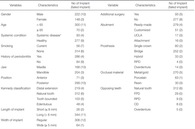
defined as failed. The following nominal and ordinal vari- ables were used to assess implant failure and their associa- tions with implant failure: gender, age (≥ 65 years and < 65 years), systemic chronic disease (diabetes mellitus and/or cardiovascular disease), smoking status (current or none), history of periodontitis, implant location (maxilla, mandible, anterior, posterior), Kennedy classification, implant length and diameter, additional surgery, the kind of abutment, prosthesis type, occlusal materials, and opposing dentition (Table 1).
For statistical analysis, SPSS software version 22.0 was used. The CSR of implants was determined using Kaplan Meier analysis. Difference in CSR according to risk factor was assessed by log rank test (Mantel-Cox) (P = .05). Risk factor variables were included in multiple Cox proportional regression analysis, if preliminary Cox analysis demonstrat- ed at least a statistically borderline significance (P < .15).
RESULTS
A total of 370 implants were placed in 121 patients (66 males and 55 females) during the observation period. The average observation period was 7.2 years (S.D. = 2.4 years).
The average age of these patients was 56.1 ± 10.5 years (range, 19 to 75 years). The distributions of implants according to factor are summarized in Table 1.
Of the 370 implants placed during the observation peri- od, 13 failed. The 10-year CSR of these implants was 94.8%
(Fig. 2).
According to the preliminary analysis, smoking (current vs none) and arch (maxilla vs mandible) were included in multiple Cox proportional regression analysis (P < .15). As a result, smoking and maxillary implant were significant risk factors of implant failure with a hazard ratio of 7.48 and 3.92, respectively (Table 2, P < .05).
Fig. 1. Implantium implant (Dentium, Seoul, Korea). (A) Design of implant, (B) microstructure of implant surface (SEM MAG: 3.00 kx).
A B
Table 1. Distribution of implants according to risk factor variables Variables Characteristics No of Implant
(failed implant) Variable Characteristics No of Implant (failed implant)
Gender Male 222 (10) Additional surgery Yes 93 (5)
Female 148 (3) No 277 (8)
Age < 65 300 (11) Abutment Ready-made 279 (4)
≥ 65 70 (2) Customized 51 (2)
Systemic condition Systemic disease* 93 (4) UCLA 17 (0)
Healthy 277 (9) Attachment 16 (0)
Smoking Current 56 (7) Prosthesis Single crown 60 (2)
None 314 (6) Bridge 252 (2)
History of periodontitis Yes 286 (4) Hybrid 33 (2)
No 84 (9) RPD 4 (0)
Jaw Maxilla 166 (10) Overdenture 14 (0)
Mandible 204 (3) Occlusal material Metal/gold 270 (5)
Position Anterior 71 (3) Porcelain 63 (1)
Posterior 299 (10) Resin 30 (0)
Kennedy classification Distal extension 219 (4) Opposing teeth Natural tooth 312 (6)
Natural tooth 312 (6) FPD 29 (0)
Tooth bounded 103 (5) RPD 9 (0)
Edentulous 48 (4) CD 8 (0)
Length of implant Short (≤ 8 mm) 26 (2) Overdenture 5 (0)
Long (> 8 mm) 344 (11)
Width of implant Regular 306 (12)
Wide (≥ 5 mm) 64 (1)
Systemic disease included diabetes mellitus and/or cardiovascular disease.
Fig. 2. Cumulative survival rate (CSR) of Implantium implants by Kaplan-Meier method.
Table 2. Multivariate associations with cumulative survival rate of implants B (regression
coefficient) P value Exp (B)
(hazard ratio)
Exp (B) 95.0% CI
Lower Upper
Smoking (current vs none) 2.012 .000 7.478 2.492 22.436
Jaw (maxilla vs mandible) 1.365 .038 3.916 1.077 14.235
Significant difference (P < .05).
DISCUSSION
This study showed a 10-year CSR of Implantium implant of 94.8%. Although this result is relatively low compared to that of the Straumann dental implant system (Straumann, Basel, Switzerland) with an SLA surface,4,5 it was a clinically acceptable result. Seven of the 13 failed implants failed before loading, and the other six implants failed after load- ing. Of the 13 failed implants, re-implantation was per- formed for eight cases with consent from the patients. For the remaining five cases, restoration was completed by mod- ifying the prosthetic treatment plan followed by mainte- nance without implant replacement.
A previous 5-year study with similar researchers found various significant factors in implant failure. According to the log-rank test, the significant factors influencing implant failure were occlusal material, prosthesis design, Kennedy classification, arch, reason of tooth loss, smoking and sys- temic diseases.7 However, in this 10-year follow-up study via multiple regression analysis, the results obtained were nar- rowed down and presented high specificity. Hence, the results of this 10-year study showed smoking and implant placement in the maxilla as significant factors in implant failure.
Although some clinical reports have suggested that the advent of rough implant surface has resolved the differenc- es between smokers and non-smokers,10,11 recent meta-anal- ysis studies have shown that smoking is still associated with a higher risk of dental implant failure.12,13 Smoking may inhibit both innate and adaptive responses in a variety of ways. The increase in implant failure caused by smoking is mainly attributed to the effects of nicotine on osteogenesis and angiogenesis.13 Nicotine inhibits gene expression including BMP-2, PDGF-AA, TGF-β, and VEGF, which play important roles in osteoblast proliferation, differentia- tion, and apoptosis.14 Moreover, it induces hyperemia through vasoconstriction and chronic reduction of blood flow and vascularity,15,16 leading to low oxygen and ischemia as well as inadequate blood flow, which ultimately inhibit the normal and/or the healing processes of skeletal struc- tures.17 In this study, current smoking was also found to have significant association with implant failure. Therefore, pre-operative smoking status needs to be addressed for implant treatment. A few investigators have suggested smoking cessation.15,16,18 However, only one clinical study has reported the effectiveness of smoking cessation on implant treatment outcomes.18 More research is needed to draw definitive conclusions regarding the matter.
Previous studies showed contradicting results regarding the association of implant survival rate with arch location of implant.7,19-21 According to a meta-analysis of 54 clinical studies with at least 3-years observation period, the annual implant failure rate of maxillary implants is significantly higher than that of mandibular implants.22 This might be due to the fact that the quality and quantity of jaw bone are more often compromised in the maxillary region than in the mandibular region.23,24 In this present study, the survival rate
of maxillary implants was found to be significantly lower than that of mandibular implants, and it also was revealed in multivariate regression analysis.
This retrospective study has a limitation in the evalua- tion of marginal bone loss with consistency and in its reproducibility due to non-standardized radiographic mea- surements. For this reason, only the survival rate of implants was analyzed without assessing the success rate of implants.
CONCLUSION
Within the limitations of this study, the 10-year CSR of Implantium implants was 94.8%, and smoking and maxillary implant were identified as a significant risk factors of implant failure.
ORCID
Jin-Hong Park https://orcid.org/0000-0002-3220-9912 Sang-Wan Shin https://orcid.org/0000-0002-3100-2020 Jae-Jun Ryu https://orcid.org/0000-0001-6903-5955 Jeong-Yol Lee https://orcid.org/0000-0003-3079-0376 REFERENCES
1. Saghiri MA, Asatourian A, Garcia-Godoy F, Sheibani N. The role of angiogenesis in implant dentistry part I: Review of ti- tanium alloys, surface characteristics and treatments. Med Oral Patol Oral Cir Bucal 2016;21:e514-25.
2. Adell R, Lekholm U, Rockler B, Brånemark PI. A 15-year study of osseointegrated implants in the treatment of the edentulous jaw. Int J Oral Surg 1981;10:387-416.
3. Ekelund JA, Lindquist LW, Carlsson GE, Jemt T. Implant treatment in the edentulous mandible: a prospective study on Brånemark system implants over more than 20 years. Int J Prosthodont 2003;16:602-8.
4. Buser D, Janner SF, Wittneben JG, Brägger U, Ramseier CA, Salvi GE. 10-year survival and success rates of 511 titanium implants with a sandblasted and acid-etched surface: a retro- spective study in 303 partially edentulous patients. Clin Implant Dent Relat Res 2012;14:839-51.
5. van Velzen FJ, Ofec R, Schulten EA, Ten Bruggenkate CM.
10-year survival rate and the incidence of peri-implant disease of 374 titanium dental implants with a SLA surface: a pro- spective cohort study in 177 fully and partially edentulous pa- tients. Clin Oral Implants Res 2015;26:1121-8.
6. Rasmusson L, Roos J, Bystedt H. A 10-year follow-up study of titanium dioxide-blasted implants. Clin Implant Dent Relat Res 2005;7:36-42.
7. Lee JY, Park HJ, Kim JE, Choi YG, Kim YS, Huh JB, Shin SW. A 5-year retrospective clinical study of the Dentium im- plants. J Adv Prosthodont 2011;3:229-35.
8. Park W, Park Y, Park H, Yoo S, Chung S, Han J, Kim SW, Kim DM. A 10-year retrospective radiographic study of implan- tium dental implants. Int J Periodontics Restorative Dent 2015;35:49-54.
9. Albrektsson T, Zarb GA. Current interpretations of the os- seointegrated response: clinical significance. Int J Prosthodont 1993;6:95-105.
10. Balshe AA, Eckert SE, Koka S, Assad DA, Weaver AL. The effects of smoking on the survival of smooth- and rough- surface dental implants. Int J Oral Maxillofac Implants 2008;
23:1117-22.
11. Maló P, de Araújo Nobre M, Gonçalves Y, Lopes A. Long- term outcome of implant rehabilitations in patients with sys- temic disorders and smoking habits: a retrospective clinical study. Clin Implant Dent Relat Res 2016;18:649-65.
12. Renvert S, Quirynen M. Risk indicators for peri-implantitis. A narrative review. Clin Oral Implants Res 2015;26:15-44.
13. Chrcanovic BR, Albrektsson T, Wennerberg A. Smoking and dental implants: A systematic review and meta-analysis. J Dent 2015;43:487-98.
14. Ma L, Zwahlen RA, Zheng LW, Sham MH. Influence of nic- otine on the biological activity of rabbit osteoblasts. Clin Oral Implants Res 2011;22:338-42.
15. Morozumi T, Kubota T, Sato T, Okuda K, Yoshie H.
Smoking cessation increases gingival blood flow and gingival crevicular fluid. J Clin Periodontol 2004;31:267-72.
16. Nair P, Sutherland G, Palmer RM, Wilson RF, Scott DA.
Gingival bleeding on probing increases after quitting smok- ing. J Clin Periodontol 2003;30:435-7.
17. Wang Y, Wan C, Deng L, Liu X, Cao X, Gilbert SR, Bouxsein ML, Faugere MC, Guldberg RE, Gerstenfeld LC, Haase VH, Johnson RS, Schipani E, Clemens TL. The hypoxia-inducible factor alpha pathway couples angiogenesis to osteogenesis during skeletal development. J Clin Invest 2007;117:1616-26.
18. Bain CA. Smoking and implant failure-benefits of a smoking cessation protocol. Int J Oral Maxillofac Implants 1996;11:
756-9.
19. Shigehara S, Ohba S, Nakashima K, Takanashi Y, Asahina I.
Immediate loading of dental implants inserted in edentulous maxillas and mandibles: 5-year results of a clinical study. J Oral Implantol 2015;41:701-5.
20. Moy PK, Medina D, Shetty V, Aghaloo TL. Dental implant failure rates and associated risk factors. Int J Oral Maxillofac Implants 2005;20:569-77.
21. Del Fabbro M, Ceresoli V. The fate of marginal bone around axial vs. tilted implants: a systematic review. Eur J Oral Implantol 2014;7:S171-89.
22. Kern JS, Kern T, Wolfart S, Heussen N. A systematic review and meta-analysis of removable and fixed implant-supported prostheses in edentulous jaws: post-loading implant loss. Clin Oral Implants Res 2016;27:174-95.
23. Truhlar RS, Orenstein IH, Morris HF, Ochi S. Distribution of bone quality in patients receiving endosseous dental implants.
J Oral Maxillofac Surg 1997;55:38-45.
24. He J, Zhao B, Deng C, Shang D, Zhang C. Assessment of im- plant cumulative survival rates in sites with different bone density and related prognostic factors: an 8-year retrospective study of 2,684 implants. Int J Oral Maxillofac Implants 2015;
30:360-71.